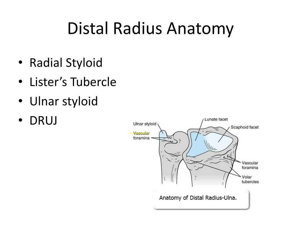
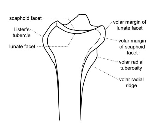
A cadaveric study of the volar distal radius was performed to better understand the anatomy relevant to the volar approach for distal radius fractures. Anatomic features of the distal radius include the styloid process the dorsal tubercle and four surfaces.
 Distal End Radius Fracture
Distal radius fractures broken wrist the radius is the larger of the two bones of the forearm.
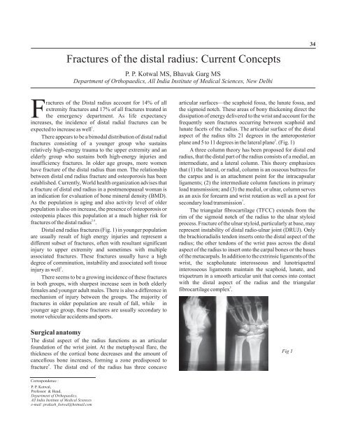
Anatomy of distal radius. General features of distal radius anatomy. A fracture of the distal radius occurs when the area of the radius near the wrist breaks. A distal radius fracture also known as wrist fracture is a break of the part of the radius bone which is close to the wrist.
The bone also forms an ellipsoidal joint with the proximal carpal row that. Drfs in the times of hippocrates and galen were thought to be wrist dislocations. Many papers have been written about them with more than 200 having been published in the first 6 months of 2018 alone.
The wrist may be deformed. About 50000 occur each year in the united states. Three investigators separately or collectively dissected six cadavers and examined nine dried bones.
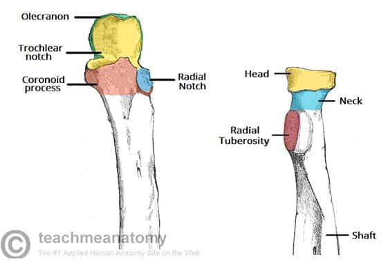
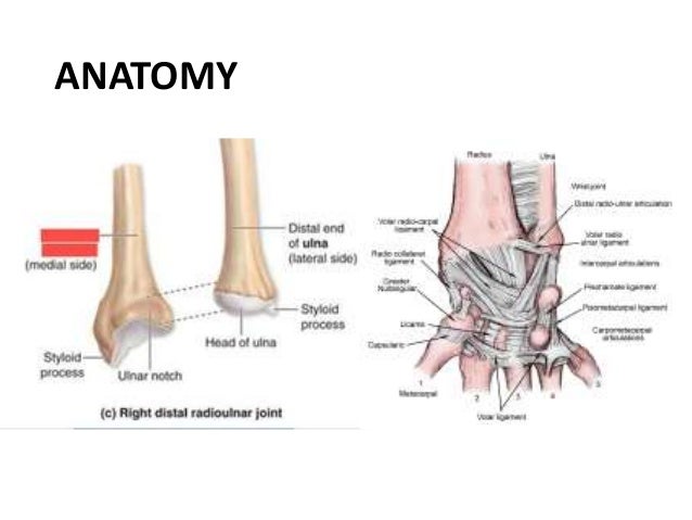
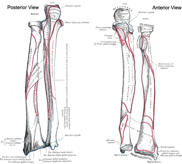
Distal end of radius. The lateral side projects distally as the styloid process. The break usually happens due to falling on an outstretched or flexed hand.
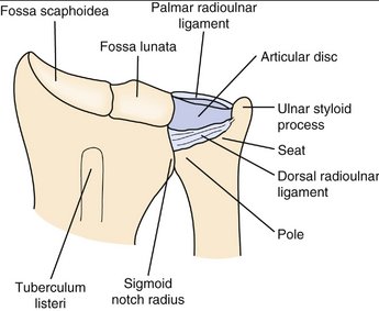
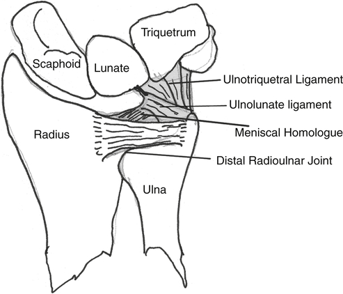
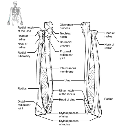
The end toward the wrist is called the distal end. In the distal region the radial shaft expands to form a rectangular end. In the medial surface there is a concavity called the ulnar notch which articulates with the head of ulna forming the distal radioulnar joint.
The distal end which tends to be turned slightly forwards has a somewhat triangular form. The distal portion of the radius has a quadrilateral cross section and includes the metaphyseal and epiphyseal regions. In younger people these fractures typically occur during sports or a motor vehicle collision.
The part of the radius connected to the wrist joint is called the distal radius. Anterior lateral posterior and medial. Symptoms include pain bruising and rapid onset swelling.
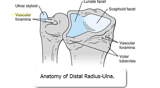
When the radius breaks near the wrist it is called a distal radius fracture. Distal radius fractures drf are common injuries. The inferior distal surface of the lower end of the radius bone provides a lateral triangular area for articulation along with the scaphoid and a medial quadrangular area for articulation with the lateral components of the lunate.
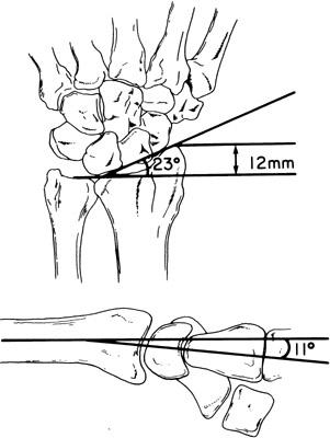
Functions of the radius it forms a hinge joint with the humerus bone which allows us to flex and extend the elbow 7. The radius moves around the ulna at the wrist enabling us to turn our hands palm up and palm down 8. Its distal carpal articular surface concave from before backwards and slightly so from side to side is divided into two facets by a slight antero posterior ridge.
Distal radius fractures are very common. The ulna bone may also be broken. In fact the radius is the most commonly broken bone in the arm.
What is a distal radius fracture. Distal region of the radius. The radius is one of two forearm bones and is located on the thumb side.
 Ppt Distal Radius Fractures Powerpoint Presentation Free
 Anatomy And Biomechanics Of Forearm Rotation Clinical Gate
 X Wrist Startradiology
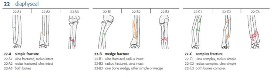
 Distal Forearm Decision Support Ao Surgery Reference

 Fcr Approach To Distal Radius Approaches Orthobullets
 The Radioulnar Joints Teachmeanatomy
 Management Of Wrist Fractures Grabb And Smith S Plastic
 Distal Forearm Decision Support Ao Surgery Reference
 Distal Radius Fractures Everything You Need To Know Dr Nabil Ebraheim
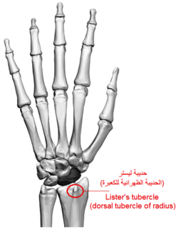 Lister S Tubercle Wikipedia
 Distal Radius Fractures Trauma Orthobullets
Fractures Of The Distal Radius Punjab Orthopaedic Association
 Wrist Fractures Musculoskeletal Key
 Imaging Findings Of The Distal Radio Ulnar Joint In Trauma
 Radius Radiology Reference Article Radiopaedia Org

 Anatomy Of Distal Radius Bone And Spine
 Common Types Of Distal Radius Fractures Nabil Ebraheim
 Anatomy Of Distal Radius Bone And Spine
_moved.gif) Distal Radius Fractures Trauma Orthobullets
Distal Radius Fractures Trauma Orthobullets
 Distal Radius Fracture
 Fracture Of Distal Radius
 Galeazzi Fractures Trauma Orthobullets
 Bones Of The Upper Limb Anatomy And Physiology
 Radius Bone Anatomy Bone And Spine
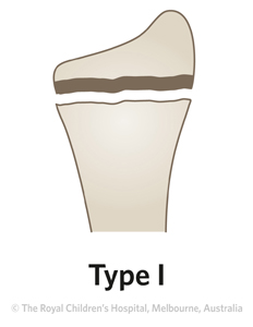 Clinical Practice Guidelines Distal Radial Physeal
 Distal Radius Fracture S52 539a 813 41 Eorif


Posting Komentar
Posting Komentar