Radiographic imaging pearls and pitfalls david e. Gross anatomy articulations the elbow joint is made up of three articulations 23.
 Elbow Xray Anatomy Stock Photos Page 1 Masterfile
This anatomy is increasingly important in evaluating abnormalities such as osteonecrosis of the capitellum panners disease osteochondral defects and medial apophysitis little league elbow for example.

Elbow x ray anatomy. Normal elbow x ray appearances on the lateral image there is often a visible triangle of low density lying anterior to the humerus. This is a normal structure. Play this quiz called elbow xray anatomy and show off your skills.
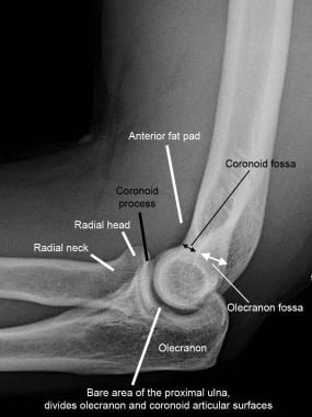
The elbow is a complex synovial joint formed by the articulations of the humerus the radius and the ulna. Grashey shoulder x ray anatomy. Normal radiographic anatomy of the elbow.
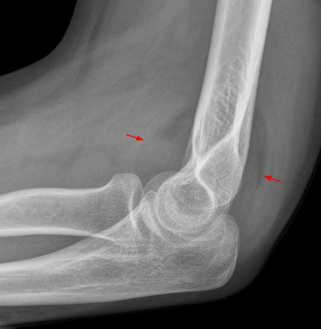
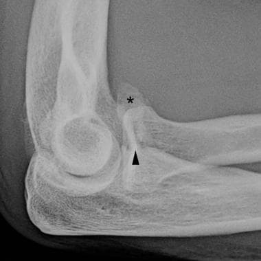
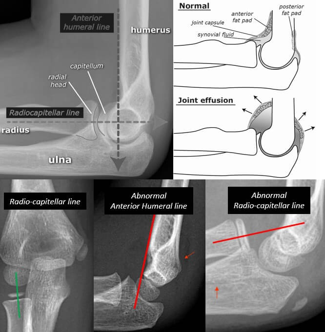
Injuries around the joint can produce a joint effusion which will displace the fat pads making them more visible. Both anterior and posterior fat pad signs exist and both can be found on the same x ray. This is the anterior fat pad which lies within the elbow joint capsule.
Grayson md major usaf mc d iagnostic imaging of the elbow has seen remarkable advances in the last several years. Other games by same author. Position of patient the patient should be seated sideways at the end of the the table.
In order to establish treatment algorithms and evaluate outcomes common and reliable methods of measurement and assessment are necessary. Lateral knee x ray anatomy. Normal radiographic anatomy of the elbow.
Capitellum of the humerus with the ra. Systematic review whenever you look at an adult elbow x ray review. On a normal elbow x ray only a small stripe of an anterior fat pad should be visible.
Normal radiographic anatomy of the elbow. The multiplanar capabilities and increasing availability of magnetic reso nance mr and volumetric multislice computed tomo graphicct. Alignment fat pads bone cortex alignment check the anterior humeral line.
It is caused by displacement of the fat pad around the elbow joint. Drawn down the anterior surface of the humerus should intersect the middle 13 of the capitellu. On an elbow x ray a fat pad sign suggests an occult fracture.
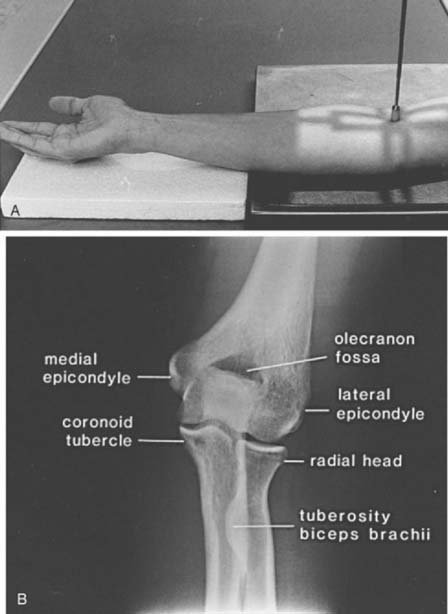
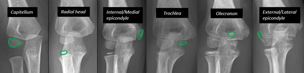
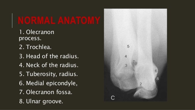
The radioanatomy of the elbow is studied via an ap x ray image and one in profile showing the medial and lateral epicondyles the olecranon the head and neck of the radios the radial and olecranon fossae the humeral trochlea and allow of the anatomical structures composing the humeroulnar joint humeroradial joint and proximal radioulnar joint. No posterior fat pad should be seen. Anatomy humerus ulna radius xray elbow olecranon process elbow xray lateral elbow ap elbow.
 Radiographic Anatomy Of The Skeleton Elbow
 Fat Pad Sign Wikipedia
 File X Ray Of Normal Elbow By Lateral Projection Jpg Wikipedia
 Diagnostic Imaging Of The Elbow Clinical Gate
 Imaging Of Elbow Fractures And Dislocations In Adults
 Elbow X Ray Labeled Anatomy Radiology Case Radiopaedia Org
 Mnemonic Approach To Elbow Xray Fool Epomedicine
 Elbow Radiology Key
Positioning In X Ray Elbow Joint Ap Lat Special Edition Anatomy And Physiology Part 15
 Elbow Ap Projection Radtechonduty
 Fracture Elbow Forearm X Rays Image Showing Plate And
 Radiological Anatomy Of The Shoulder Arm Elbow Forearm
 Epicondyle Medial Epicondyle Of The Humerus Wikipedia
 Startradiology

 Mnemonic Approach To Elbow Xray Fool Epomedicine
 Elbow Radiology Key
 The Radiology Assistant Elbow Fractures In Children
 Anatomical Position X Ray Whole Body Head Neck Thorax Heart
 Medical Imaging Technology Radiographic Anatomy Of Elbow
 Elbow Xray Positioning Ap Oblique Projection Lateral

 Imaging Of Elbow Fractures And Dislocations In Adults

 Elbow X Ray Images Stock Photos Vectors Shutterstock
 Anatomy Of The Elbow Ct Arthrography
 Elbow Xray Anatomy Stock Photos Page 1 Masterfile
 Elbow Xray Interpretation
 Film Critique Of The Upper Extremity Part 2 Elbow And Forearm

 X Ray Of Elbow Joint


Posting Komentar
Posting Komentar