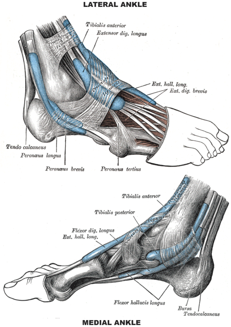
There are several bones that make up the ankle. Mnemonics that can be used to remember the anatomy of the ankle tendons from anterior to posterior as they pass posteriorly to the medial malleolus under the flexor retinaculum in the tarsal tunnel include.
 Gross Anatomy Of The Ankle Joint Clinical Gate
Tom dick and very nervous harry.

Medial ankle anatomy. The calcaneofibular ligament cfl which connects the calcaneus or heel bone to. The medial malleolus felt on the inside of your ankle is part of the tibias base. Medial ankle view showing the ligamentous anatomy of the deltoid ligament and related structures.
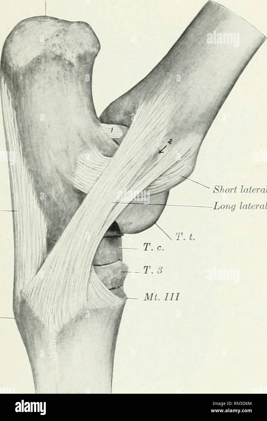
The aim of this pictorial review on the anatomy of the ankle ligaments is to provide a guide to those who are involved in diagnosing and treating ligament injury around the ankle. Medial ankle stability is provided by the strong deltoid ligament the anterior tibiofibular ligament and the bony mortise. Three ligaments on the outside of the ankle that make up the lateral ligament complex as follows.
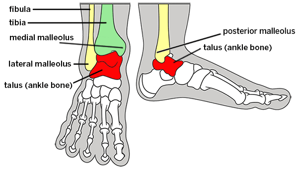
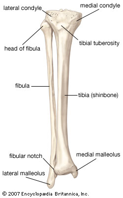
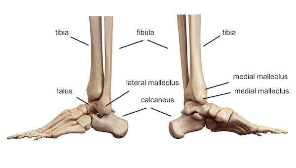
The ankle joint is formed where the talus the uppermost bone in the foot and the tibia shin meet. Upper ankle joint tibiotarsal talocalcaneonavicular and subtalar joints. The lateral malleolus felt on the outside.
Because of the bony articulation between the medial malleolus and the talus medial ankle sprains are less common than lateral sprains. The posterior malleolus felt on the back of your ankle is also part of the tibias base. The bones of the foot and ankle begin with the ankle joint itself.
The bony bumps or protrusions seen and felt on the ankle have their own names. Posterior ankle tendons mnemonic dr daniel j bell and dr jeremy jones et al. Medial injury is probably more influenced by the rotating component of the subtalar joint to which the capsule and the mcl are subject.
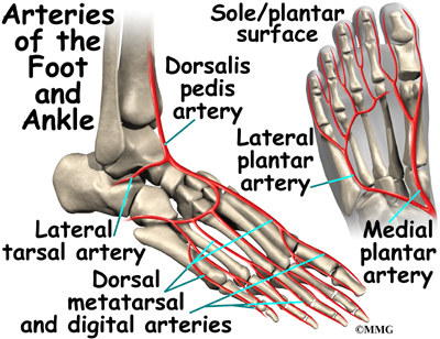
The last two together are called the lower ankle joint. The ankle joint also known as talocrural joint is an example of a synovial joint and is formed by the bones tendons and ligaments found in the leg and the foot 1 2. The tibia the fibula the talus and the calcaneus.
View media gallery the superficial deltoid ligament originates from an anterior bony prominence of the medial malleolus referred to clinically as the anterior colliculus. Ankle anatomy the ankle joint also known as the talocrural joint allows dorsiflexion and plantar flexion of the foot. Tom dick and harry.
The anterior talofibular ligament atfl which connects the front of the talus bone to the fibula or shin bone. In medial ankle sprains the mechanism of injury is excessive eversion and dorsiflexion. The ankle joint talocrural joint is formed where the distal end of the leg meets the foot.
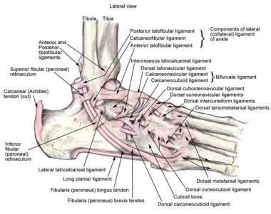
Lateral side of the ankle joint capsule. It is made up of three joints.
 Broken Ankle Types Of Fractures Diagnosis Treatments
 Bursitis Ankle Bursa Care And Prevention
 This Trial Exhibit Depicts A Bimalleolar Left Ankle Fracture
 This Trial Exhibit Depicts A Bimalleolar Right Ankle

 Image Result For Tenderness Posterior To The Medial
 Ankle Foot Anatomy
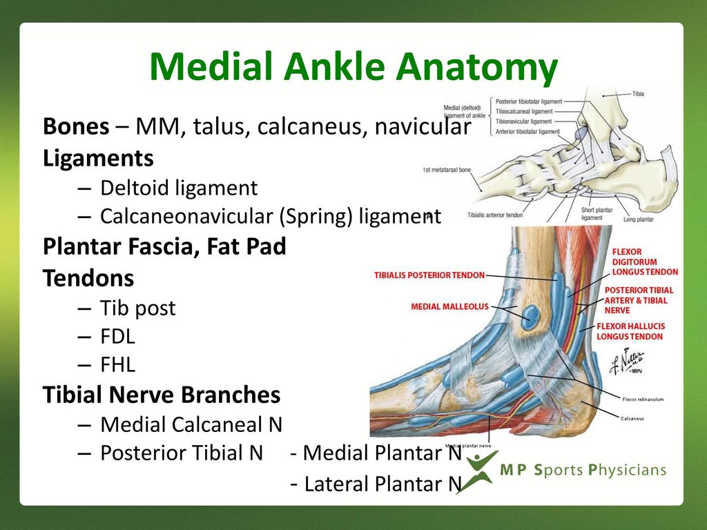 Medial Ankle And Heel Pain Ppt Download
 The Radiology Assistant Ankle Mri Examination
 Ankle Anatomy

 Medial Malleolus Stock Photos Medial Malleolus Stock
 Normal Anatomy Of The Medial Ankle Download Scientific
 Pin On Foot Issues
 Ankle Block Landmarks And Nerve Stimulator Technique Nysora
 Pin On Aaa
 Ankle Anatomy Eorthopod Com
 Ankle Joint Anatomy Overview Lateral Ligament Anatomy And
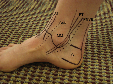
 11 Surface Anatomy Of The Medial Ankle Mm Medial Malleolus
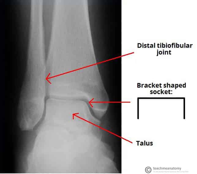 The Ankle Joint Articulations Movements Teachmeanatomy

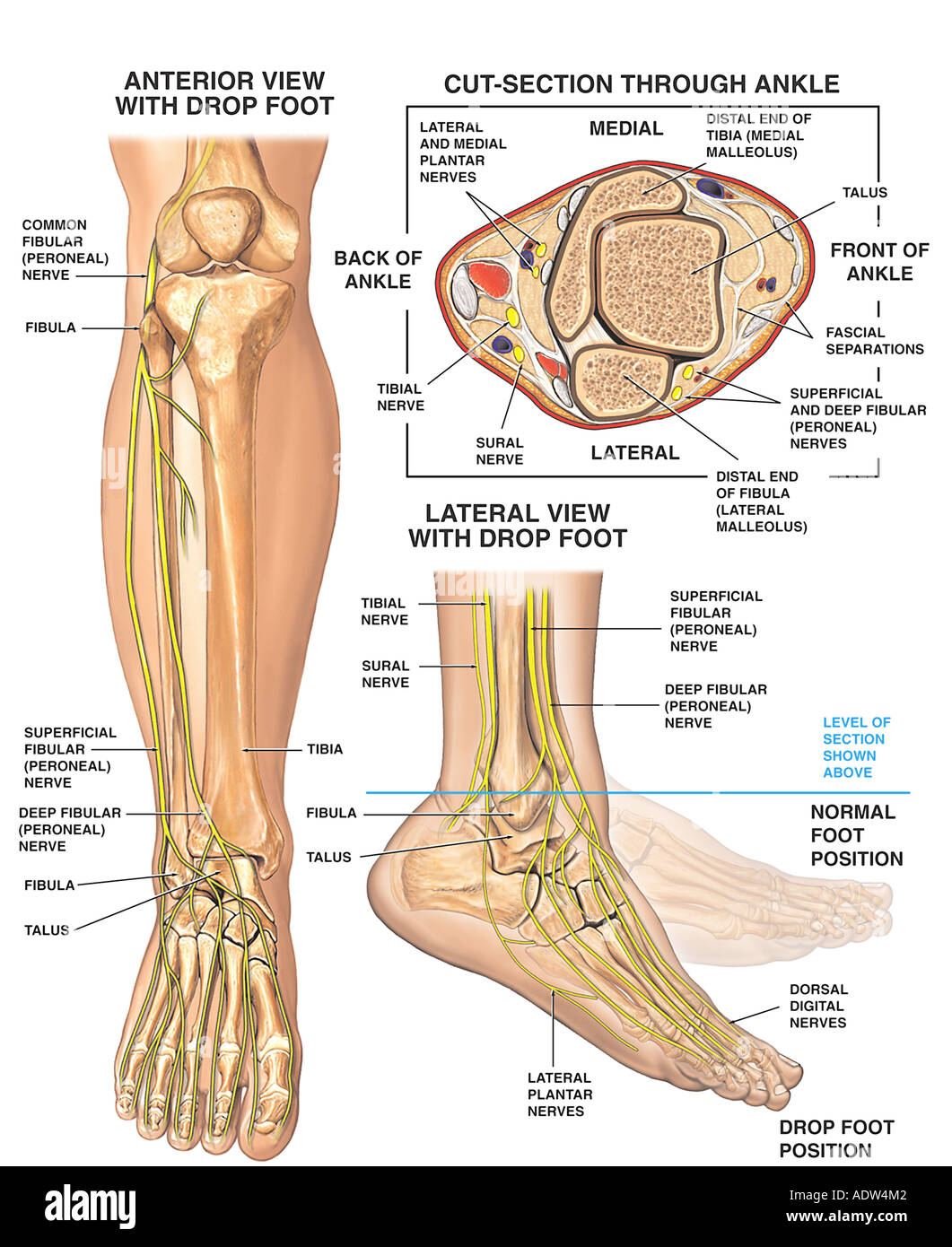
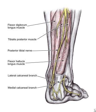 What Is The Anatomy Of The Tibial Nerve Relevant To A
 Medial Malleolus Stock Photos Medial Malleolus Stock


Posting Komentar
Posting Komentar