Each myosin has multiple z lines are at both ends of a sarcomere. A sarcomere extends from one are held by direct attachment to z lines.
 13 Which Of
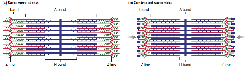
A sarcomere is the complicated unit of striated muscle tissue.

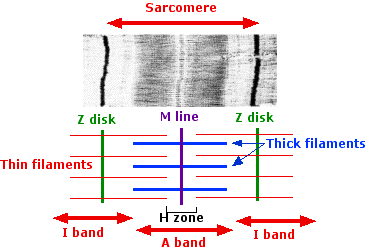
Sarcomere anatomy. Myosin is the protein that makes protein strands that project outward. Myofibrils are composed of repeating sections of sarcomeres which appear under the microscope as alternating dark and light bands. Herein lies the sarcomeres main purpose.
Due to the striated nature of both skeletal muscle and cardiac muscle is observed by microscope slides. Skeletal muscles are composed of tubular muscle cells which are formed in a process known as myogenesis. Are at both ends of a sarcomere.
A sarcomere extends from one are held by direct attachment to z lines. Start studying sarcomere anatomy physiology. Actin is the protein are not attached to z lines.
This unit is distinctive in some types of muscle tissue. Are at both ends of a sarcomere. Knowing all of the features and landmarks of the sarcomere will help us to.
Anatomical is said to be the term of microanatomy. Myosin heads bind and form a cross bridge with actin molecules. This unit is distinctive in some types of muscle tissue.
The sarcomeres are the individual contractile units of the myofibrils tiny rod like elements within our muscle cells. Sarcomeres are able to initiate large sweeping movement by contracting in unison. Actin is the protein are not attached to z lines.
Each myosin has multiple z lines are at both ends of a sarcomere. It is the repeating unit between two z lines. The sarcomere is the basic unit function with muscle fiber cells.
Skeletal muscle is the muscle type that initiates all of our voluntary movement. Sarcomeres are composed of long fibrous proteins as filaments that slide past each other when a muscle. Myosin is the protein that makes protein strands that project outward.
The myosin heads then pull on the actin molecules causing them to slide along the myosin filaments. A sarcomere is the functional unit of striated muscle. 0 0000 a shoutout is a way of letting people know of a game you want them to play.
Muscle fibers contain numerous tubular myofibrils. Learn vocabulary terms and more with flashcards games and other study tools. It can be observed on microscope slides due to the striated nature of both skeletal muscle and cardiac muscle.
This means it is the most basic unit that makes up our skeletal muscle. This is a distinguishing unit in some types of muscle tissue. A sarcomere is the basic functional within muscle cells.
Just pick an audience or yourself and itll end up in their incoming play queue.
 Muscle Fiber Contraction And Relaxation Anatomy And
 Try It Out Ch 9 Sarcomere Anatomy Diagram Quizlet
 The Sarcomere And Sliding Filaments In Muscular Contraction Definition And Structures
 Structure And Function Of A Sarcomere Physiology Of Sport And Exercise Seventh Edition
 10 2 Skeletal Muscle Anatomy Physiology
 Muscle Contraction
 Solved Review The Two Figures Below And Compare The

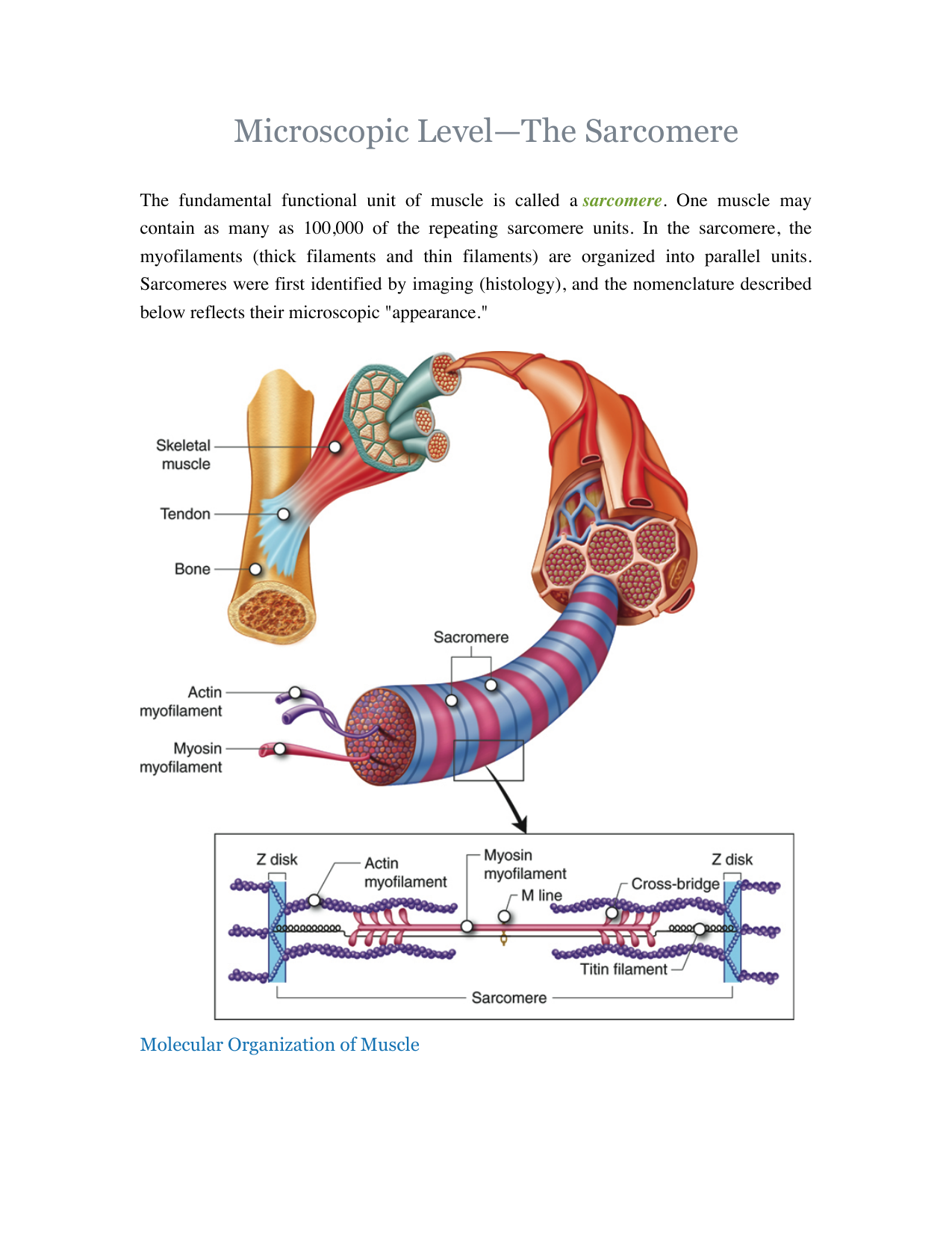 Microscopic Level The Sarcomere
 Muscles
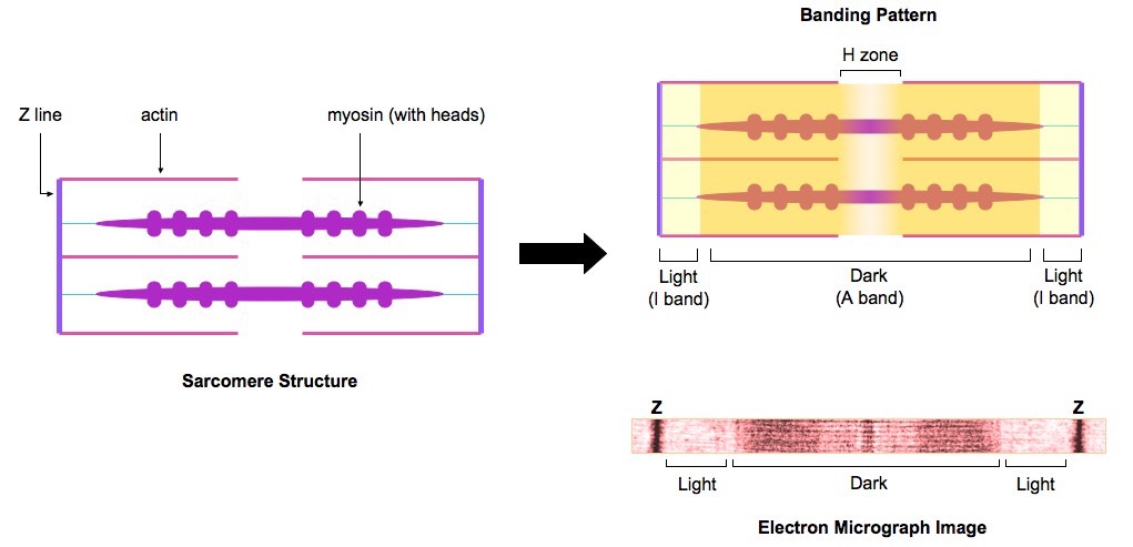
Anatomy Of A Skeletal Muscle And A Sarcomere A From Seer
 Skeletal Muscle Contraction Analogy A P Physiology Worksheet Nmj Sarcomere Key
 Sarcomere Structure Anatomy Physiology Diagram Anatomy
Ch 9 Sarcomere Worksheet Bio 220 Anatomy And Physiology
Sarcomere Structure Anatomy Physiology Physiology Anatomy
 Ch 09 Sarcomere Composition
 Sarcomere Wikipedia
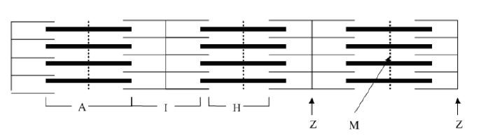 Solved 1 How Many Sarcomeres Are Shown In The Above Diag
 Diagram Of Sarcomere Wiring Schematic Diagram 5 Laiser
 Sarcomeres Bioninja
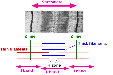
 Difference Between A Band And I Band Sarcomere
 Sarcomere Anatomy Diagram Quizlet
 Sarcomere Anatomy Physiology 120 With Lavender At


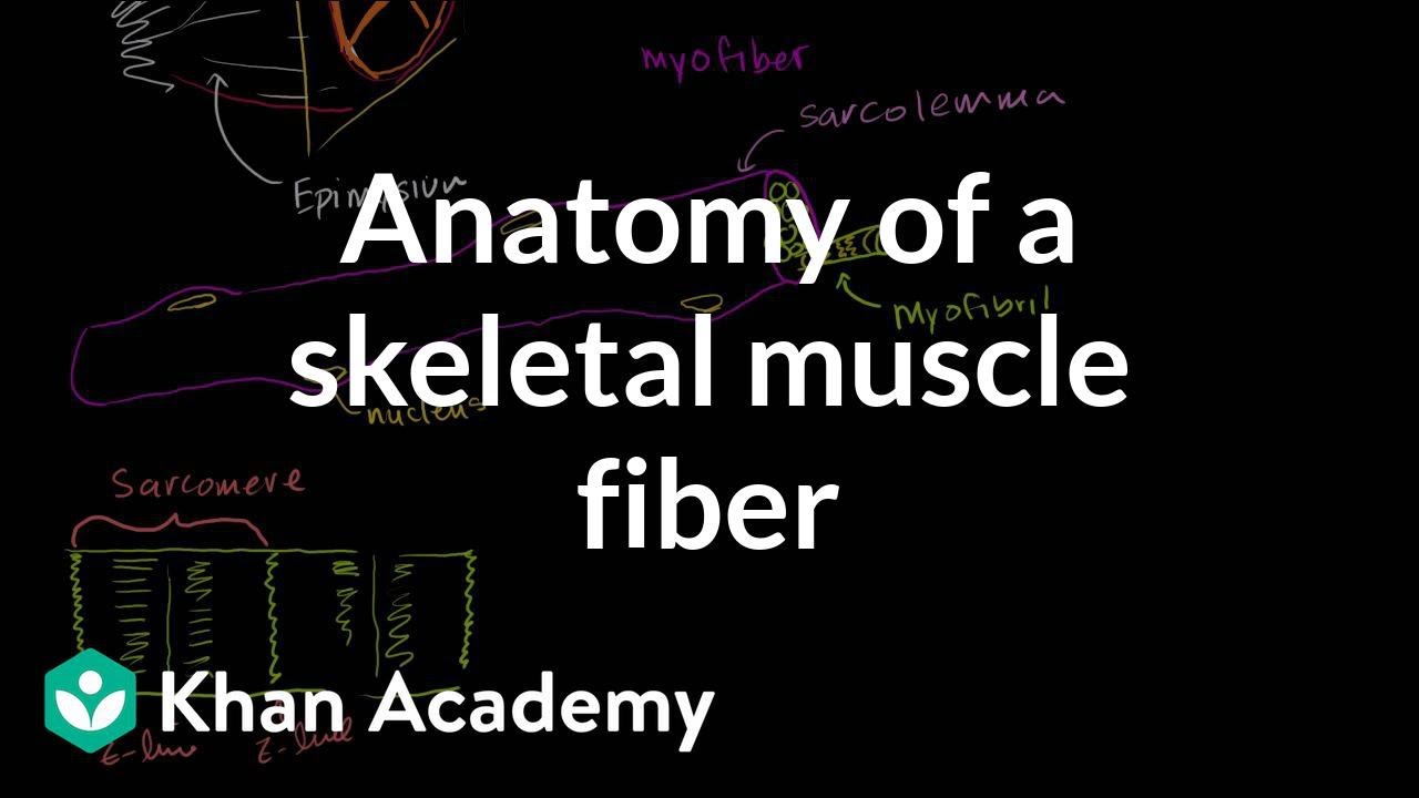 Anatomy Of A Skeletal Muscle Fiber Video Khan Academy


Posting Komentar
Posting Komentar