Foot and leg anatomy the foot and leg muscletendon connection to better understand foot and leg muscletendon injuries it is important to appreciate the basic elements that enable your body parts to move. This part of the foot consists of bones that make the arch of the foot.
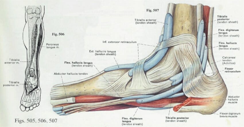 Foot Anatomy Bones Ligaments Muscles Tendons Arches
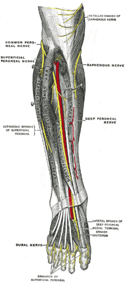
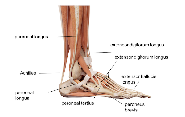
The major muscles of the lower leg other than the gastrocnemius which is cut away are shown in figure 9 12.

Foot and leg anatomy. This occurs during the phase of walking called toe off which allows you to propel yourself forward without your foot collapsing beneath you. The hindfoot consists of bone from the leg and the ankle joint. The leg muscles acting on the foot are called the extrinsic foot muscles whilst the foot muscles located in the foot are called intrinsic.
Having two separate bones instead of one connecting the foot to the leg gives the foot and leg extra balance and maneuverability. The lower ends of the tibia and fibula the calcaneus and talus bones are located at the hindfoot. To ensure your body moves smoothly with a minimum of friction muscles are enveloped in a slippery skin like tissue called fascia.
The balls of your feet are five joints that together act as a hinge when you go on tiptoe. The muscles of the lower leg called simply the leg by anatomists largely move the foot and toes. The end of the leg on which a person normally stands and walks.
When the muscles of the foot and leg twist the foot in a particular direction the tarsal bones lock together to form a rigid post. Supporting balancing and propelling the body is the work of the muscular system of the legs and feet. You can also see the thick web of ligaments on the top of the foot where the bones of the foots core are connected on the top side.
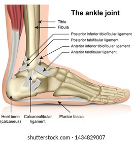
The ankle is a joint that connects the lower leg to the foot. The midfoot is just in front of the hindfoot. The foot is an extremely complex anatomic structure made up of 26 bones and 33 joints that must work together with 19 muscles and 107 ligaments to execute highly precise movements.
The feet and legs are constructed as a series of hinge joints known as single degree of freedom joints alternating with rotational multiple degree of freedom joints. From the large strong muscles of the buttocks and legs to the tiny fine muscles of the feet and toes these muscles can exert tremendous power while constantly making small adjustments for balance whether the body is at rest or in motion. Its main function is to allow for plantar flexion and dorsiflexion of the foot.
In a similar way mid means the middle of the foot. Dorsiflexion extension and plantar flexion occur around the transverse axis running through the ankle joint from the tip of the medial malleolus to the tip of the lateral malleolus.
 Leg And Foot Musculature Google Search Muscle Anatomy
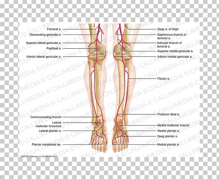 Thumb Foot Human Leg Artery Crus Png Clipart Abdomen
 Anatomy Of Leg And Foot Anatomy And Physiology Of The Leg
 Lower Limb Tips In Plastic Surgery
 Foot Picture Image On Medicinenet Com
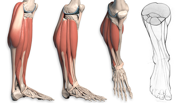 How To Draw The Lower Leg Anatomy For Artists Proko
 Muscles Of The Foot Part 1 3d Anatomy Tutorial
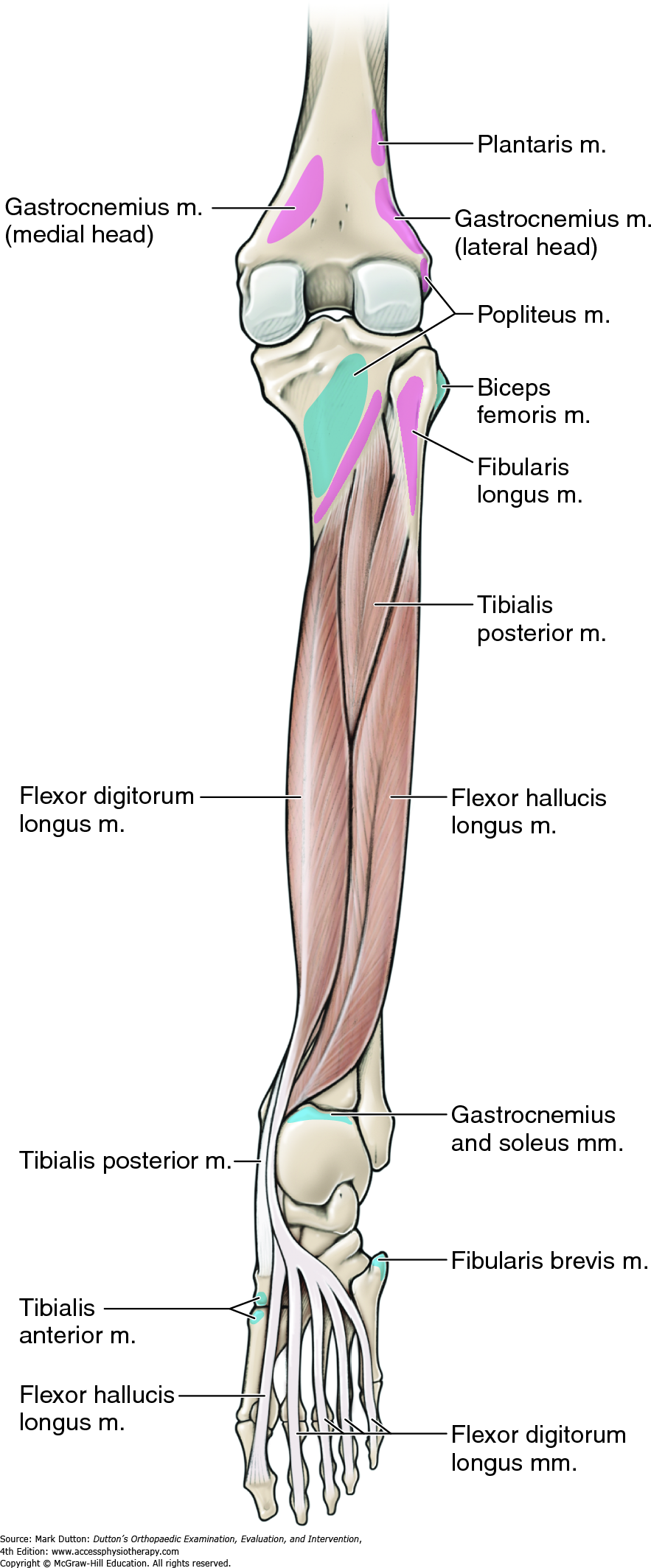 Lower Leg Ankle And Foot Dutton S Orthopaedic
 Bone Fracture Human Leg Anatomy And Skeleton Stock Vector
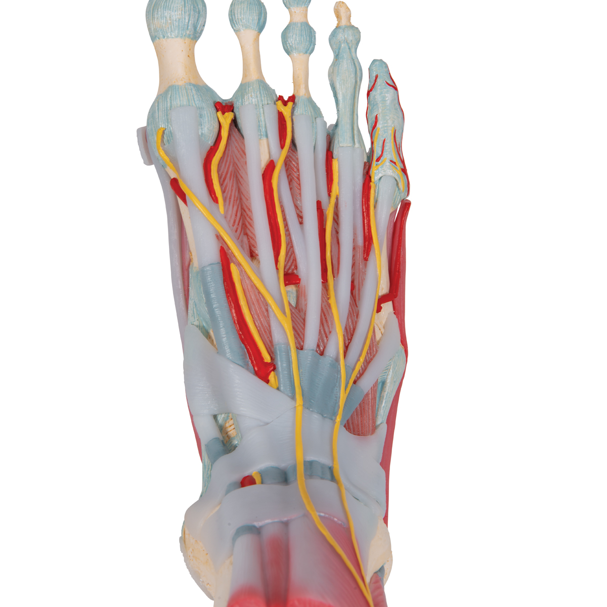 Anatomical Teaching Models Plastic Human Joint Models
 Leg Muscles Anatomy Support Movement
 3d Anatomy Of The Foot And Ankle Cl3ver

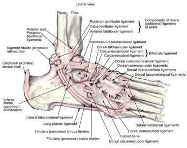 Ankle Joint Anatomy Overview Lateral Ligament Anatomy And
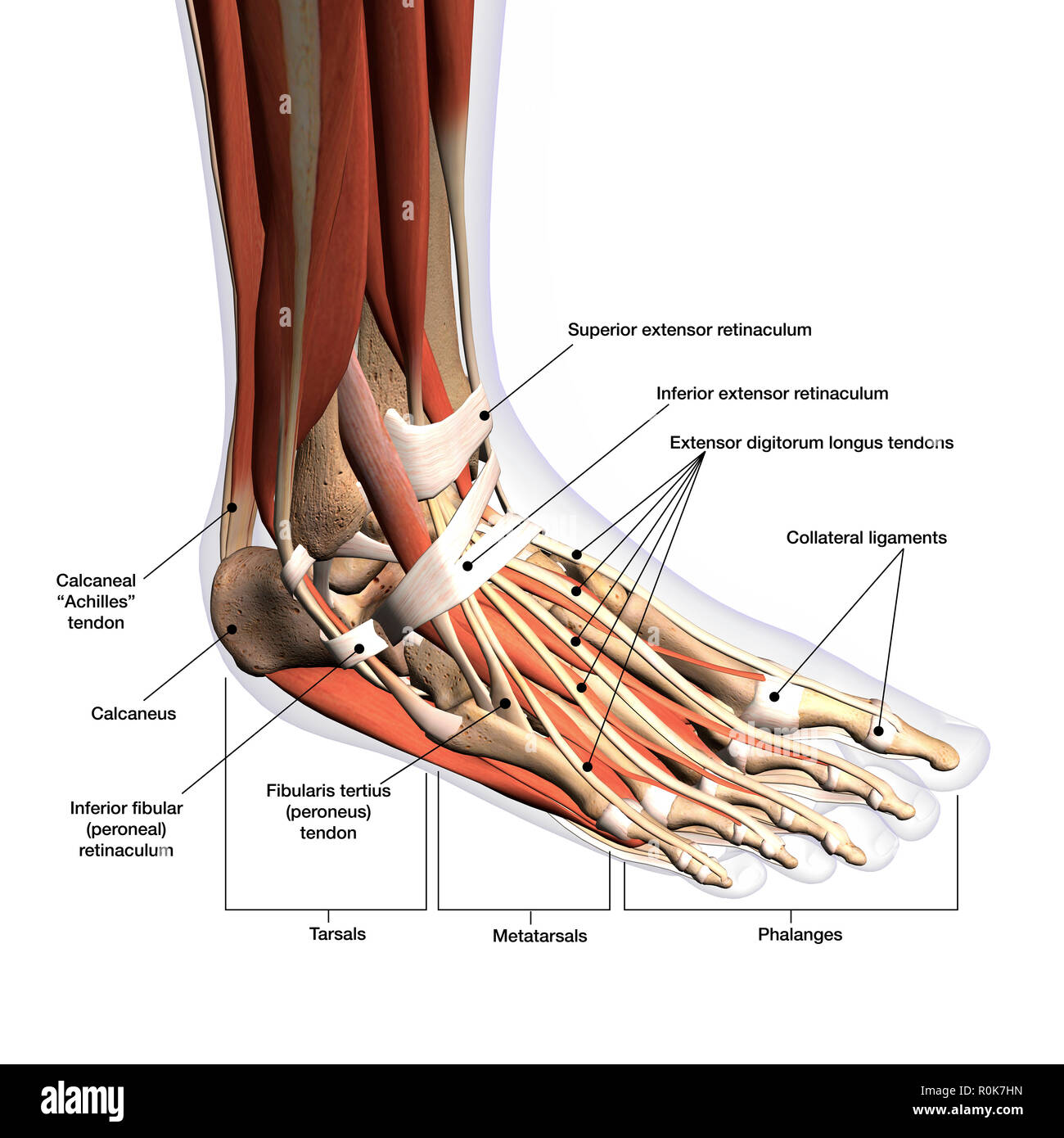 Human Foot Anatomy Stock Photos Human Foot Anatomy Stock
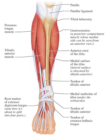
 Ankle Images Stock Photos Vectors Shutterstock
:background_color(FFFFFF):format(jpeg)/images/library/11153/muscles-tibia-fibula_english__2_.jpg) Leg And Knee Anatomy Bones Muscles Soft Tissues Kenhub
Leg And Knee Anatomy Bones Muscles Soft Tissues Kenhub
 7 Anatomy Of The Leg And Dorsum Of The Foot
Foot Anatomy Foot Ankle Lower Leg Orthopedic Assessment


Posting Komentar
Posting Komentar