In human digestive system. Each lobule contains a bronchiole and affiliated branches a thin wall and clusters of alveoli.
 Liver Lobules Human Physiology 78 Steps Health Journal
A subdivision of the four main liver lobes the basic functional unit of the liver.
Liver lobule anatomy. The liver also has two minor lobes the quadrate lobe and the caudate lobe. Deflned as a lobule figure 23. In lung further subdivides into hundreds of lobules.
A fibrous capsule encloses the liver and ligaments divide the organ into a large right lobe and a smaller left lobe figure 2. Each lobe is separated into many tiny hepatic lobules the livers functional units figure 3. The falciform ligament runs inferiorly from the diaphragm across the anterior edge of.
A small subunit of the liver composed of cells hepatocytes that process blood from an incoming portal venule and send the resulting blood to an outgoing hepatic venule. The lobules are held together by a fine dense irregular fibroelastic connective tissue layer extending from the fibrous capsule covering the entire liver known as glissons capsule. They vary in size from 12 to 25µ in diameter.
The blood in the capillary plexus around the liver cells is brought to the liver principally by the portal vein. Ligaments of the liver falciform ligament this sickle shaped ligament attaches the anterior surface of the liver to the anterior abdominal wall and forms a natural anatomical division. The lobules of liver or hepatic lobules are small divisions of the liver defined at the microscopic histological scale.
Located on the lateral borders of the left and right lobes respectively the left and right triangular ligaments. Each lobule is made up of millions of hepatic cells hepatocytes which are the basic metabolic cells. The hepatic cells are polyhedral in form.
The wide coronary ligament connects the central superior portion of the liver to the diaphragm. Lobules are the functional units of the liver. The liver the human liver is both the largest internal organ the skin being the largest organ overall and the largest gland in the human body.
They contain one or sometimes two distinct nuclei. Coronary ligament anterior and posterior folds attaches the superior surface of the liver to the inferior. The hepatic lobule is a building block of the liver tissue consisting of a portal triad hepatocytes arranged in linear cords between a capillary network and a central vein.
The lobule largely consists of hepatocytes liver cells which are arranged as interconnected plates usually one or two hepatocytes. There are two types of liver lobule which look at the same cluster of liver cells from opposite ends. Microscopic anatomy of clusters of cells called lobules where the vital functions of the liver are carried out.
 Royalty Free Liver Lobule Stock Images Photos Vectors
 Liver Lobule Definition Of Liver Lobule By Medical Dictionary
 Liver Sectional View Liver Anatomy Digestive System
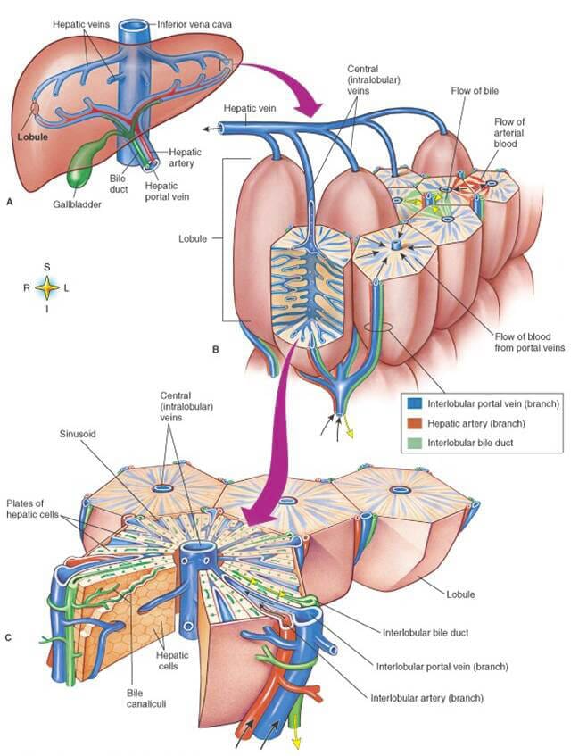 Liver On Chip Liver Lobule Microfluidics Elveflow
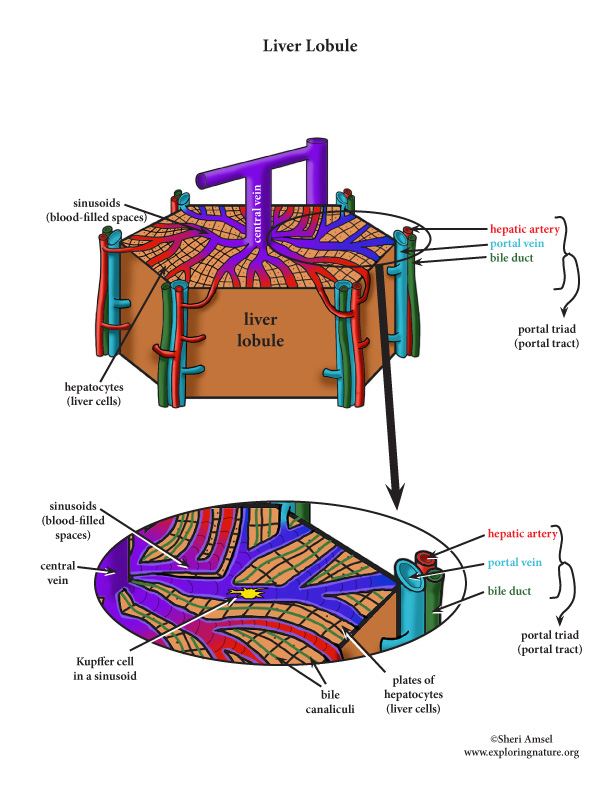 Liver Structures And Functions A Closer Look Advanced
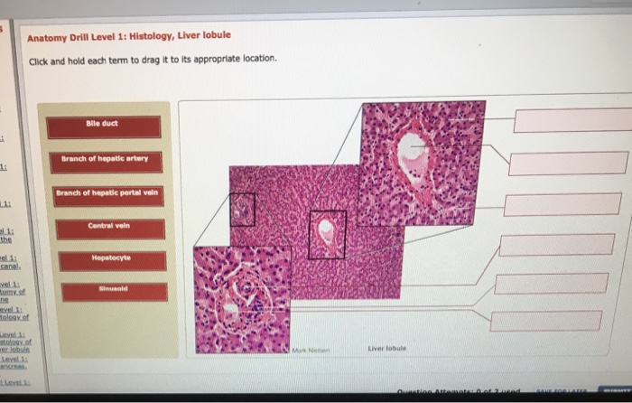 Solved Anatomy Drill Level 1 Histology Liver Lobule Cli
 Figure 1 From Construction Of Hepatic Lobule Like Vascular
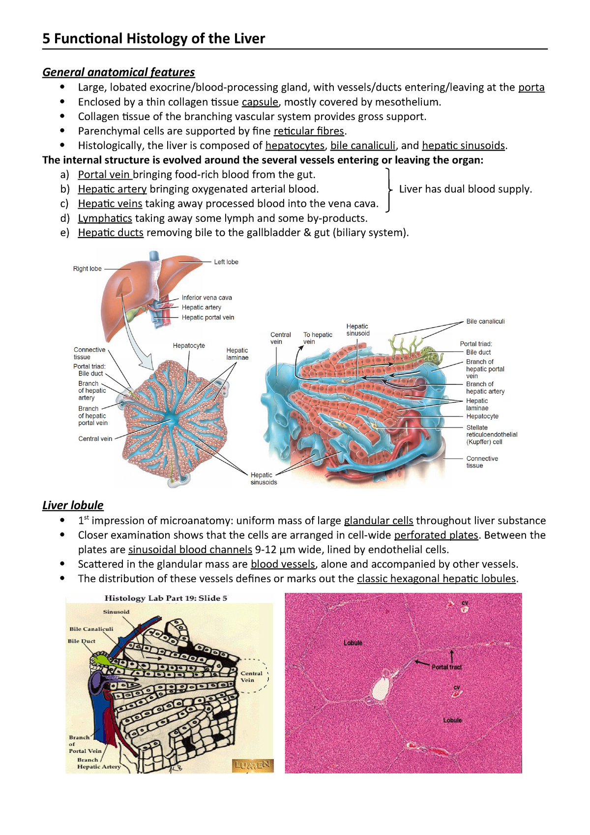 5 Anatomy And Functional Histology Of The Liver Medicine
 Figure 1 From A 3d Porous Media Liver Lobule Model The
 Structure Of A Liver Lobule Liver Anatomy Anatomy
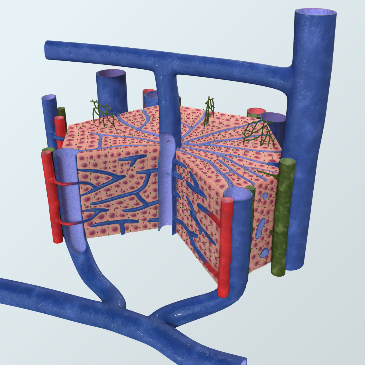 Realistic Liver Lobule Anatomy
 Classical Vs Functional Liver Lobules Student Doctor Network
Structure Of A Liver Lobule Eclinpath
 Exercise 34 Figure 34 12 Flow Of Blood And Bile Through
 Liver Lobule An Overview Sciencedirect Topics
 Structure Of One Liver Lobules Download Scientific Diagram
 Liver Anatomy Physiology Wikivet English
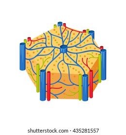 Royalty Free Liver Lobule Stock Images Photos Vectors
 Liver Lobule An Overview Sciencedirect Topics
 Dr Das Liver1 Law Civil Procedure I With Baron At


Posting Komentar
Posting Komentar