Click on a link to get t1 axial view t2 fatsat axial view t1 coronal view t2 fatsat coronal view t2 fatsat sagittal view. This webpage presents the anatomical structures found on shoulder mri.
 Shoulder Anatomy And Normal Variants
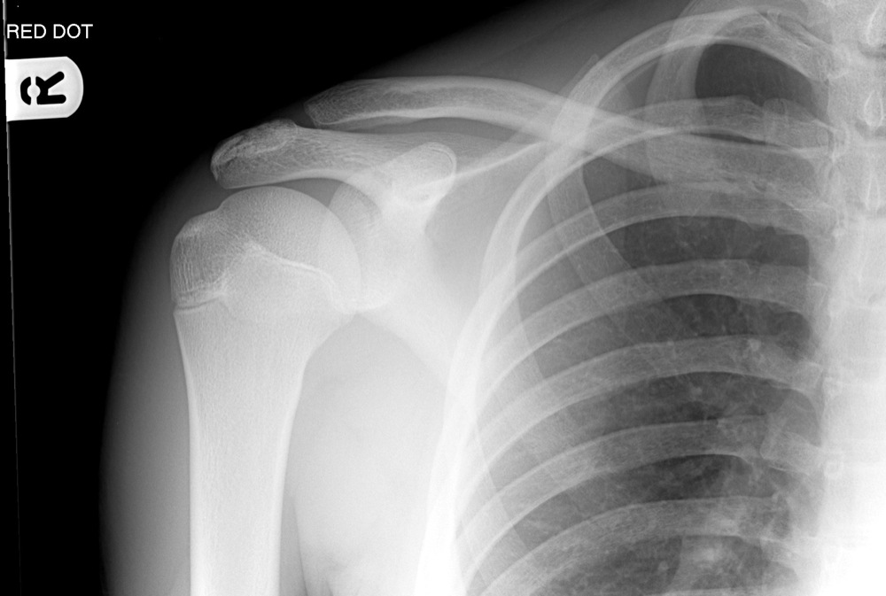
The shoulder series is fundamentally composed of two orthogonal views of the glenohumeral joint including the entire scapula.
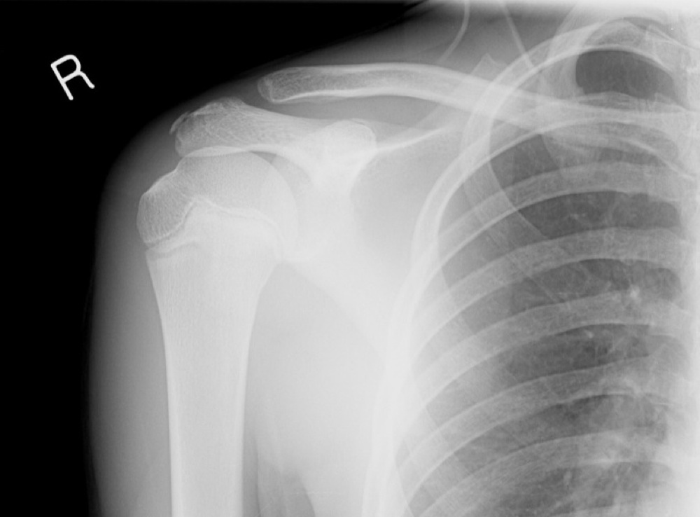
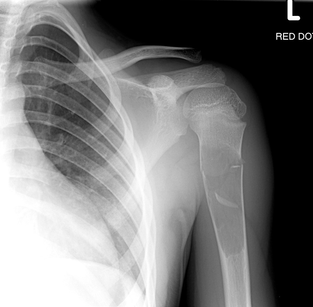
Shoulder anatomy xray. Do not confuse with dislocation. A plain x ray film of the shoulder may show dislocation osteoarthritis or a fracture of the humerus. Shoulder radiographs are performed for a variety of indications including.
Usually secondary to trauma. The extension of the shoulder series depends on the radiography department protocols and the clinical indications for imaging. Mr is the best imaging modality to examen patients with shoulder pain and instability.
X ray films cannot diagnose muscle or tendon injuries. Anteroposterior shoulder view allows assessment of especially the humeral head lesions and clavicular fractures. In part ii we will discuss shoulder instability.
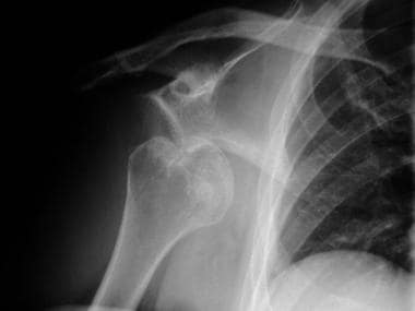
Click here to load quiz. This effusion suggests intra articular fracture. An effusion or haemorrhage into the joint displaces the humeral head inferiorly.
X ray shoulder x ray pelvis x ray hand pa ct head. The shoulder can dislocate posteriorly but anterior dislocation is approximately 50 times more common. It is the most complete reference of human anatomy available on web ipad iphone and android devices.
While achieving anteroposterior shoulder x ray in neutral position the patient is erect or in supine position. Stanford bone tumor bayesian network issssr msk lectures for residents ocad msk cases from around the world stanford msk mri atlas has served almost 800000 pages to users in over 100 countries. E anatomy is an award winning interactive atlas of human anatomy.
Explore over 5400 anatomic structures and more than 375 000 translated medical labels. Atlas of shoulder mri anatomy. In part iii we will focus on impingement and rotator cuff tears.
Ct mri radiographs anatomic diagrams and nuclear images. Shoulder dislocation is a term often used loosely to indicate dislocation of the head of the humerus from the glenoid of the scapula. In shoulder mr part i we will focus on the normal anatomy and the many anatomical variants that may simulate pathology.
Opening the quiz in incognito mode will prevent answers becoming pop up suggestions for future attempts. Central x ray should be directed to 25 cm inferior to the coracoid process. Quizzes about radiology anatomy quiz.
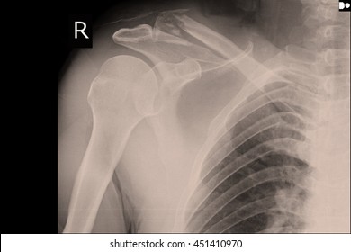
 X Ray Of Both Human Shoulders Stock Photo Image Of
 The Radiology Assistant Shoulder Mr Anatomy
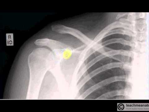
 Radiology In Ped Emerg Med Vol 4 Case 12
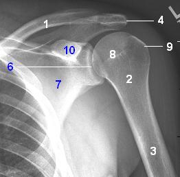
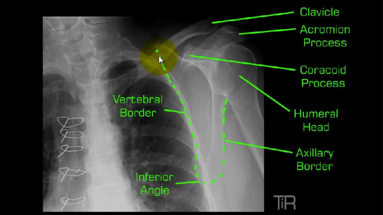
 Anatomically Labelled Ap Shoulder X Ray Radiology Student

 Shoulder Dislocation Imaging Practice Essentials
 X Schouder Startradiology
 X Schouder Startradiology
 Ap Of The Shoulder Radiology Student Shoulder Anatomy
 Interpreting X Rays Of The Shoulder Joint

 Frozen Shoulder Anatomy Stock Photos Images Photography
 The Shoulder
The Shoulder
 Y View Shoulder Mp4
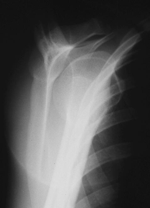
Anteroposterior A And Scapular Y B Shoulder Radiographs
 Shoulder Anatomy And Normal Variants
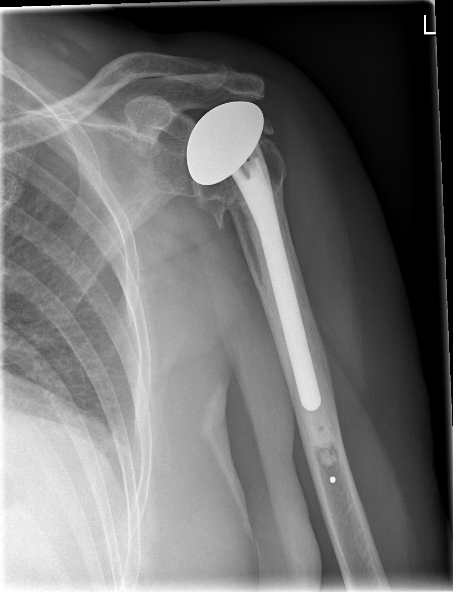 Shoulder Replacement Wikipedia
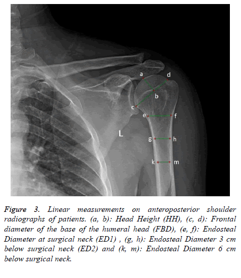
 Linear Measurements On Anteroposterior Shoulder Radiographs

 Ap Shoulder Internal Rotation
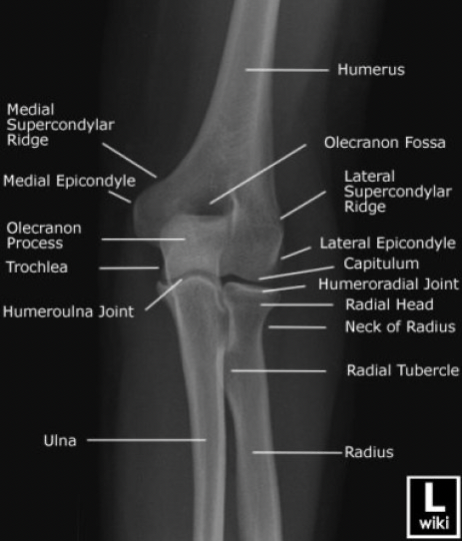 X Ray Vision Shoulders And Elbows Taming The Sru
 Medical Imaging Technology Radiographic Anatomy Of Shoulder
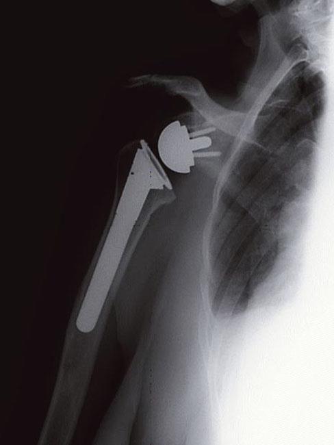 Reverse Total Shoulder Replacement Orthoinfo Aaos
 Acromio Clavicular Injuries Ac Separation Shoulder
 Posterior Shoulder Dislocation Litfl Medical Blog Trauma
 The Shoulder

 X Schouder Startradiology


Posting Komentar
Posting Komentar