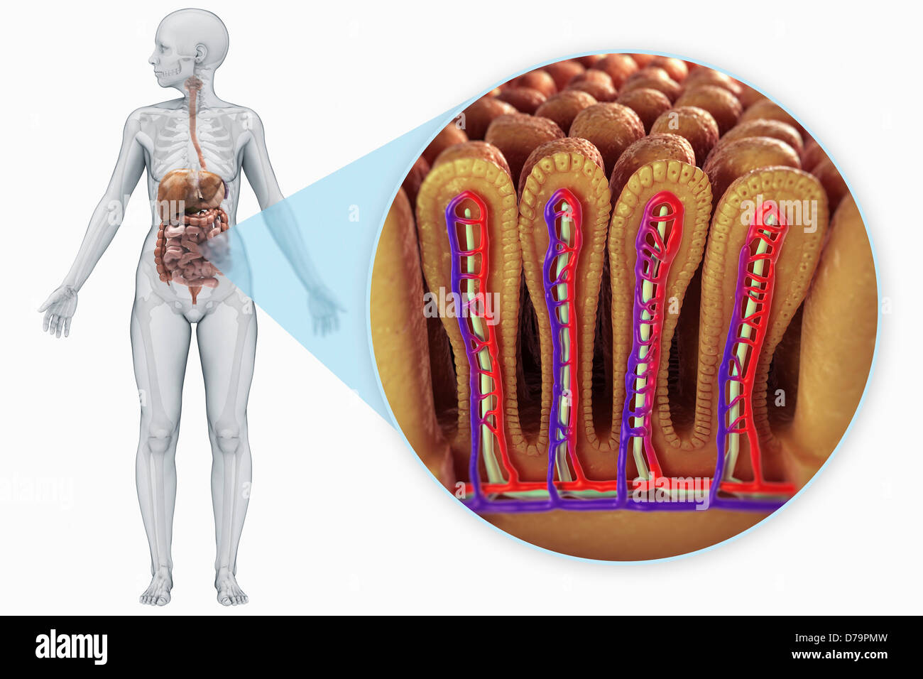
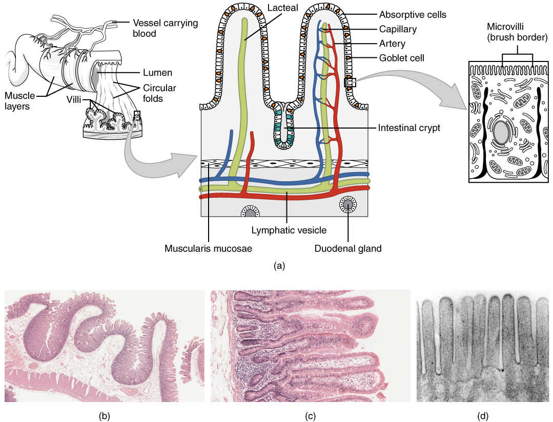
It is thicker more vascular and has more developed mucosal folds than the jejunum. The villi usually vary from 05 to 1 mm in height.
 Villi Stock Photos Villi Stock Images Alamy
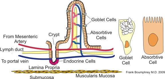
Each villus is approximately 0516 mm in length in humans and has many microvilli projecting from the enterocytes of its epithelium which collectively form the striated or brush border.

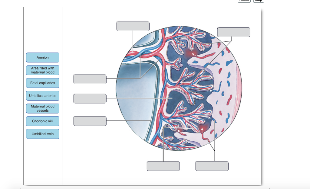
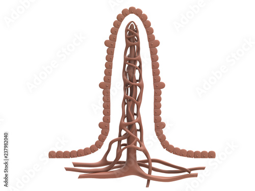
Villi anatomy. Branches of the umbilical arteries carry embryonic blood to the villi. Chorionic villi are villi that sprout from the chorion to provide maximal contact area with maternal blood. Each of these villi is covered in even smaller finger like structures called microvilli.
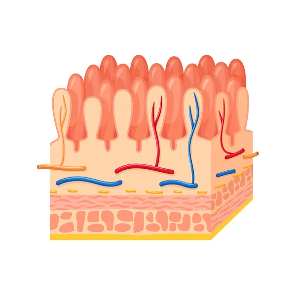
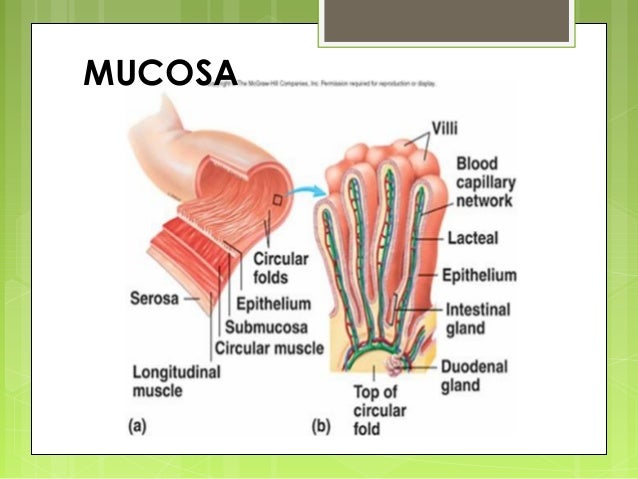
After circulating through the capillaries of the villi blood returns to the embryo through the umbilical vein. The villi are covered by a single layer of tall columnar cells called goblet cells because of their rough resemblance to empty goblets. Villus are small finger like projections that extend into the lumen of the small intestine.
The intestinal villi villi intestinales are highly vascular processes projecting from the mucous membrane of the small intestine throughout its whole extent and giving to its surface a velvety appearance. The ileum is the longest part of the small intestine measuring about 18 meters 6 feet in length. Villus in anatomy any of the small slender vascular projections that increase the surface area of a membrane.
Skip trial 1 month free. The villi of the small intestine project into the intestinal cavity greatly. Find out why close.
Development of small and large intestine the highest villi height was related to the 5 g kgsup 1 thymolinar treatment and the lowest one is related to the 20 g kgsup 1 thymolinar treatment. The ileum joins the cecum the first portion of the large intestine at the ileocecal sphincter or valve. Each of these microvilli are much smaller than a single villus.
Get youtube without the ads. The internal walls of the small intestine are covered in finger like tissue called villi. Important villous membranes include the placenta and the mucous membrane coating of the small intestine.
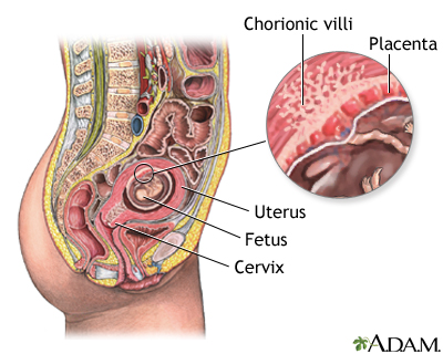
They are an essential element in pregnancy from a histomorphologic perspective and are by definition a product of conception. Villi develop at the duodenum at first as a result of the proliferation of mesenchymal tissue beneath the epithelium 9. Microscopic anatomy of intestinal villi yoyis88godoy.
Thus villi are part of the border between maternal and fetal blood during pregn. Noun plural villi ˈvɪlaɪ usually plural zoology anatomy any of the numerous finger like projections of the mucous membrane lining the small intestine of many vertebrates. Villi epithelium and glands.
Their diameters vary from approximately one eighth to one third their height. Any similar membranous process such as any of those in the mammalian placenta. They are largest and most numerous in the duodenum and jejunum and become fewer and smaller in the ileum.
 Celiac Disease Detailed Anatomy Healthy Intestinal Villi And
Chorionic Villi Google Search Chorionic Google Search
 Intestinal Villi Small Intestine Lining Haneen
 ᐈ Villi Stock Images Royalty Free Villi In Small Intestine
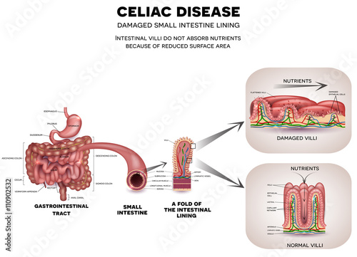 Gastrointestinal Tract Anatomy And Celiac Disease Affected
 Intestine Intestinal Villi Anatomy Digestive System Organs Small Intestine Medical Art Clinic Decor 1656
 Digestion S Cutest Heroes A Dive Into The Intestinal Villi
 Intestinal Villi Blue Technology Background

 Gastrointestinal Tract Anatomy Intestinal Villi Small Intestine
 Solved Art Labeling Activity Anatomy Of The Placenta Aft
 What Would Be The Result If There Were No Villi In The
 23 5 The Small And Large Intestines Anatomy And Physiology
 Intestine
 Intestinal Villus Wikipedia
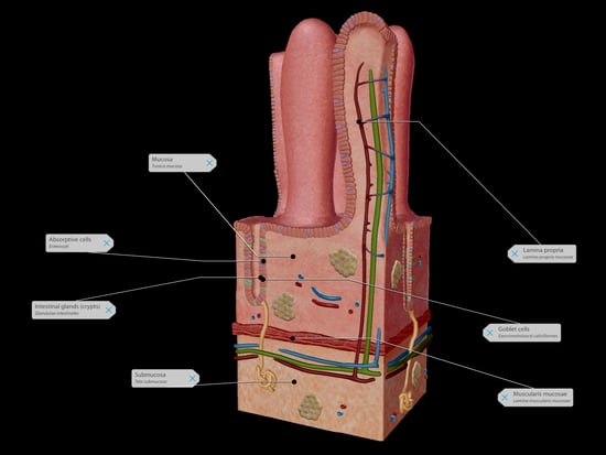
 Anatomy Of Intestinal Tissue A Intestinal Section Showing
 Intestinal Anatomy And Physiology

 Chorionic Villus Sampling Series Normal Anatomy
 Celiac Disease Affected Small Intestine Villi Damaged Cells
 Healthy Villi Microvilli Arteries Veins Biology Healthy
 Structure Of The Small Intestine Functions Of The Small Intestine What Are Villi
 Intestinal Villi Anatomy Small Intestine Lining Villi And Epithelial
 Villi Model Human Anatomy Physiology Anatomy Physiology
 Medical Physiology Gastrointestinal Physiology Anatomy
 General Embryology Iii Anatomy Qa
 Ans 312 Applied Animal Nutrition Feedstuffs And Ration
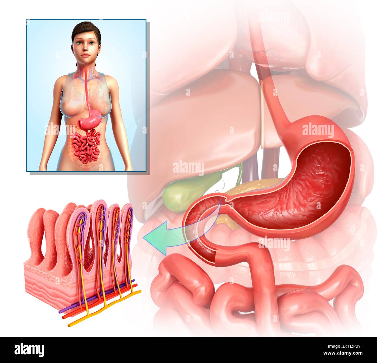 Illustration Of Stomach Anatomy And Intestinal Villi Stock
 The Wall Of The Small Intestine Showing Numerous Villi At
 Amazon Com Antique Anatomy Print Microscope Intestinal
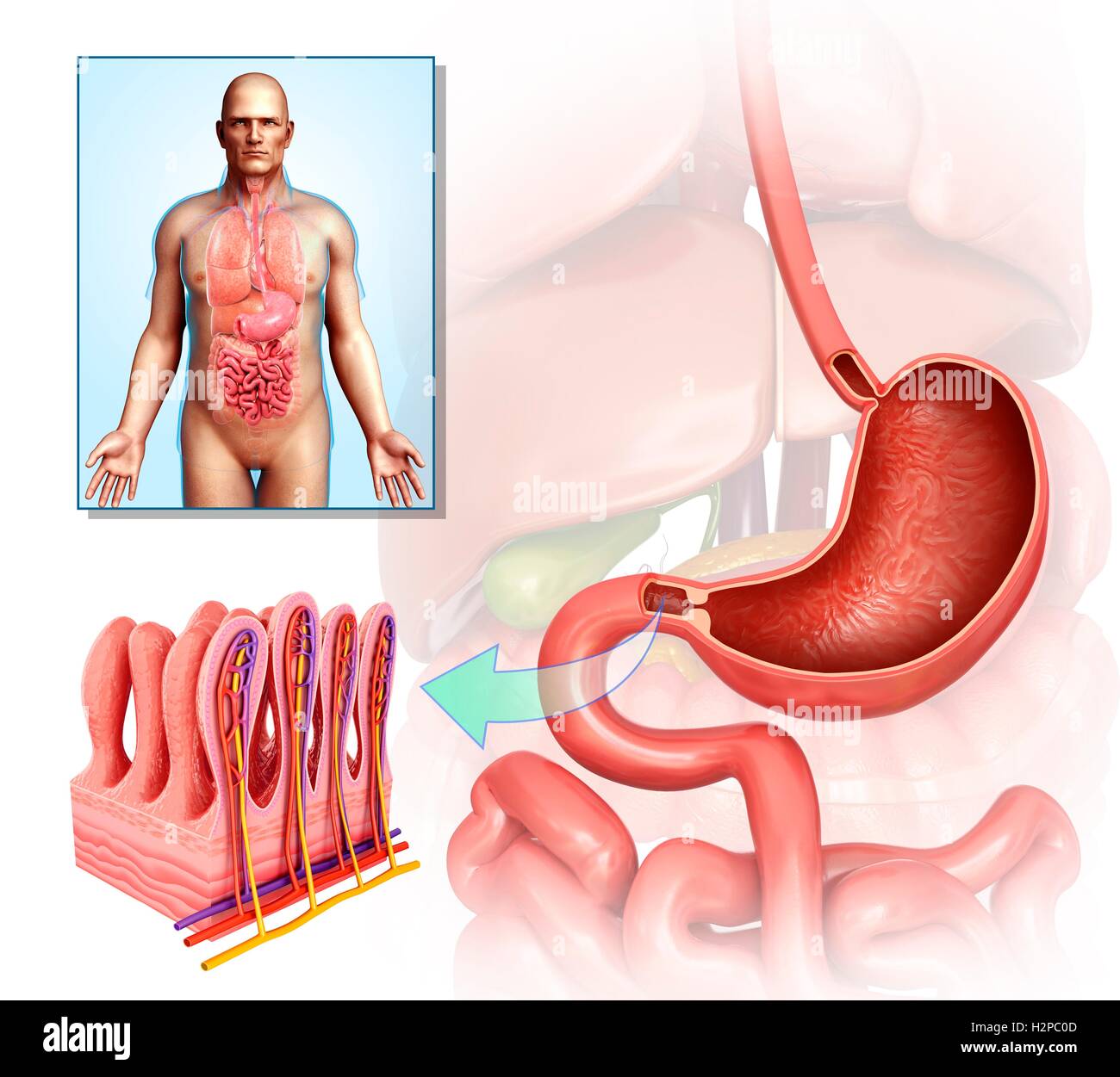 Illustration Of Stomach Anatomy And Intestinal Villi Stock
 Intestinal Villi Anatomy Epithelial Cells With Micro Villi


Posting Komentar
Posting Komentar