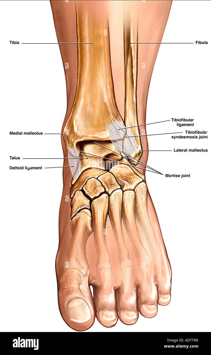
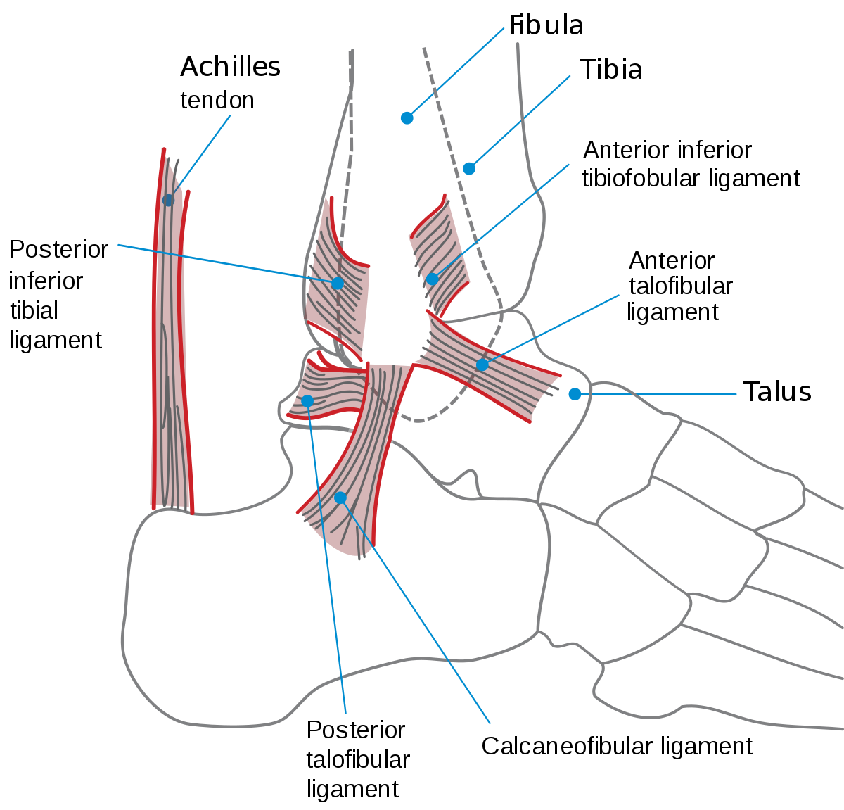
Ligaments are strong dense and flexible bands of fibrous connective tissue. The ankle joint proper or talocrural joint the subtalar joint and the inferior tibiofibular joint.
 Ankle Posterolateral Approach Approaches Orthobullets
There are nineteen bones in the forefoot.
Anatomy of ankle. This joint is a main contributor of stability in the lower limbs and it allows humans to perform actions such as running jumping and walking 1 2. Fascia is a broad fibrous. It is made up of three joints.
A foot bone that sits above the heel bone talus. Upper ankle joint tibiotarsal talocalcaneonavicular and subtalar joints. The hindfoot comprises of the ankle joint found at the bottom of the leg and is where the end.
Foot bones the hindfoot. The shin bone tibia. In common usage the term ankle refers exclusively to the ankle region.
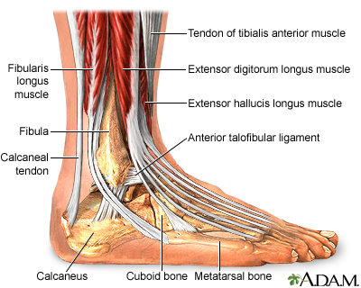
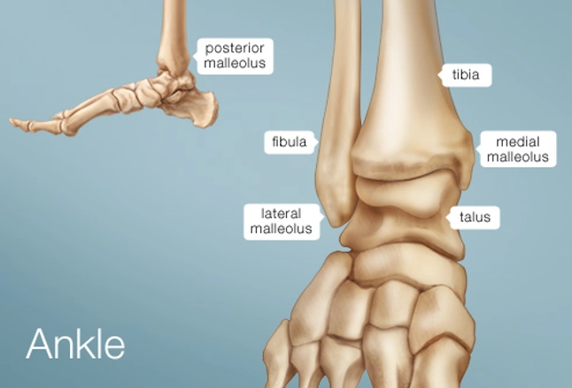
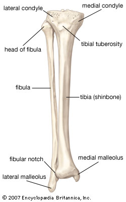
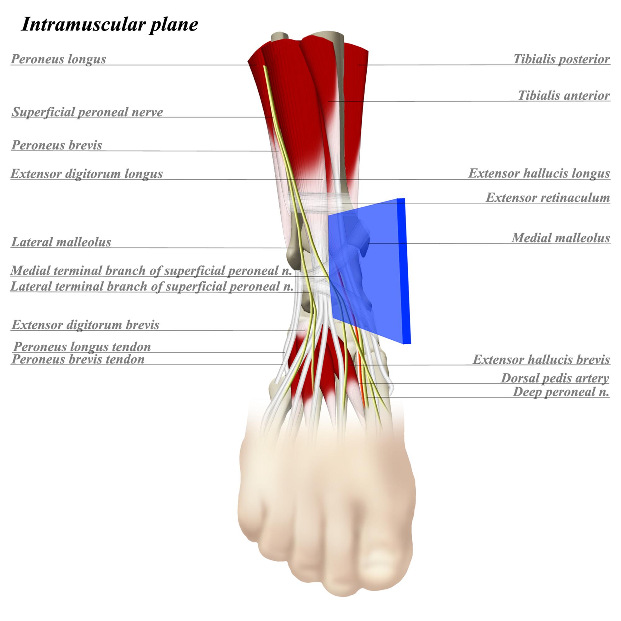
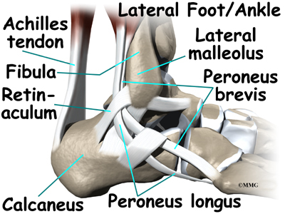
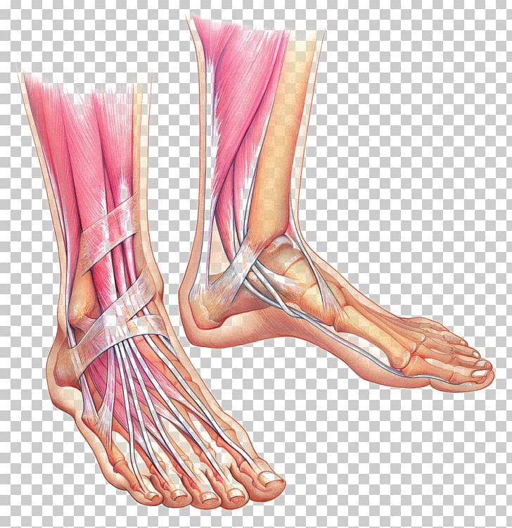
There are many muscles that help to move and support the ankle and foot. The thinner bone running next to the shin bone fibula. The inner bone is the tibia or shinbone which supports most of a persons weight when standing.
The ankle or the talocrural region is the region where the foot and the leg meet. The syndesmosis of the ankle refers to the membrane connecting the tibia to the fibula. Medically reviewed by healthline medical team on april 8 2015.
Tendons are elastic tissues made up of collagen. The tibia and fibula are connected throughout their length by an interosseous membrane. Biomechanically a certain amount of motion is allowed in all planes with respect to the distal ends of the tibia and fibula.
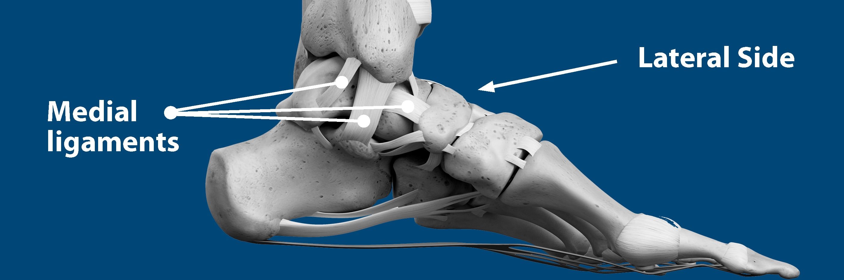
The ankle includes three joints. The ankle is a large joint made up of three bones. Soft tissues of the foot and ankle ligaments.
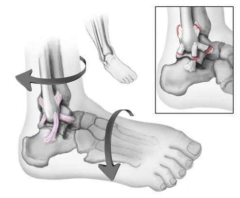
The ankle is the joint between the foot and leg composed of three separate bones. The movements produced at this joint are dorsiflexion and plantarflexion of the foot. The ankle is the joint that is located between the leg and the foot.
Ankle anatomy the ankle joint also known as the talocrural joint allows dorsiflexion and plantar flexion of the foot. The five bones of the midfoot are what make up our foot arches. The outer bone is the fibula or calf bone.
 Normal Anatomy Of The Ankle Stock Photo 7710327 Alamy
 Ankle Anatomy Diagram Quizlet
 Ankle Anatomy Medlineplus Medical Encyclopedia Image
 Ankle Joint Anatomy Overview Lateral Ligament Anatomy And

 Ankle Human Anatomy Image Function Conditions More
 Get To Know The Ankle Joint Yoga Journal
 Mri Anatomy Of Ankle
 Doctor Macc Ankle Sprain Ankle Ligaments Sprained Ankle
 Ankle Bones Anatomy Ankle Anatomy Foot Anatomy Ankle

 Anatomy Of An Ankle Sprain Bouldercentre For Orthopedics
Ankle Fractures Broken Ankle Orthoinfo Aaos
![]() Ankle Joint Anatomy Bones Ligaments And Movements Kenhub
Ankle Joint Anatomy Bones Ligaments And Movements Kenhub
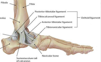 Medial Ankle Ligament Physiopedia

 Ankle Anterior Approach Approaches Orthobullets
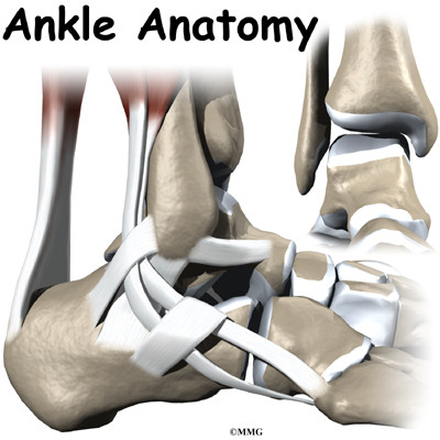 Ankle Anatomy Orthogate
 Ankle Wikipedia
![]() Ankle Joint Anatomy Bones Ligaments And Movements Kenhub
Ankle Joint Anatomy Bones Ligaments And Movements Kenhub
 Sprained Ankle Florida Orthopaedic Institute
![]() Ankle And Foot Anatomy Bones Joints Muscles Kenhub
Ankle And Foot Anatomy Bones Joints Muscles Kenhub
 Ankle Anatomy Orthogate
 Foot And Ankle Anatomical Chart
 Foot Anatomy Muscle Ankle Bone Png Clipart Anatomy Ankle
 Anatomy Of The Ankle Stock Illustration Illustration Of


Posting Komentar
Posting Komentar