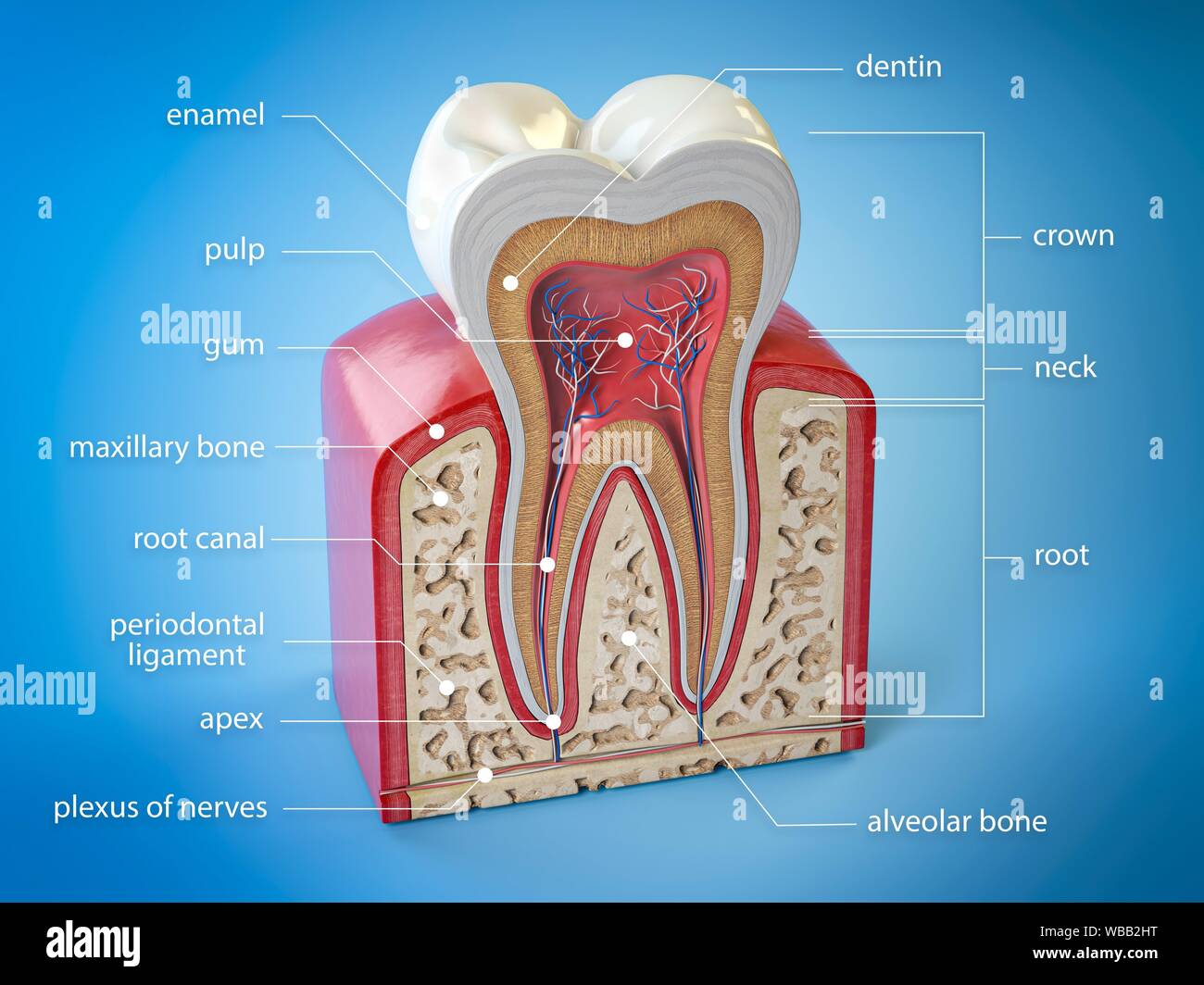
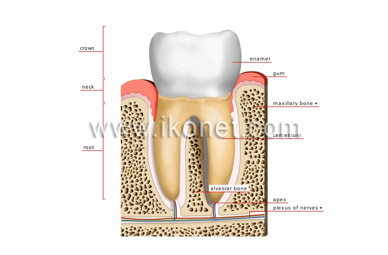
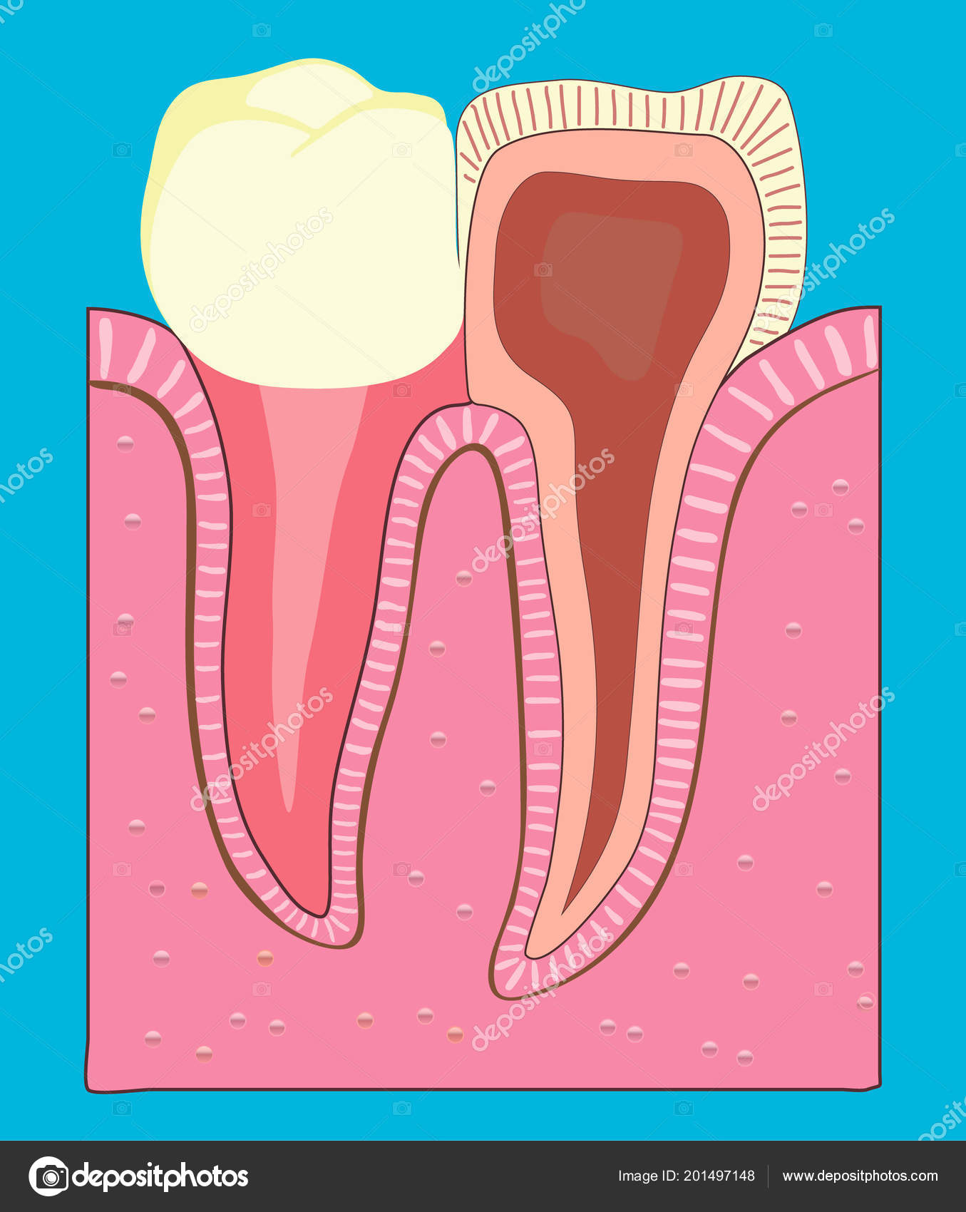
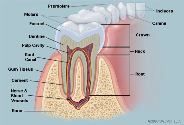
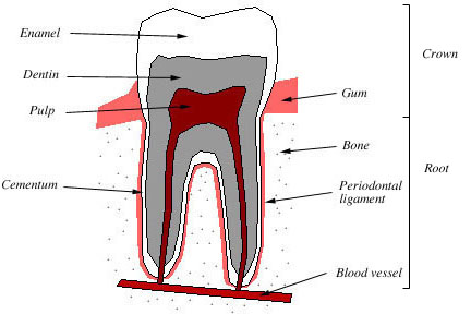
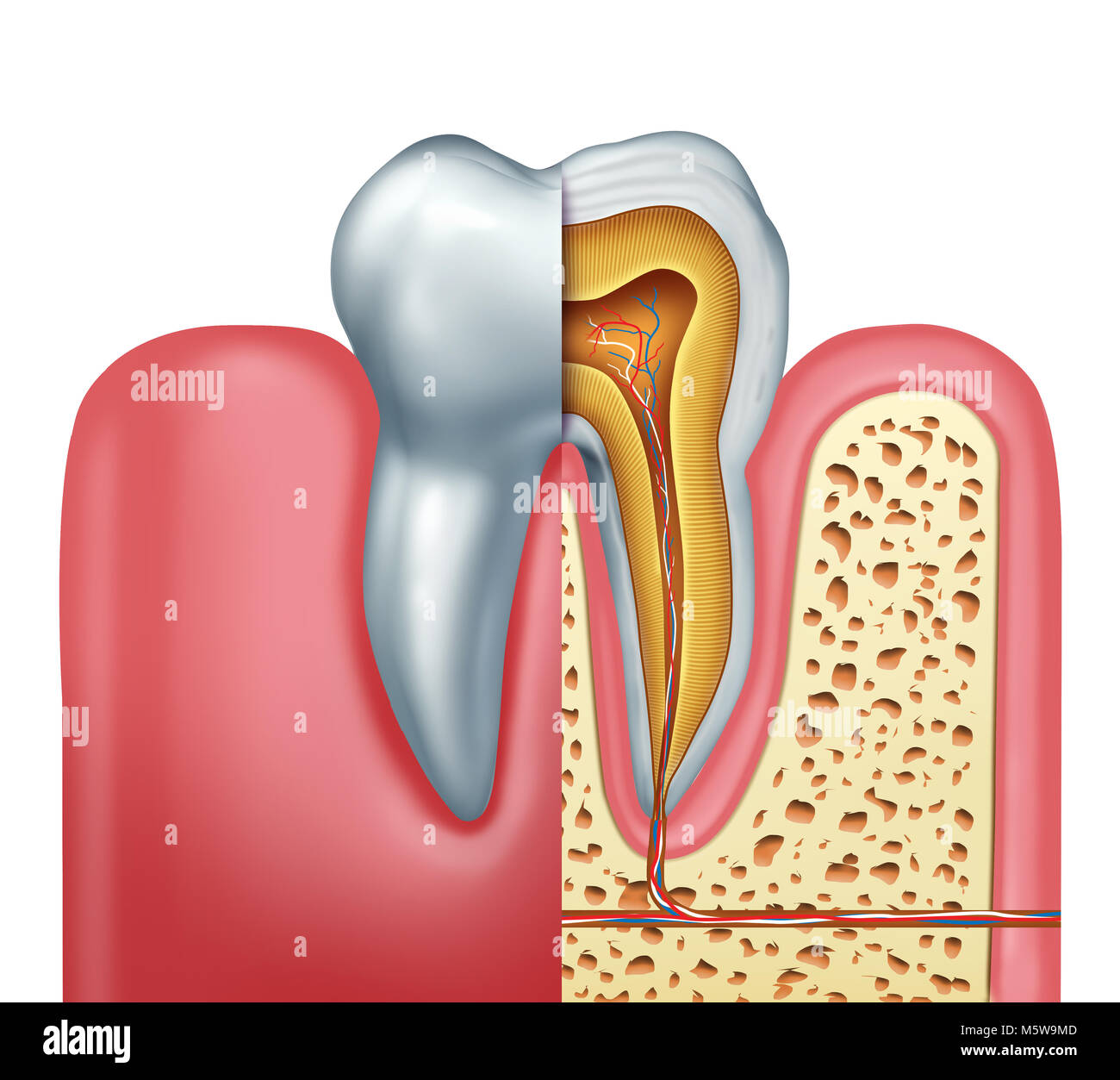
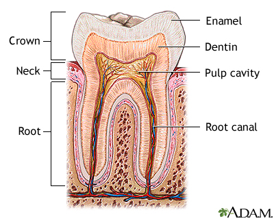
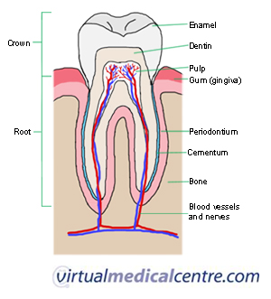
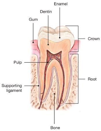
Pulp this is the soft tissue found in the center of all. The hardest white outer part of the tooth.
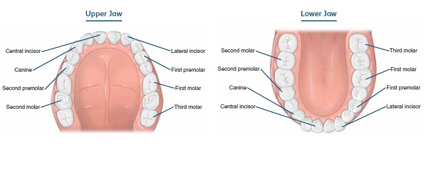 Tooth Types Dental Health Foundation
The softer living inner structure of teeth.

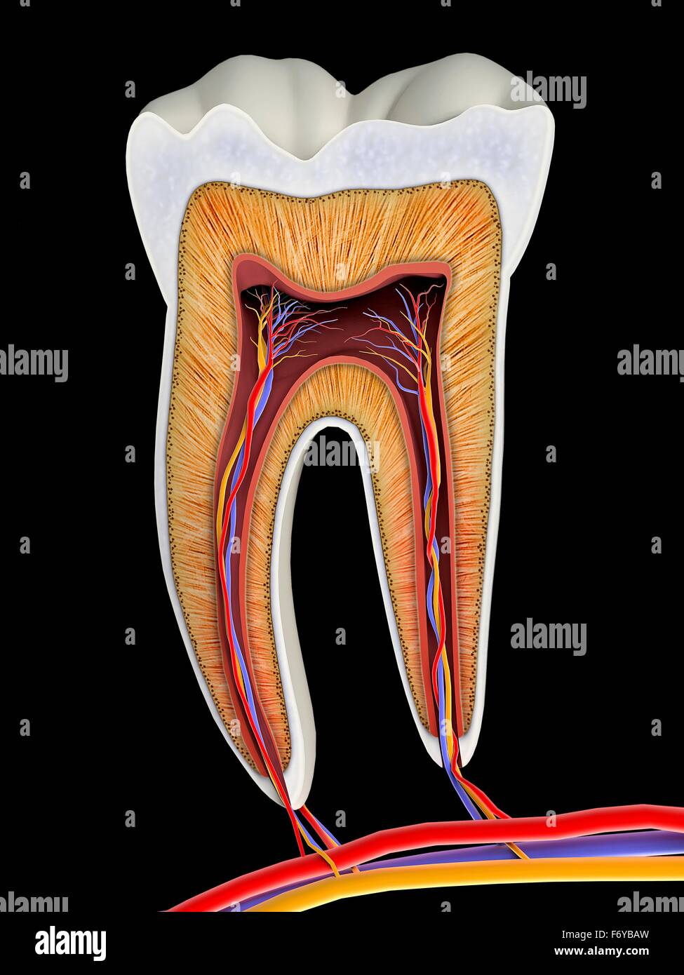
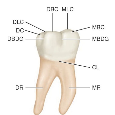
Anatomy of molar tooth. These complexities include multiple canals isthmuses lateral canals and apical ramifications. Enamel this is the outer and hardest part of the tooth that has the most mineralized tissue in. They are more developed in mammals.
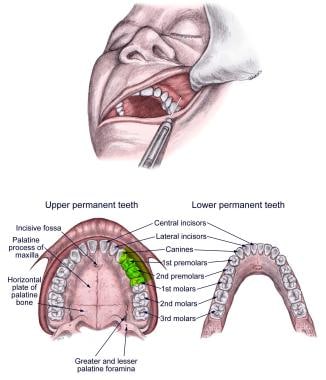
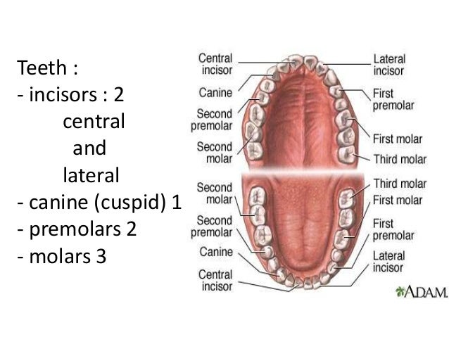
There are twenty deciduous primary teeth in young children with ten per jaw and five in each quadrant which consist of distal to mesial. The function of teeth as they contact one another falls elsewhere under dental occlusion tooth formation begins before birth. A layer of connective tissue that binds the roots of the.
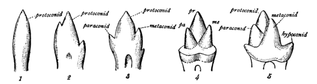
The deciduous primary teeth start erupting at six months lower central incisor and are completely erupted by around 3 years of age. Each premolar has two cusps hence the name bicuspid. The development appearance and classification of teeth fall within its purview.
The mandibular molars in particular the mandibular first molar are the most frequently endodontically treated teeth. Picture of the teeth enamel. It is a hard tissue that contains microscopic tubes.
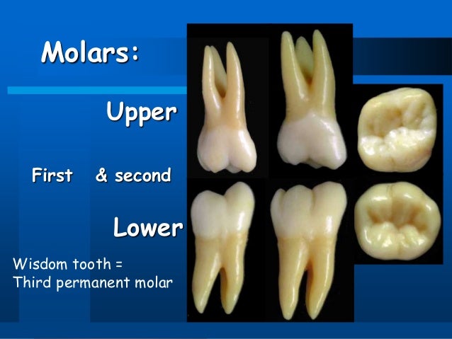
A layer underlying the enamel. They are the teeth farthest back in the mouth. Most people start off adulthood with 32 teeth not including the wisdom teeth.
They are more developed in mammals. The root is the part of the tooth that extends into the bone and holds. Their treatment offers a variety of anatomical challenges.
Each tooth has several distinct parts. Dentin this is the layer of the tooth under the enamel. They are used primarily to grind food during chewing.
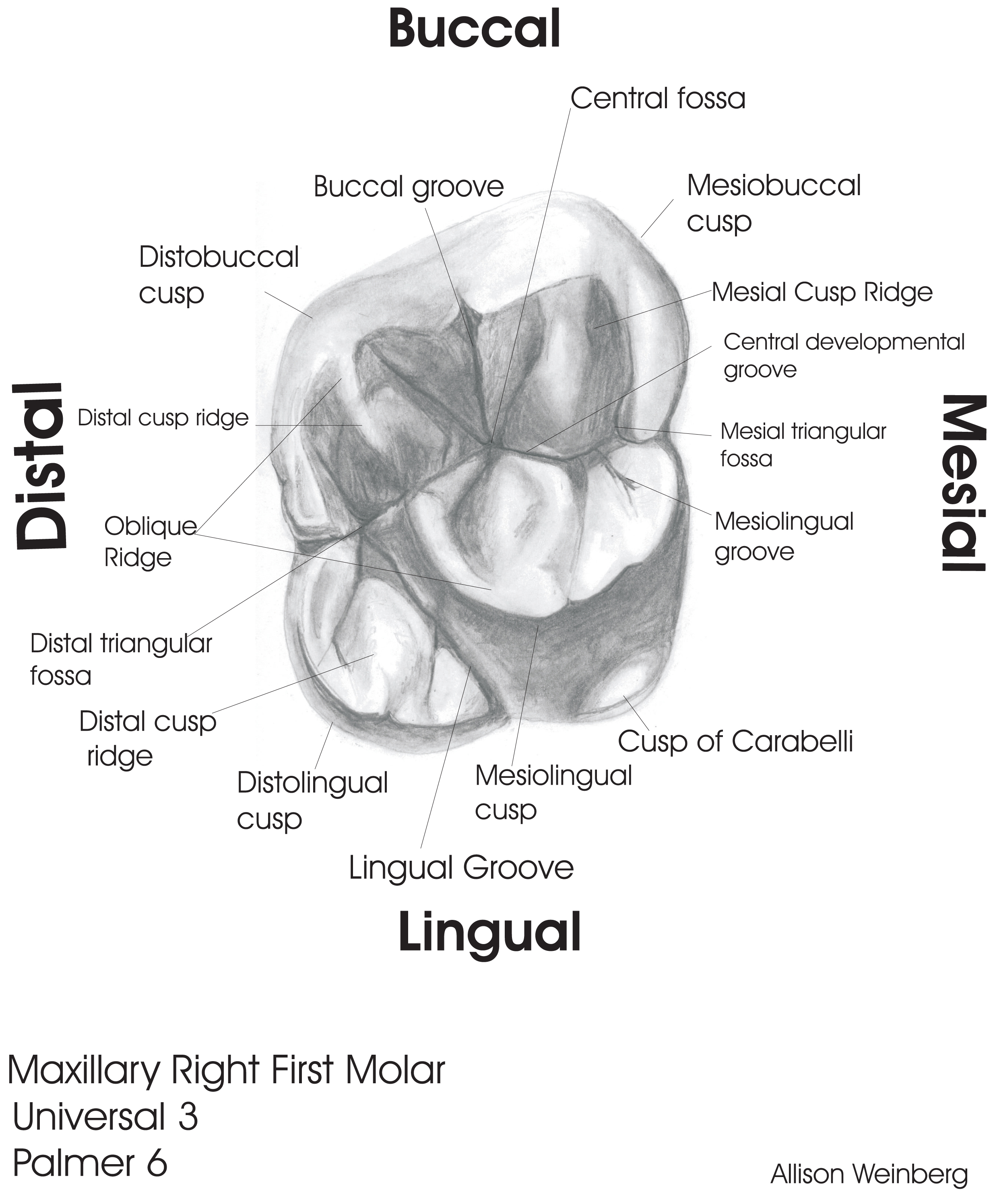
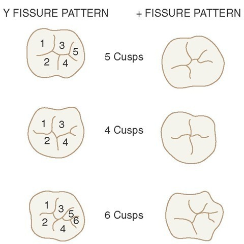
The molars or molar teeth are large flat teeth at the back of the mouth. The molars by contrast are used exclusively for crushing and grinding. Each molar typically has four or five cusps.
Anatomy of the tooth the tooth is one of the most individual and complex anatomical as well as histological structures in the body. Dental anatomy is a field of anatomy dedicated to the study of human tooth structures. Explore the interactive 3 d diagram below to learn more about teeth.
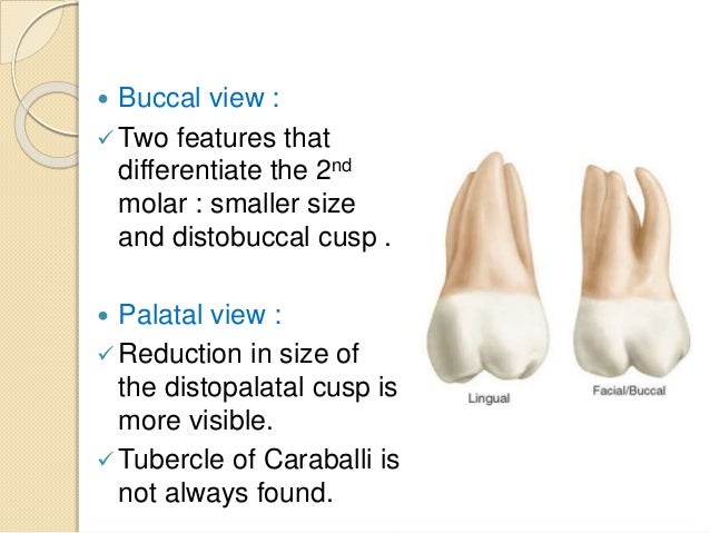
Tooth anatomy types of teeth. The tissue composition of a tooth is only found within the oral cavity and is limited to the dental structures. The third molar in humans tends to be variable in size number of roots cusp pattern and eruption.
Here is an overview of each part.
 Dental Anatomy Maxillary Molars
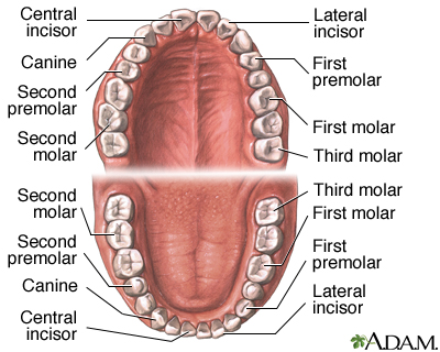 Dental Anatomy Medlineplus Medical Encyclopedia Image
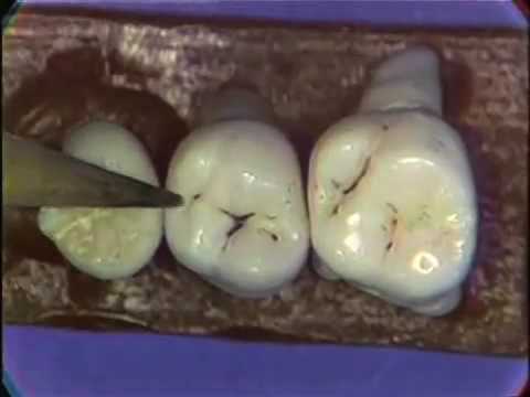
 Somso Molar Tooth With Caries
 Molar Tooth Cross Section Stock Photos Molar Tooth Cross
 Dental Anatomy
 Molar Tooth Britannica
 Premolar Wikipedia
 Oral Nerve Block Overview Indications Contraindications
 Sample Drawings Tooth Morphology
 Anatomy Of The Molar Tooth Stock Vector C Ashpin Tyev List
 External And Internal Root Canal Anatomy Of The First And
 Human Molar Tooth 3d Model In Anatomy 3dexport

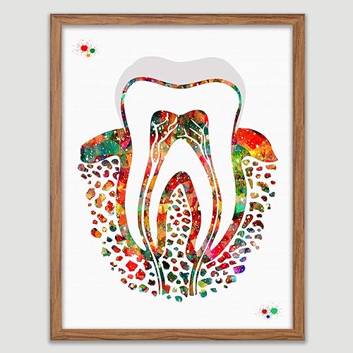 Amazon Com Molar Tooth Watercolor Poster Tooth Anatomical
 The Teeth Human Anatomy Diagram Names Number And
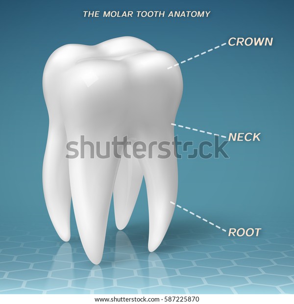 Molar Anatomy Crown Neck Root Tooth Stock Vector Royalty

 Molar Tooth Wikipedia
 Molar Tooth Cross Section Stock Photos Molar Tooth Cross
 Anatomy And Morphology Of Teeth
 Dental Anatomy
 Tooth Bone And Gingiva Watercolor Print Abstract Dental
 Molar Tooth Cross Section Stock Photos Molar Tooth Cross
 Tooth Anatomy Medlineplus Medical Encyclopedia Image
 Teeth Anatomy Adult Teeth Permanent Dentition
 Cracked Teeth American Association Of Endodontists


Posting Komentar
Posting Komentar