A vein can range in size from 1 millimeter to 1 15 centimeters in diameter. Veins are thin walled being thinner than the arteries.
 Leg Dvt Normal Ultrasoundpaedia
The deep femoral profunda femoris vein joins the femoral vein to form the common femoral vein at about 9 cm below the inguinal ligament27 the common femoral vein is medial to the common femoral artery and it becomes the external iliac vein at the level of the inguinal ligament.
Anatomy veins. Veins are the large return vessels of the body and act as the blood return counterparts of arteries. There are three main deep veins in the lower leg. This is a vital task to keep your body healthy and functioning.
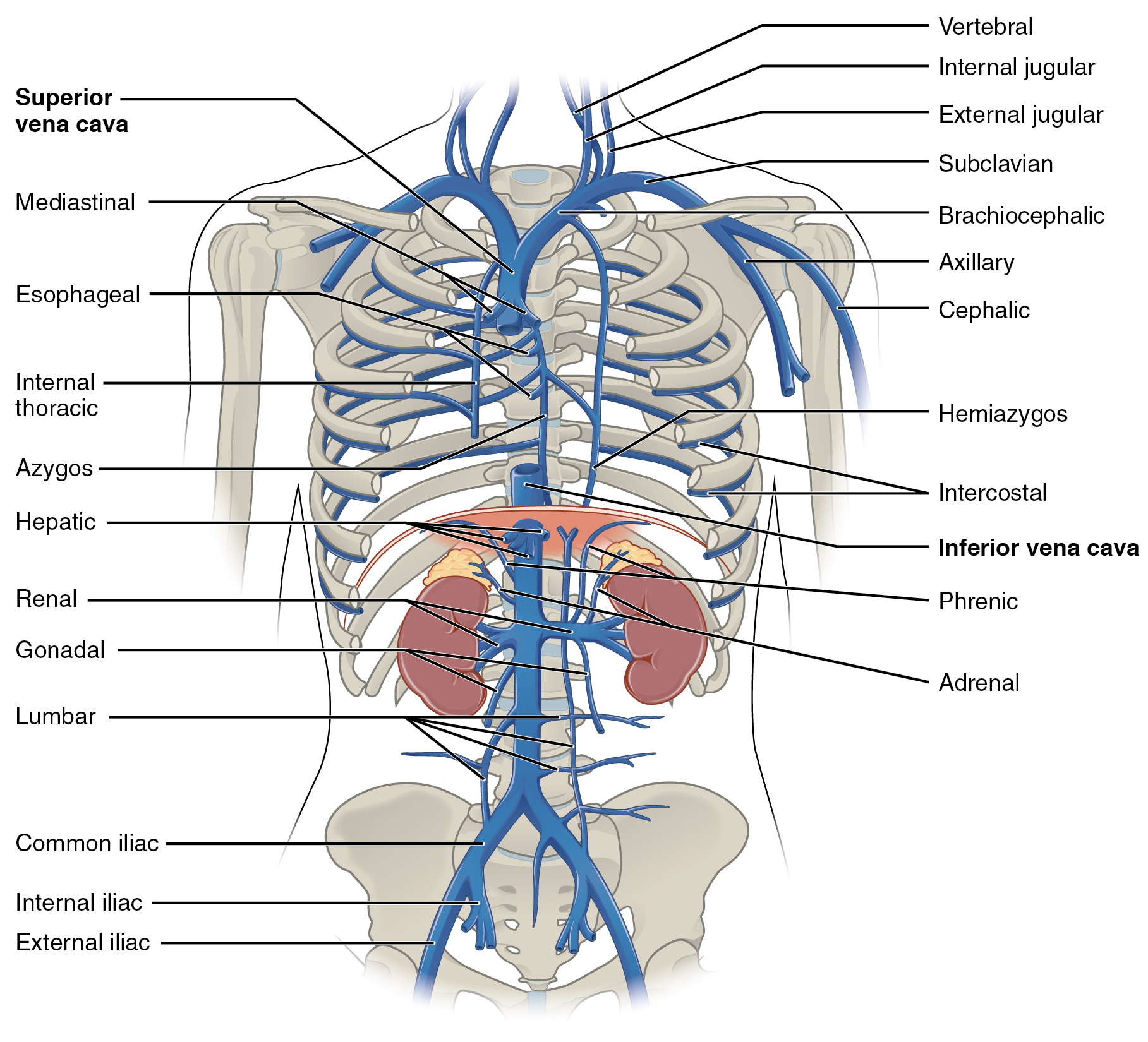
The anatomy of the inferior vena cava. The anatomy and importance of the pulmonary vein. Capillary structure and function in the body.
Because the arteries arterioles and capillaries absorb most of the force of the hearts contractions veins and venules are subjected to very low blood pressures. They carry the deoxygenated blood which is bluish in color and for the same reason veins appear blue. The pulmonary veins serve a very important purpose of delivering freshly oxygenated blood.
Anterior tibial vein which receives blood from the dorsal venous arch. Veins are responsible for carrying the de oxygenated blood back to the heart. The anatomy and function of the portal vein.
Veins are the blood vessels which carry the blood from peripheral tissues towards heart. Veins are blood vessels that carry blood towards the heart. Veins lie closer to the surface of the skin than arteries and can often be seen on various parts of the body that contain a lot of muscle mass such as your arms legs and chest area.
Their lumen is larger than that of the accompanying arteries. The pulmonary veins can be affected by. Most veins carry deoxygenated blood from the tissues back to the heart.
The pulmonary veins along with the pulmonary arteries make up the pulmonary circulation. The smallest veins in the body are called venules. The anatomy of the pulmonary vein anatomy.
Anatomy function and significance. Exceptions are the pulmonary and umbilical veins both of which carry oxygenated blood to the heart. How blood flows through the heart and lungs.
Posterior tibial vein and fibular vein also known as the peroneal vein which form from the medial and lateral plantar veins. The anatomy of jugular veins. Types of veins systemic veins.
In contrast to veins arteries carry blood away from the heart.
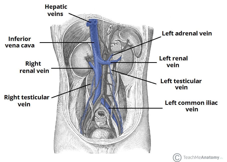 Venous Drainage Of The Abdomen Teachmeanatomy

 Human Red Eye Veins Set Anatomy Blood Vessel
 Blood Finds A Way Pictorial Review Of Thoracic Collateral
 Human Veins Diagram Click Through For The Full Circulatory
 Anatomy Veins Head Neck Brain To Heart Diagram Quizlet
 Vessel Anatomy Veins Of The Lower Extremities Diagram
 20 5 Circulatory Pathways Anatomy And Physiology
 Femoral Vein Wikipedia
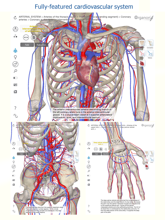
3d Organon Anatomy Heart Arteries And Veins App Price Drops
 Anatomy Of Major Abdominal Veins Inferior Vena Cava
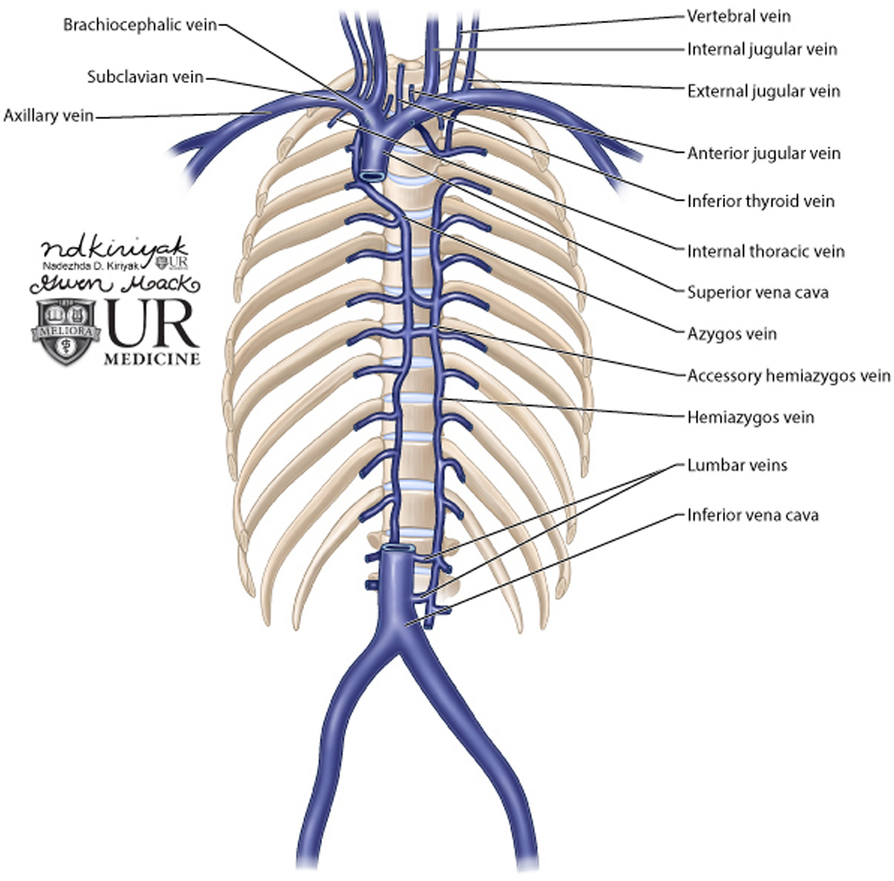
 Venous Anatomy Thoracic Key

 Nerves Blood Vessels And Lymph Advanced Anatomy 2nd Ed

 Thoracic And Abdominal Veins Course Hero
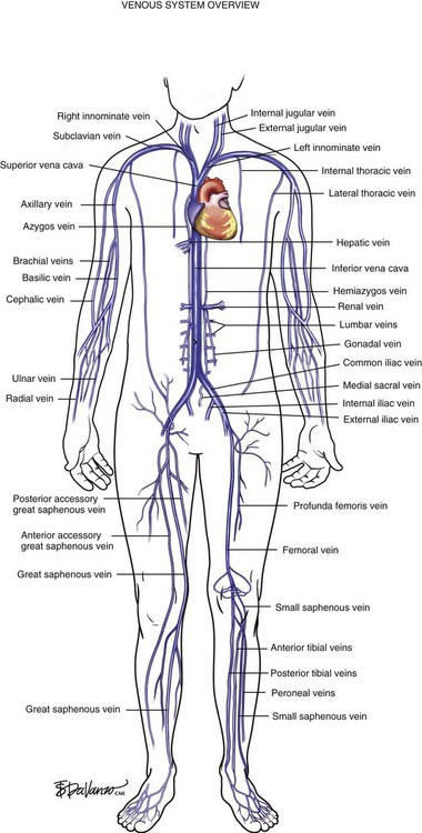
 This Diagram Shows The Major Veins In The Human Body
 Vintage Anatomy Veins And Arteries Illustration Square Sticker Zazzle Com
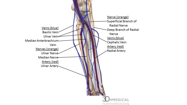
 Anatomy Veins Of The Hand And Forearm Critical Care
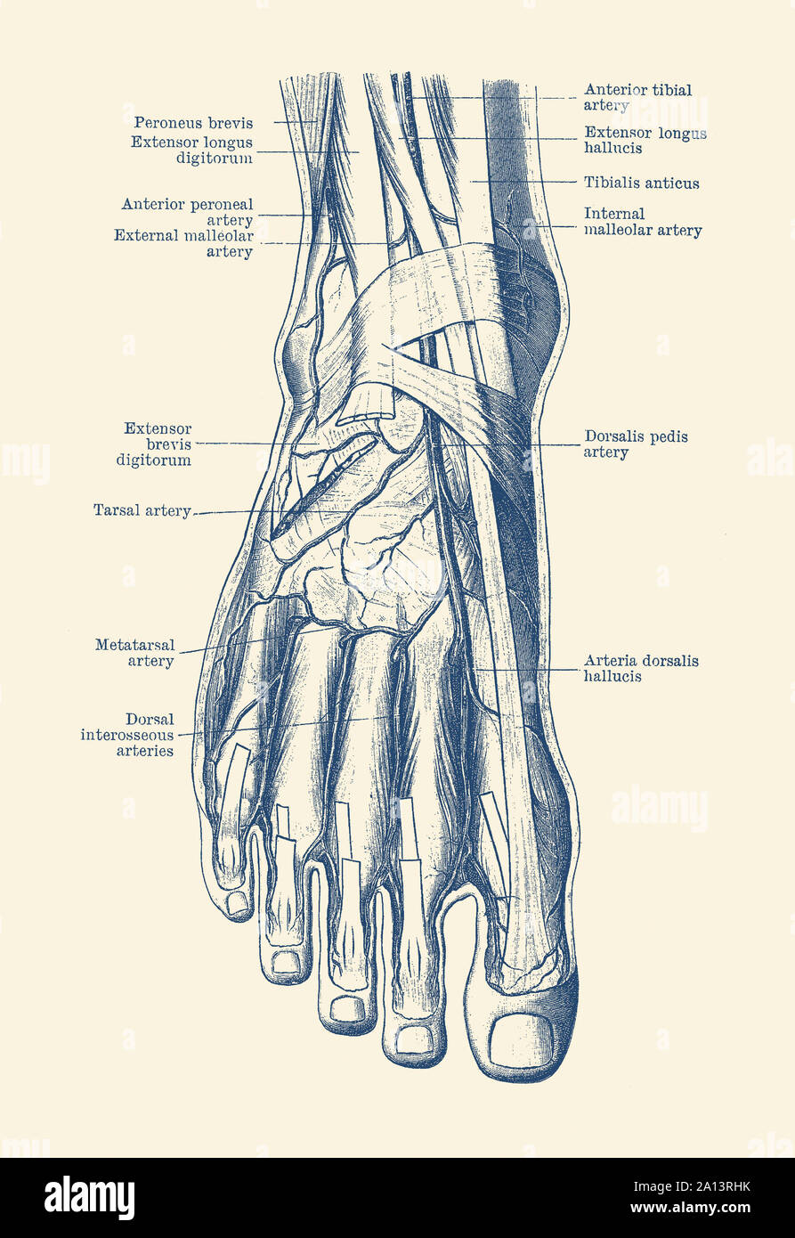 Vintage Anatomy Print Of The Human Foot Showcasing The
 Veins Of Anatomy T Shirt By Designpro44 Design By Humans
 Vintage Human Anatomy Blood Veins
 Stock Illustration
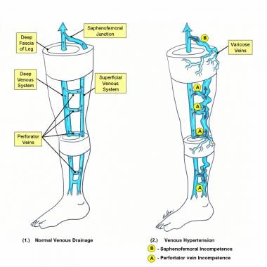 Varicose Vein Surgery Practice Essentials Anatomy
 Vein Wikipedia
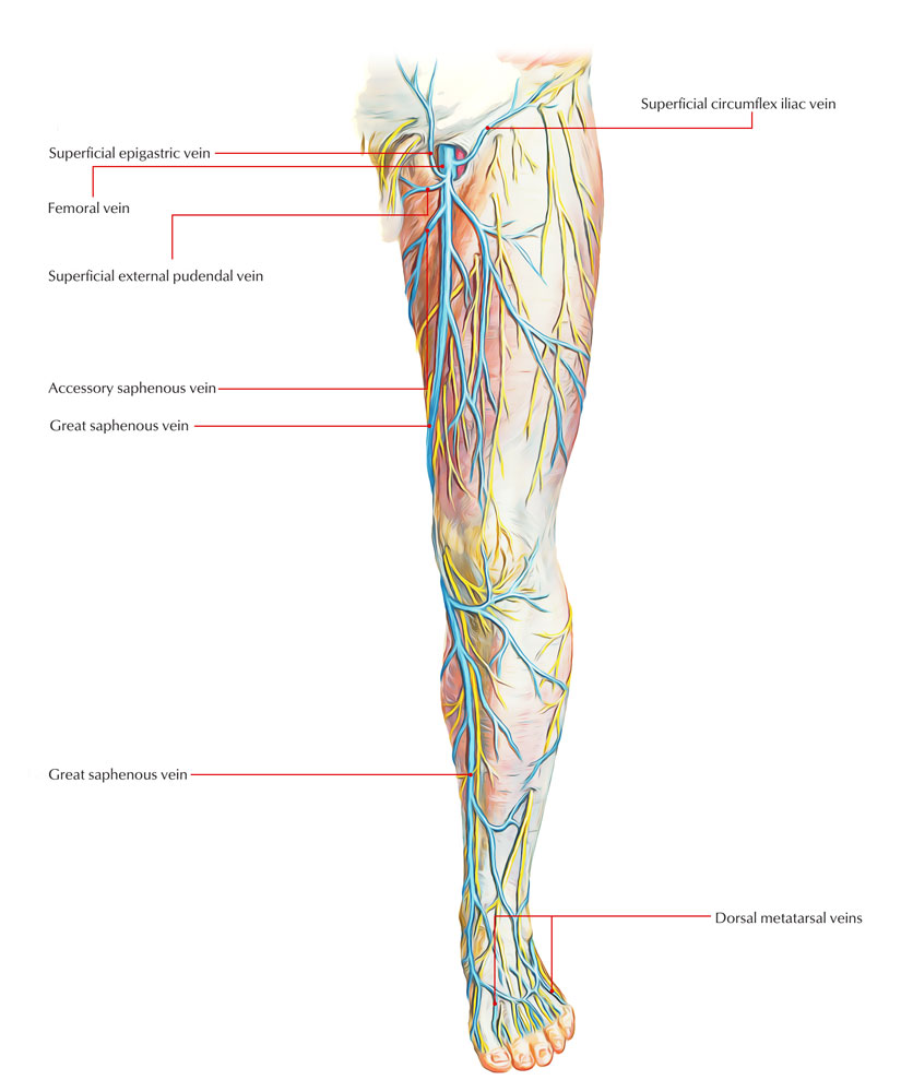 Veins Of Lower Limb Earth S Lab


Posting Komentar
Posting Komentar