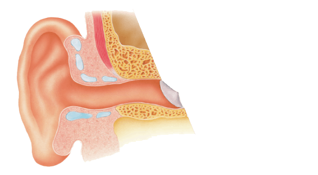
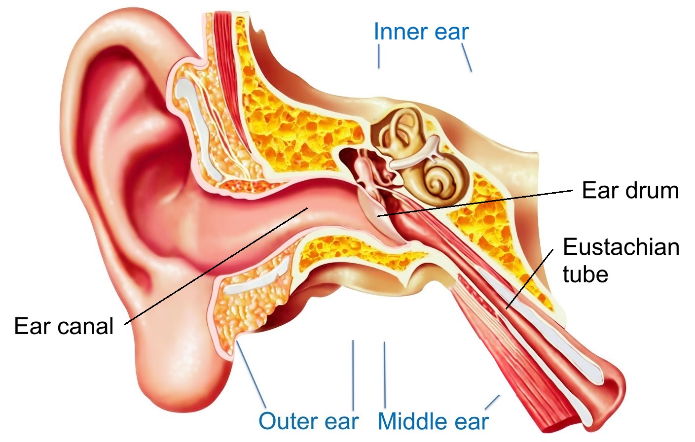
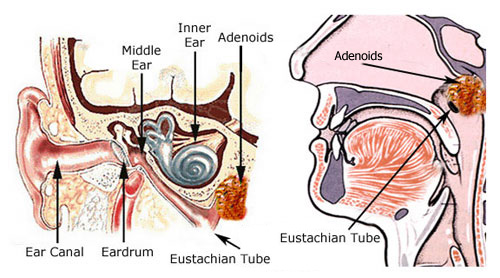
Ear canal the ear canal starts at the outer ear and ends at the ear drum. These types of agonizing conditions might be related to injury such as minor strain sprain or whiplash.
 What Is Eustachian Tube Dysfunction Sinussurgeryoptions Com
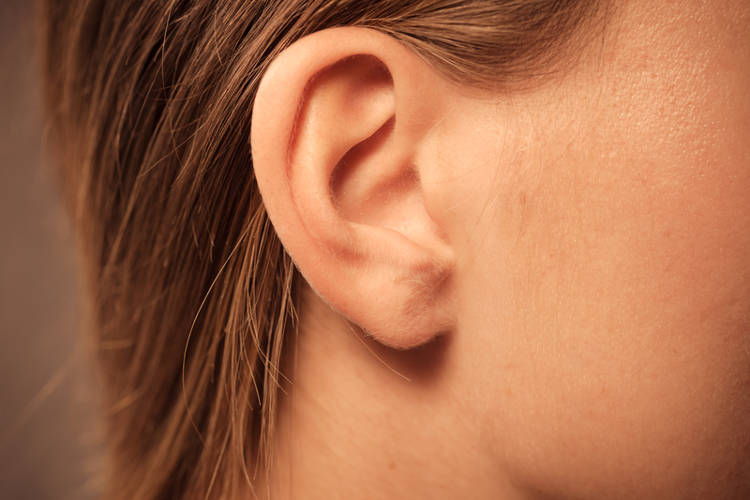
This is the outside part of the ear.
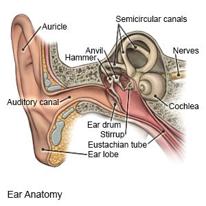
Behind the ear anatomy. The parts of the ear include. The mastoid bone is the bone behind the ear which is hollow. Ear anatomy outer ear.
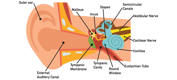
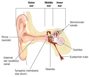
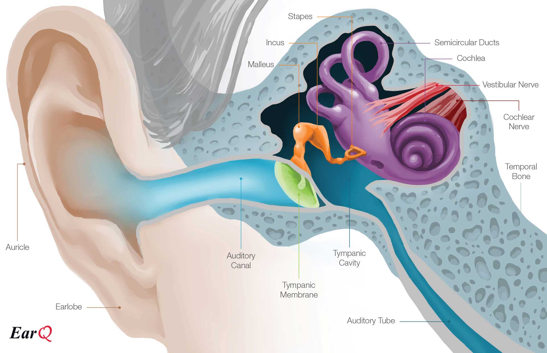
This is the tube that connects the outer ear to the inside or middle ear. This part of the ear provides protection and channels sound. External auditory canal or tube.
The outer ear includes. It is located behind the ear and is known as the c1 bone of the spinal vertebral level. Infection of the mastoid bone just behind the ear.
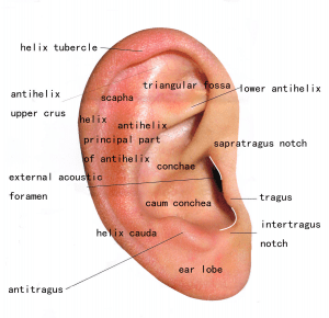
There are three different parts to the outer ear. Tympanic membrane also called the eardrum. The tragus helix and the lobule.
Mastoiditis can result from untreated middle ear infections. The ear is the organ of hearing and balance. The canal is approximately an inch in length.
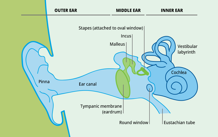
One opening is covered with the stapes bone and is called the oval window. On the inside wall of the box are two openings into the inner ear. The mastoid process anatomy comprises complex structures.
The outer ear includes an ear canal that is is lined with hairs and glands that secrete wax. Sound travels through the auricle and the auditory canal a short tube that ends at the eardrum. The mastoid process is the small bony prominence located behind each ear.
Mastoiditis can result from untreated middle ear infections. Auricle cartilage covered by skin placed on opposite sides of the head auditory canal also called the ear canal eardrum outer layer also called the tympanic membrane the outer part of the ear collects sound. The outer ear is made up of cartilage and skin.
Muscular neck pain behind ear. The other opening is covered with a thin membrane and is called the round window. The mastoid process bone itself is in the shape of a pyramid that projects behind the temporal bone.
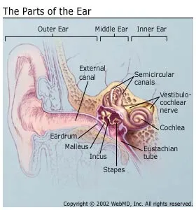
External or outer ear consisting of. Three tiny bones the malleus incus and stapes within the middle ear transfer sound vibrations from the eardrum to the inner ear. Mastoid process bone behind the ear.
The skin of the ear canal is very sensitive to pain and pressure. The soft tissues of the head anatomy can also experience or cause pain behind the ear structure.
 Swimmer S Ear Otitis Externa Causes Diagnosis Treatment
 Middle Ear An Overview Sciencedirect Topics
 Ears Pasadena Newport Beach Dr Hung
 Otitis Media Wikipedia
 Tympanic Membrane Rupture And Middle Ear Infection In Cats
 Learn How Middle Ear Infections Can Affect Hearing Ability
 Anatomy Of The Ear
 Anatomy Of The Ear Inner Ear Middle Ear Outer Ear
 Ear Anatomy Mydr Com Au
 Anatomy Of The Human Ear Diagram Chart Black Wood Framed Art Poster 14x20
Can Ear Infections Cause Hearing Loss
 Anatomy Of The Ear Medcor
:max_bytes(150000):strip_icc()/GettyImages-506836745-595d4dc05f9b58843f509ed2.jpg) Patulous Eustachian Tube Symptoms Causes And Treament
Patulous Eustachian Tube Symptoms Causes And Treament
 Deafness And Hearing Loss Causes Symptoms And Treatments
 Ear Education Tampa Bay Hearing
 Vector Illustration Of Diagram Of Human Ear Anatomy
 Introduction To Biology Of The Ears Nose And Throat Ear
 Ear Infection Middle Ear Symptoms Treatment Southern

 Microtia Congenital Ear Deformity Institute Hearing
 045 Sensory Innervation Of The Ear Anatomy For Emergency
 Demystifying The Ear Canal Consider Professional Ear
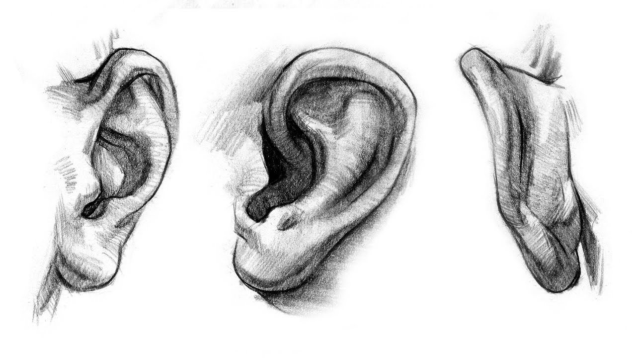 How To Draw Ears Anatomy And Structure
 Mastoiditis What You Need To Know


Posting Komentar
Posting Komentar