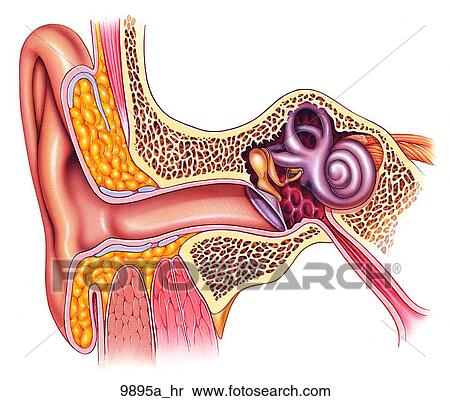
The external ear can be divided functionally and structurally into two parts. The internal ear or labyrinth human anatomy.
 Ear Functional Anatomy Ear Nose Throat Medbullets Step 2 3
In this video we take an anatomical view of the inner ear.
Ear anatomy internal. External middle and innerthis article will focus on the anatomy of the external ear its structure neurovasculature and its clinical correlations. Pain in the ear can have many causes. The outer middle and inner ear.
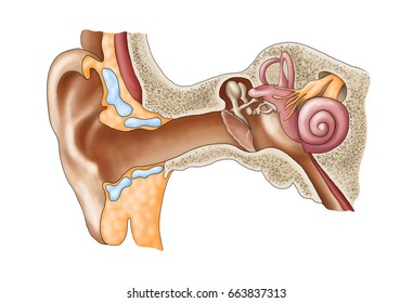
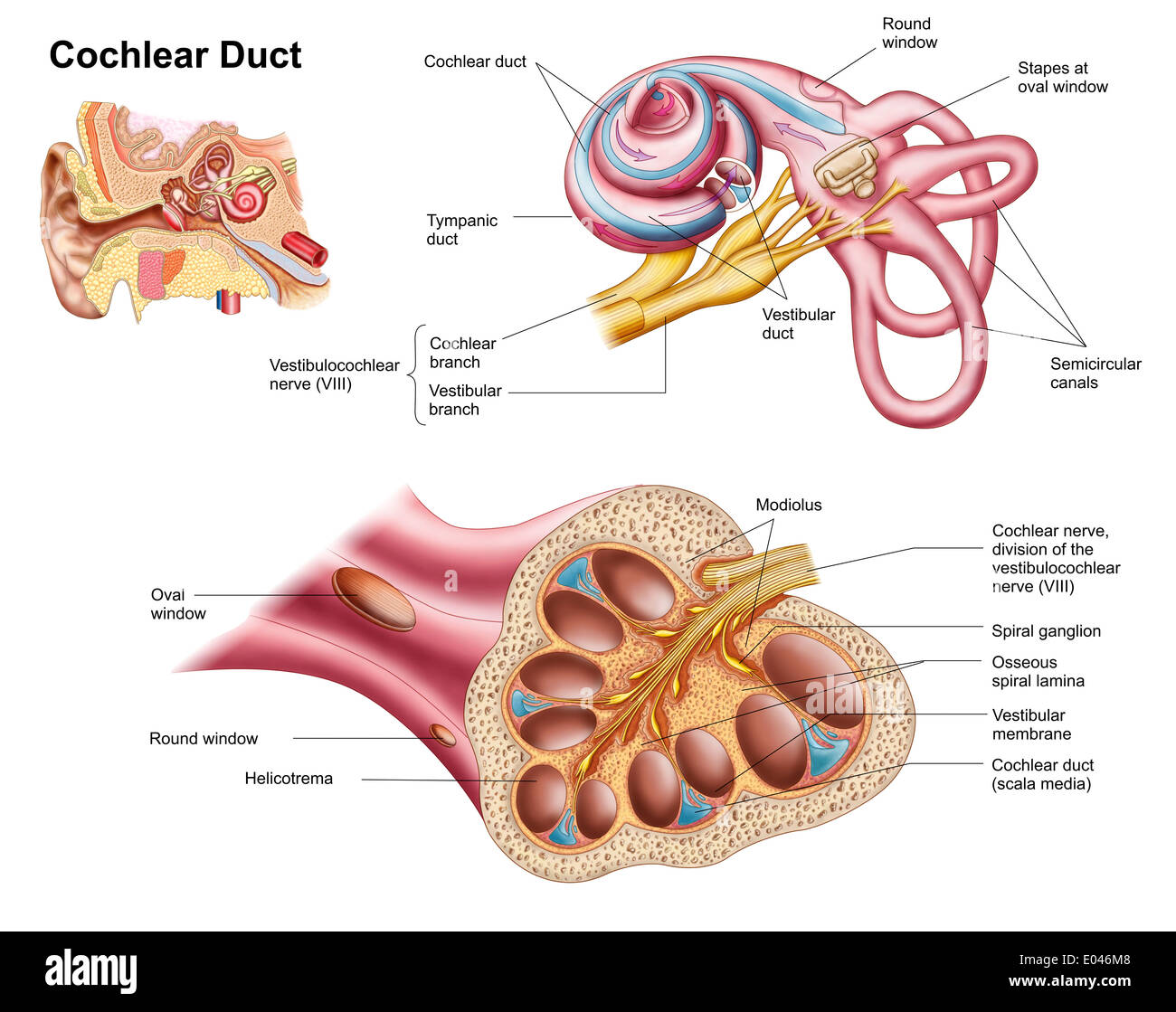
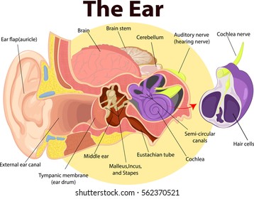
Inner ear also called labyrinth of the ear part of the ear that contains organs of the senses of hearing and equilibrium. The cochlea is shaped like a snail and is divided into two chambers by a membrane. Within the bony labyrinth is a membranous labyrinth.
I briefly discuss some physiological components to these anatomical structures. It is called the labyrinth from the complexity of its shape and consists of two parts. In vertebrates the inner ear is mainly responsible for sound detection and balance.
The inner ear internal ear auris interna is the innermost part of the vertebrate ear. The fluid filled semicircular canals labyrinth attach to the cochlea and nerves in the inner ear. The osseous labyrinth a series of cavities within the petrous part of the temporal bone and the membranous labyrinth a series.
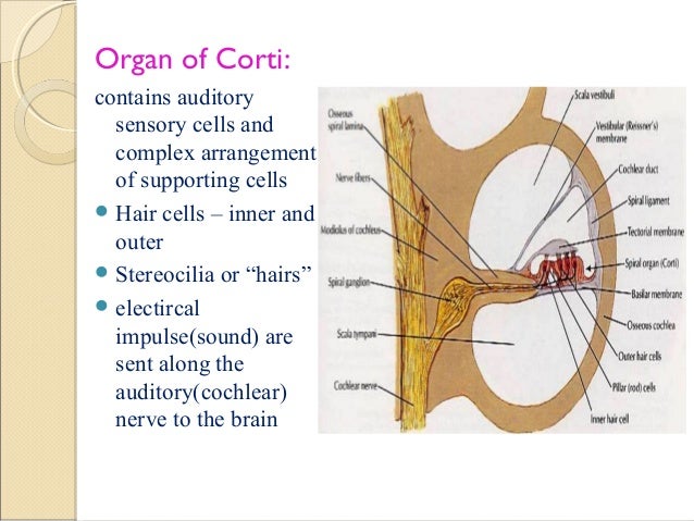
The internal ear is the essential part of the organ of hearing receiving the ultimate distribution of the auditory nerve. The bony labyrinth a cavity in the temporal bone is divided into three sections. The chambers are full of fluid which vibrates when sound comes in and causes the small hairs which line the membrane to vibrate and send electrical impulses to the brain.
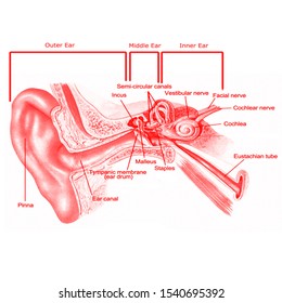
In mammals it consists of the bony labyrinth a hollow cavity in the temporal bone of the skull with a system of passages comprising two main functional parts. The inner ear has two main components the bony labyrinth and membranous labyrinth. Acquired entities can further be delineated into intrinsic processes such as cancer and extrinsic processes such as trauma.
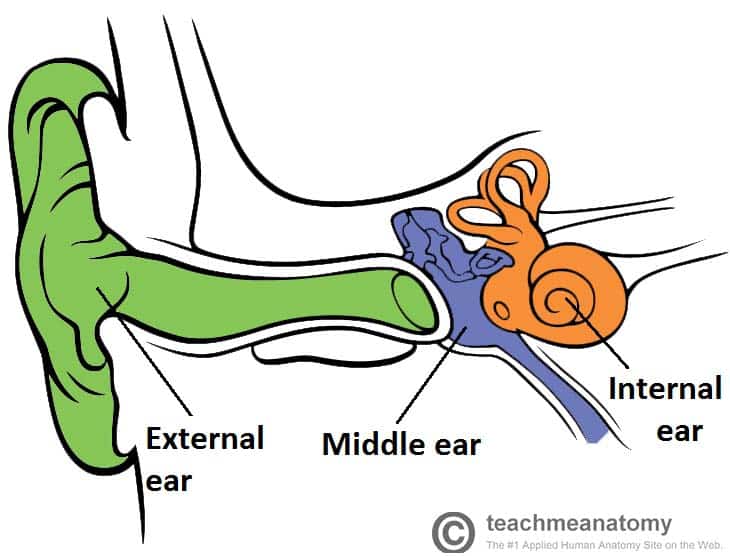
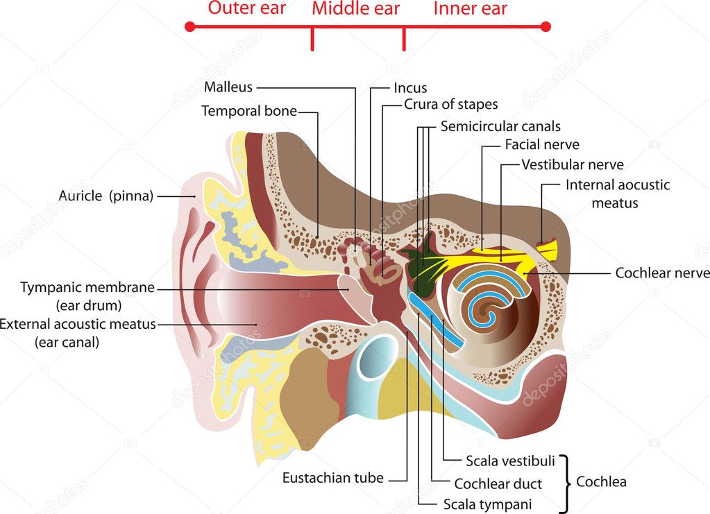
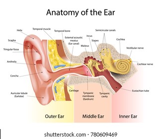
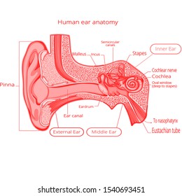
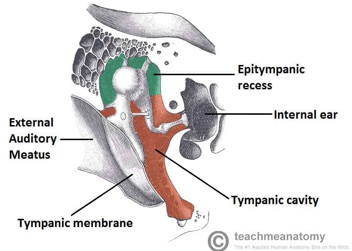
The auricle or pinna and the external acoustic meatus which ends at the tympanic membrane. All three parts of the ear are important for detecting sound by working together to move sound from the outer part through the middle and into the inner part of the ear. It lies between the middle ear and the internal acoustic meatus which lie laterally and medially respectively.
The ear is made up of three parts. The eustachian auditory tube drains fluid from the middle ear into the throat pharynx behind the nose. Ear anatomy inner ear.
Congenital abnormalities of the ear are common and largely affect the shape of the auricle. Ears also help to maintain balance. Variant anatomy of the external ear can be divided into congenital and acquired entities.
The cochlea which is the hearing portion and the semicircular canals is the balance portion. The vestibule the semicircular canals and the cochlea. The inner ear is located within the petrous part of the temporal bone.
They send information on balance and head position to the brain. The ear can be divided into three parts.
 Internal And Middle Ear Anatomy Diagram Quizlet
 Cartoon Of Human Internal Ear Anatomy
 Ear Anatomy Crosssection Right External Middle Stock Vector
 The Inner Ear Bony Labyrinth Membranous Labryinth
 Highlighting Internal And External Ear Injuries In 2019
 ᐈ The Inner Ear Royalty Free Inner Ear Images Download
 Vector Art Ear Anatomy Clipart Drawing Gg55508417 Gograph
 Anatomy Of Inner Ear
 Human Ear Anatomy Ears Inner Structure Organ Of
 Anatomy Of Inner Ear It Consists Of Six Mechanoreceptor
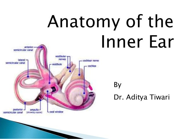 Anatomy Of Inner Ear By Dr Aditya Tiwari
 Internal Ear Anatomy Pt 2 Diagram Quizlet
 Ear The Big Picture Gross Anatomy 2e Accessmedicine
 Figure 6 1 External And Internal Structures Of The Human Ear
Inner Ear Images Stock Photos Vectors Shutterstock
 Male Human Anatomy Body Internal Organs
 Anatomy Lab Ear Biology Flashcards Quizlet
 Figure 25 1 Anatomy Of The Ear Ppt Download
 Internal Ear Stock Photos Internal Ear Stock Images Alamy
 Special Senses Figure 24 14 Anatomy Of The Internal Ear
 Inner Middle Ear Images Stock Photos Vectors Shutterstock
 Internal Ear Images Stock Photos Vectors Shutterstock
 Inner Ear Wikipedia
 Imagenes Fotos De Stock Y Vectores Sobre Internal Ear Organ
 The Middle Ear Parts Bones Muscles Teachmeanatomy
 How To Draw Human Ear Easy Way Internal Structure
 Total Ear Canal Ablation Teca In Dogs
 Ear External And Internal Anatomy Cross Section Unlabeled

 Internal Ear Images Stock Photos Vectors Shutterstock


Posting Komentar
Posting Komentar