Within the axonal terminal are many small vesicles containing a neurotransmitter substance called4. Add strength to muscle.

127 128 review sheet 11 3.
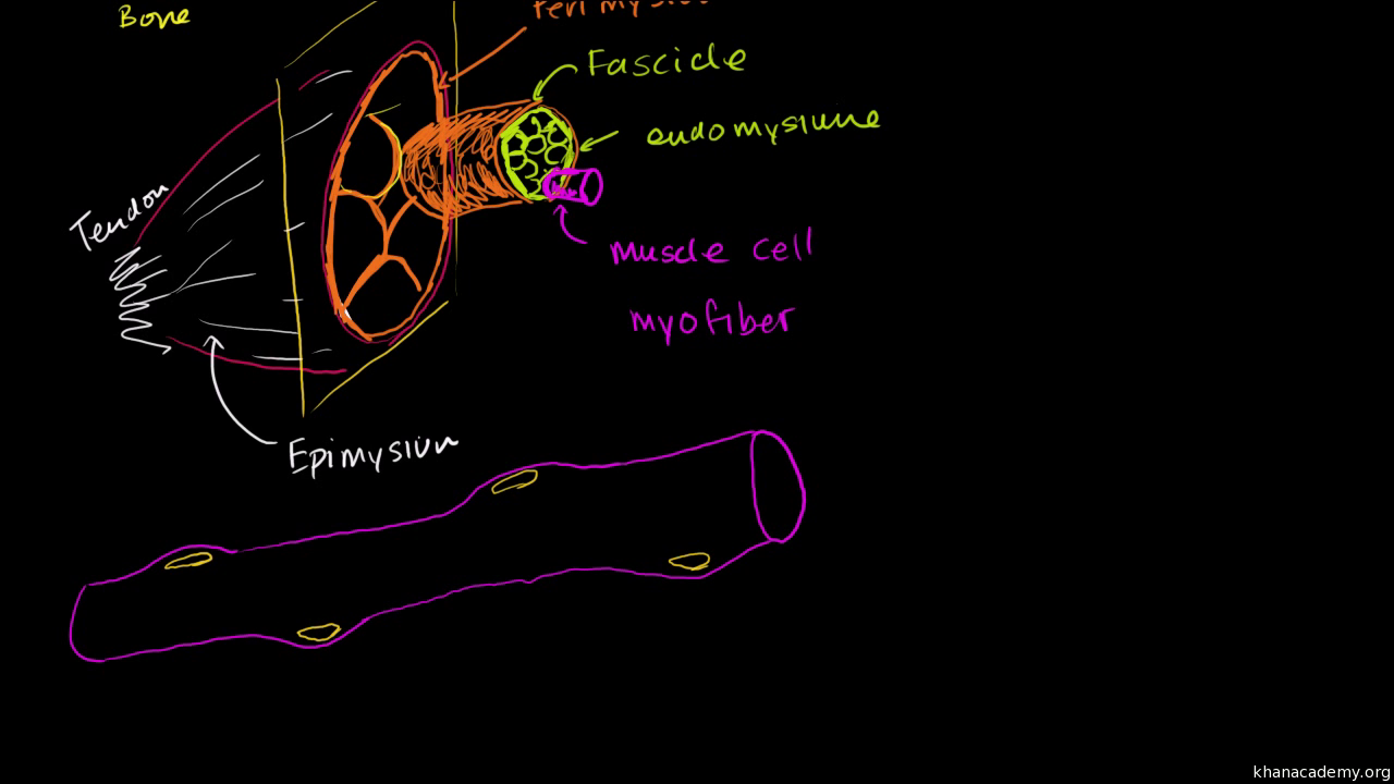
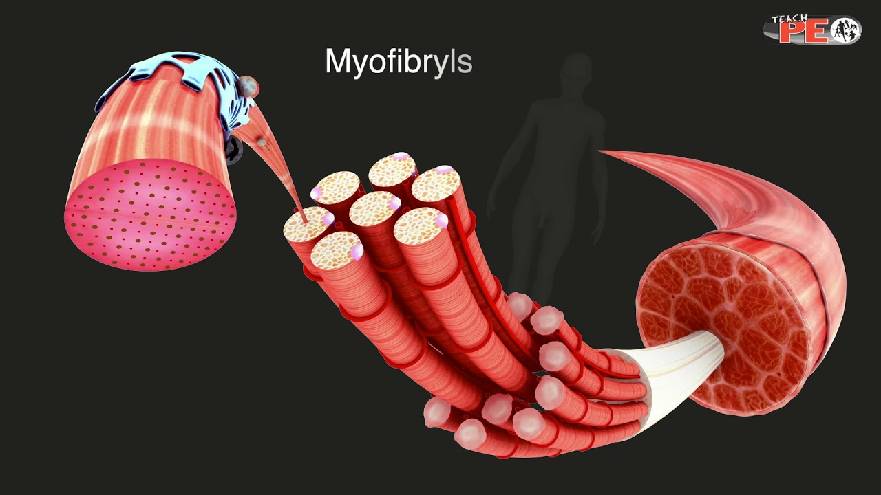
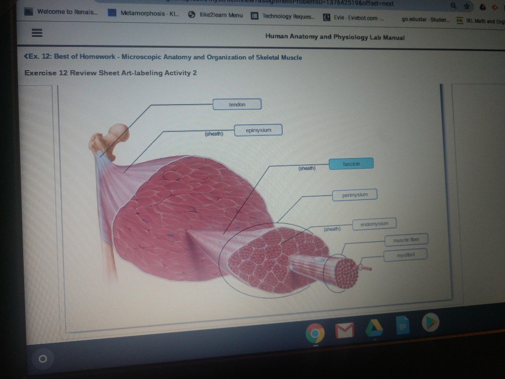
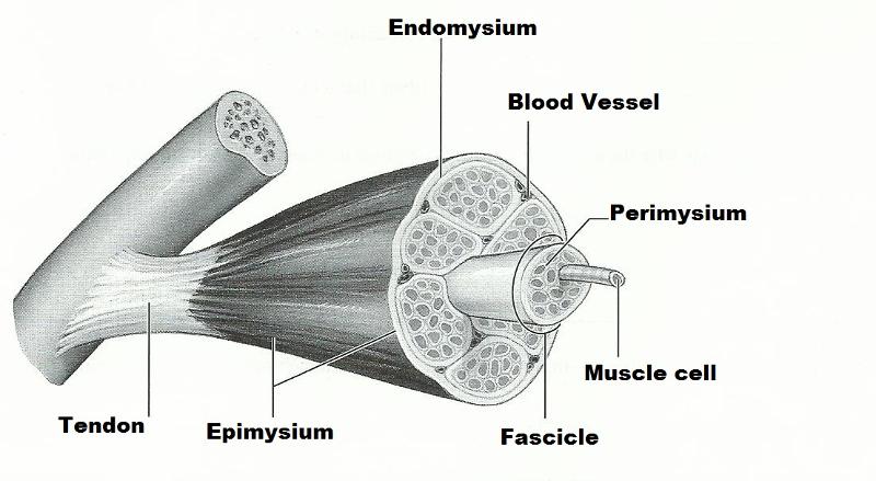
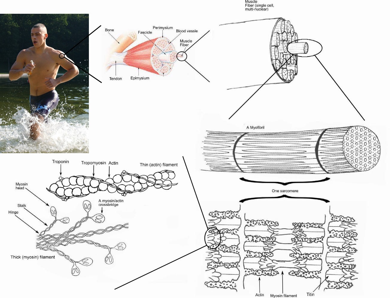
Exercise 12 microscopic anatomy and organization of skeletal muscle. Three reasons why the connective tissue wrappings of skeletal muscle are important. Provide route for entry and exit of blood vessels and nerves to muscle fibers. Aponeuroses are thick membranes that separate muscles from one another.
Why are the connective tissue wrappings of skeletal musclem 1 méortantfigaasftv give at least three reasons 1m 11. Exercise 11 microscopic anatomy and organization of skeletal muscle review sheet. Skeletal muscle cells skeletal muscle cells are commonly called skeletal muscle fibers skeletal muscle cells have peripherally located nuclei and there are many nuclei per cell multinucleated important cell structures.
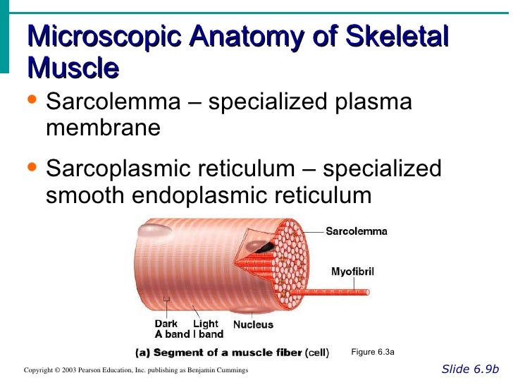
To provide a route for the entry exit of nerves blood vessels that serve muscle fibers. Sarcolemma cell membrane transverse tubule t tubule are invaginations of the sarcolemma at the i a junction 2. The combining of the neurotransmitter with the muscle membrane receptors causes a change in permeability to the membrane resulting in of the membrane.
Then contraction of the muscle fiber occurs. Cord of collagen fibers that attaches a muscle to a bone. The actual gap between the axonal terminal and the muscle cell is called a 3.
Learn vocabulary terms and more with flashcards games and other study tools. Start studying exercise 12. Microscopic anatomy and organization of skeletal muscle.
Providing strength to the muscle as a whole. Skeletal muscle cells and their organization into muscles learn with flashcards games and more for free. They are tough and resilient.
Microscopic anatomy organization of skeletal muscle. Supporting and binding the muscle fibers. Ct wrappings bundle muscle fibers together increases coordination of activity.
Amotor neuron and all of the skeletal muscle cells it stimulates is called a 2. Tendons are similar in both function and composition only they serve to connect muscles to bones. Microscopic anatomy and organization of skeletal muscle flashcards and study them anytime anywhere.
Both aponeuroses and tendons are capable of resisting considerable tension. Use the items in the key to correctly identify the structures described below.
 Structure Of Skeletal Muscle Explained In Simple Terms

Skeletal Muscle Anatomy And Physiology Openstax
 Pdf Three Dimensional Printing Of Human Skeletal Muscle
 Solved It Says 2 Are Wrong But Can T Figure Out Which Two
 Human Anatomy Physiology Laboratory Manual Pdf Free Download
 Exercise 14 Microscopic Anatomy Organization And Ppt
 Length Tension Relationship In Skeletal Muscle
 Exercise 14 Microscopic Anatomy And Organization Of Skeletal
 Exercise 14 Microscopic Anatomy And Organization Of
 Ppt Exercise 14 Powerpoint Presentation Free Download
Muscles And Muscle Tissue
 Skeletal Muscle A Review Of Molecular Structure And
Skeletal Muscle Anatomy And Physiology Openstax
 Muscle System
 Chimpanzee Super Strength And Human Skeletal Muscle
Anatomy Physiology
 B 227 Lab 8 Notes Lab 8 A Exercise 14 Microscopic Anatomy
 Skeletal Muscle Structure And Function Exercise
 Lab 11 Name Lab Time Date E X E R C I S E 11 Microscopic
 Ultrastructure Of Muscle Skeletal Sliding Filament
![]() Skeletal Muscle Organization Connective Tissue And Layers
Skeletal Muscle Organization Connective Tissue And Layers
 Muscle Tissue Junqueira S Basic Histology Text And Atlas
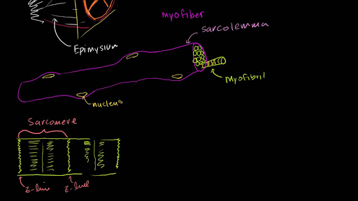
Anatomy Of A Skeletal Muscle Fiber Video Khan Academy
 Skeletal Muscle Wikipedia

 Human Anatomy Physiology Laboratory Manual Fetal Pig


Posting Komentar
Posting Komentar