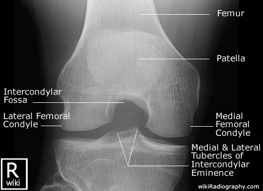
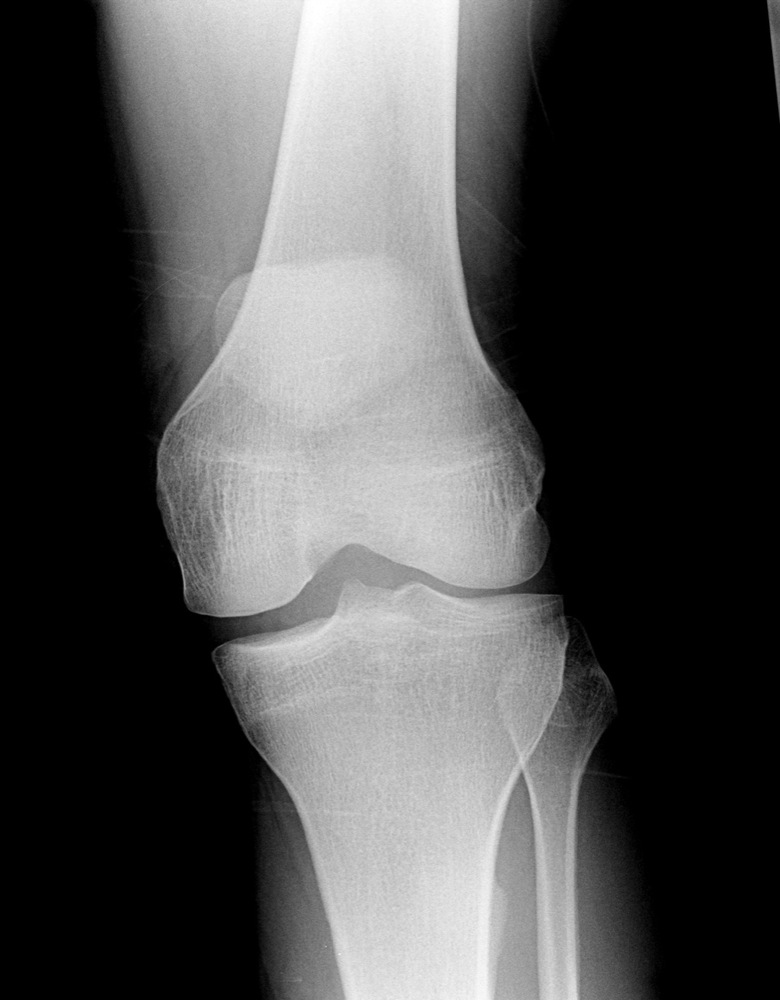
Knee normal ap. This is the insall salvatti ratio and should ideally me measure with the knee flexed at 30 degrees.
 Knee X Rays
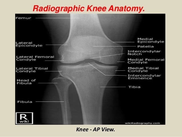
Ap stands for anteroposterior meaning the image is directed from the front to the back of the knee joint.

Knee xray anatomy. It is the largest synovial joint in the body and allows flexion and extension of the leg as well as some rotation in the flexed position. If its too long then think of a patellar tendon rupture. Use the mouse to scroll.
The knee is an important load bearing joint of the lower limb. Its length should be the same as the patellar length 20. Stanford bone tumor bayesian network issssr msk lectures for residents ocad msk cases from around the world stanford msk mri atlas has served almost 800000 pages to users in over 100 countries.
A knee x ray may appear entirely normal. An x ray is one of the most common imaging tests used to diagnose a knee problem. Atlas of knee mri anatomy.
This allows effusions to be visualised in the suprapatellar pouch. The classical radiologic picture of osgood schlatter disease is fragmentation of the tibial tubercle and local soft tissue swelling fig. Click on a link to get t1 coronal view t2 fatsat axial view t2 fatsat coronal view t2 fatsat sagittal view.
This webpage presents the anatomical structures found on knee mri. Unable to process the form. Skyline view is typically used to assess patellofemoral joint and patella condylar alignment.
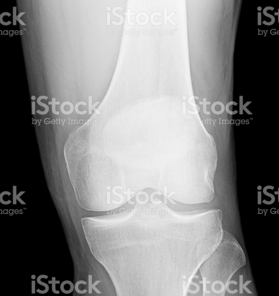
In the context of trauma the lateral view is acquired with the patient lying supine and with a horizontal x ray beam. Knee x rays are done to assess the knee joint pathology. This is a front to back view of the knee joint also called the ap view.
There may also be obliteration of the caudal portion of hoffas fat pad secondary to infrapatellar bursitis. The patellar tendon goes from the inferior pole of the patella to the tibial tuberosity. Anteroposterior and lateral views of the knee are most common knee x rays done.
The knee joint is a modified hinge joint between the femur tibia and patella. Check for errors and try again. Normal radiographic anatomy of the knee.
 Film Critique Of The Lower Extremity Part 2
 X Knee Startradiology
 Knee Anatomy Johnson Johnson Medical Devices Companies
 Vector Illustration Anatomy Front X Ray Of An Arthritic Knee
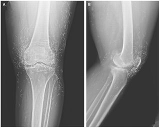
Injuries And Normal Variants Of The Pediatric Knee
Paediatric Knee Radiographs Normal Appearances Of The Knee
 Presentation1 Pptx Radiological Anatomy Of The Knee Joint
 Normal Radiographic Anatomy Of The Knee Radiology Case
 Imaging Of Tumors And Tumor Like Lesions Of The Knee
 Femoral Interference Screw Anatomical Reconstruction Of

 Redding Hospital Knee Anatomy
 Vector Illustration Anatomy Front X Ray Of An Arthritic
 X Ray Reveals Hundreds Of Gold Needles In Woman S Knees
 X Knee Startradiology
 Knee Imaging Knee Sports Orthobullets
 Debilitating Knee Pain In A Patient With Normal
 Test Yourself Regular Sets Set 27 Emergency Department
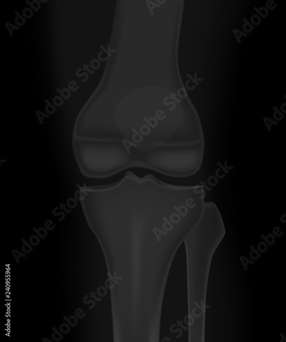 Vector Illustration Anatomy Of A Healthy Knee Joint Front
 Xray Knee Joint Anatomy Diagram Quizlet
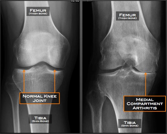 Partial Knee Replacement Austin Tx Unicompartmental Knee
 Normal Knee X Rays Bone And Spine


 The Knee
 Knee X Rays
 Radiographic Anatomy Of Adult Knee Orthopaedicsone
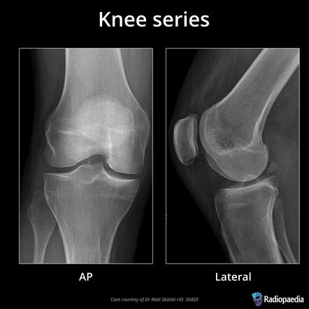 Knee Series Radiology Reference Article Radiopaedia Org
 Radiological Anatomy Of Knee Joint Radiography In Hindi


Posting Komentar
Posting Komentar