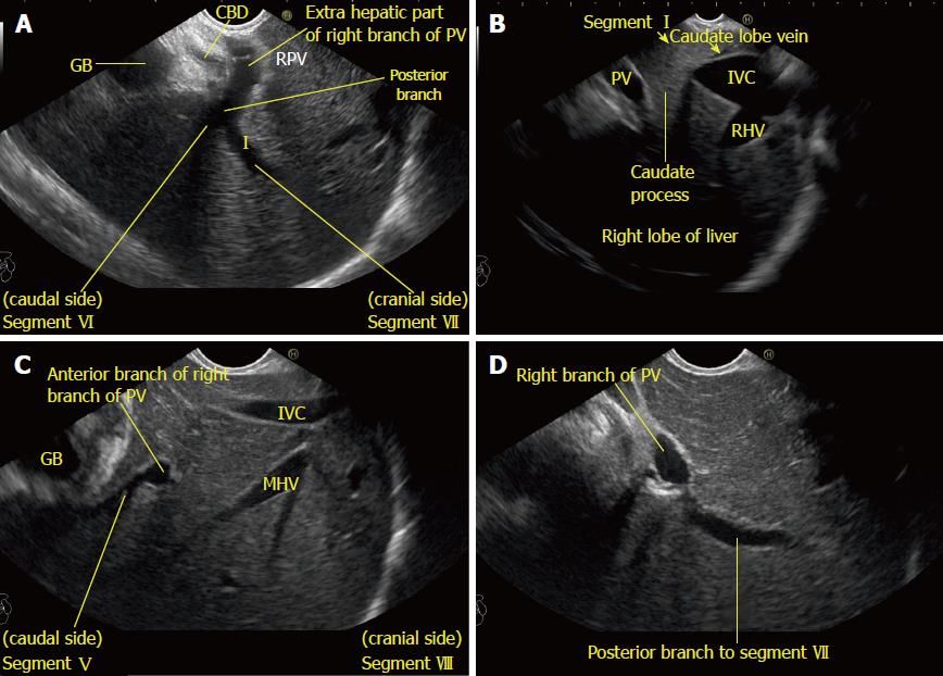
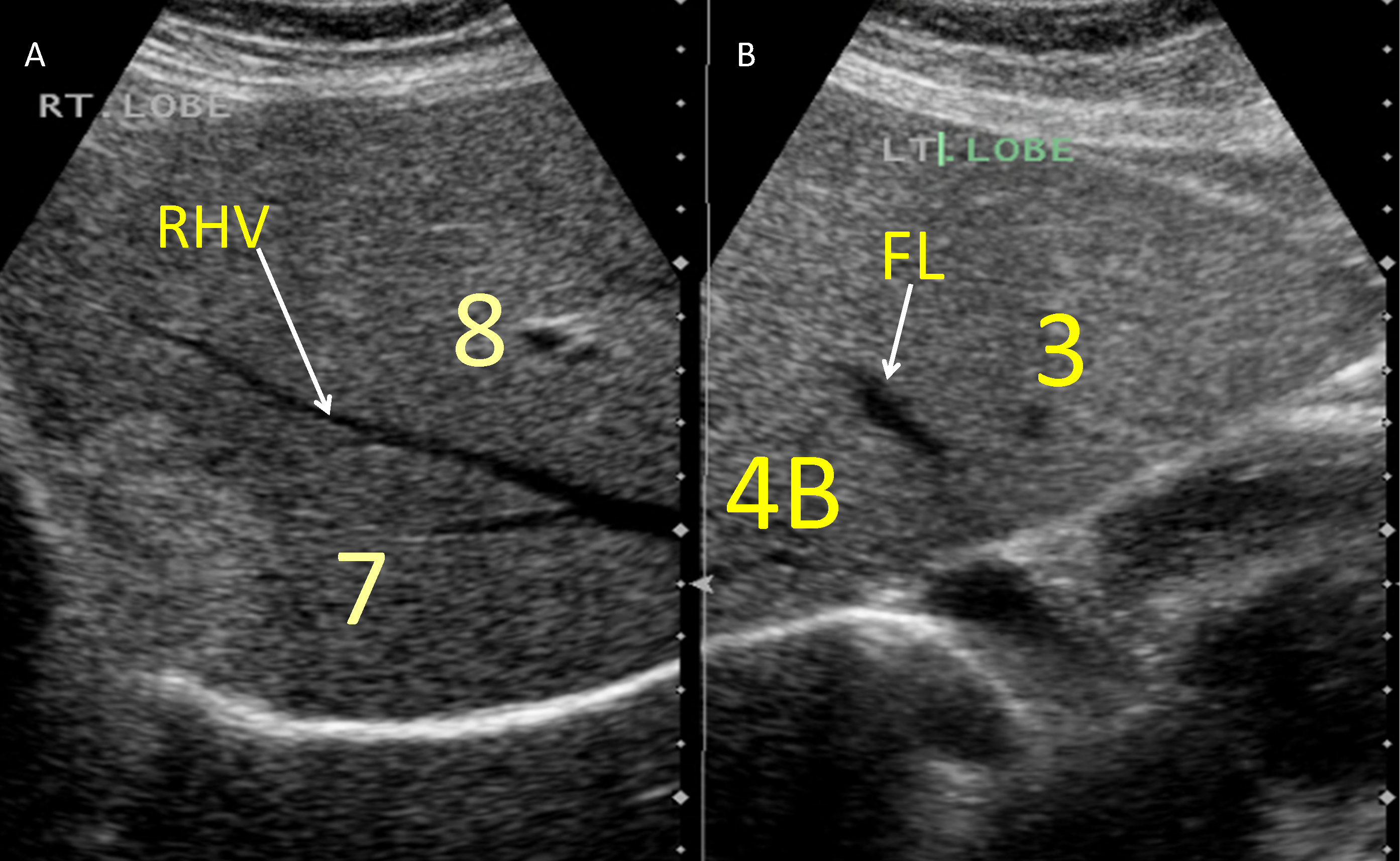
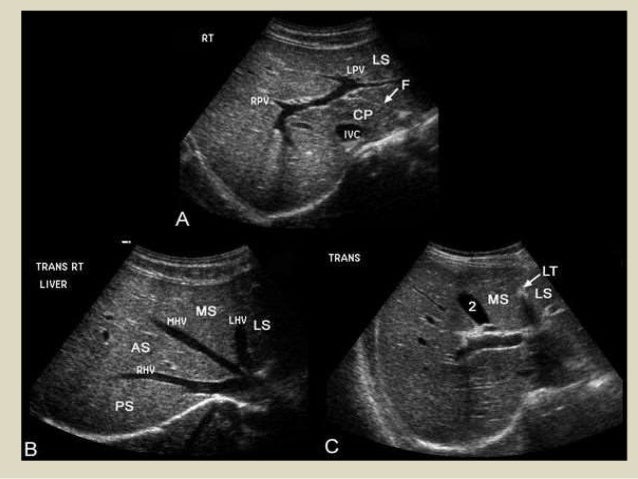
Segmental anatomy according to couinaud. Anechoic structures white arrows represent normal vesselsthe diaphragm black arrow is seen superiorly.
 Startradiology
Hover over the images for highlighted anatomy.

Liver ultrasound anatomy. The couinaud classification pronounced kwee no is currently the most widely used system to describe functional liver anatomy. It is the preferred anatomy classification system as it divides the liver into eight independent functional units ter. The liver allows for effective ultrasound imaging.
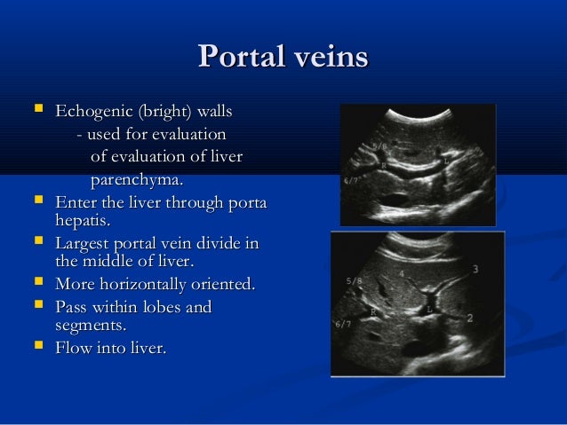
In the centre of each segment there is a branch of the portal vein hepatic artery and bile duct. Oblique left showing the ligamentum teres. A vascular ultrasound of the liver is performed to help evaluate the liver and its network of blood vessels within the liver and entering and exiting the liver.
It is largely covered by the costal cartilages. The ligamentum venosum is highlighted in orange. Each segment has its own vascular inflow outflow and biliary drainage.
A healthy liver has a homogeneous echo reflection pattern and smooth contours. The lecture discussing the basic sonographic anatomy of the hepatobiliary system including normal. Using vascular ultrasound can help physicians diagnose and review the outcome of treatments for various liver related problems and diseases.
This lecture is a part of basic radiologic anatomy series. The liver is an irregular wedge shaped organ that lies below the diaphragm in the right upper quadrant of the abdominal cavity and is in close approximation with the diaphragm stomach and the gallbladder. The couinaud classification of liver anatomy divides the liver into eight functionally indepedent segments.
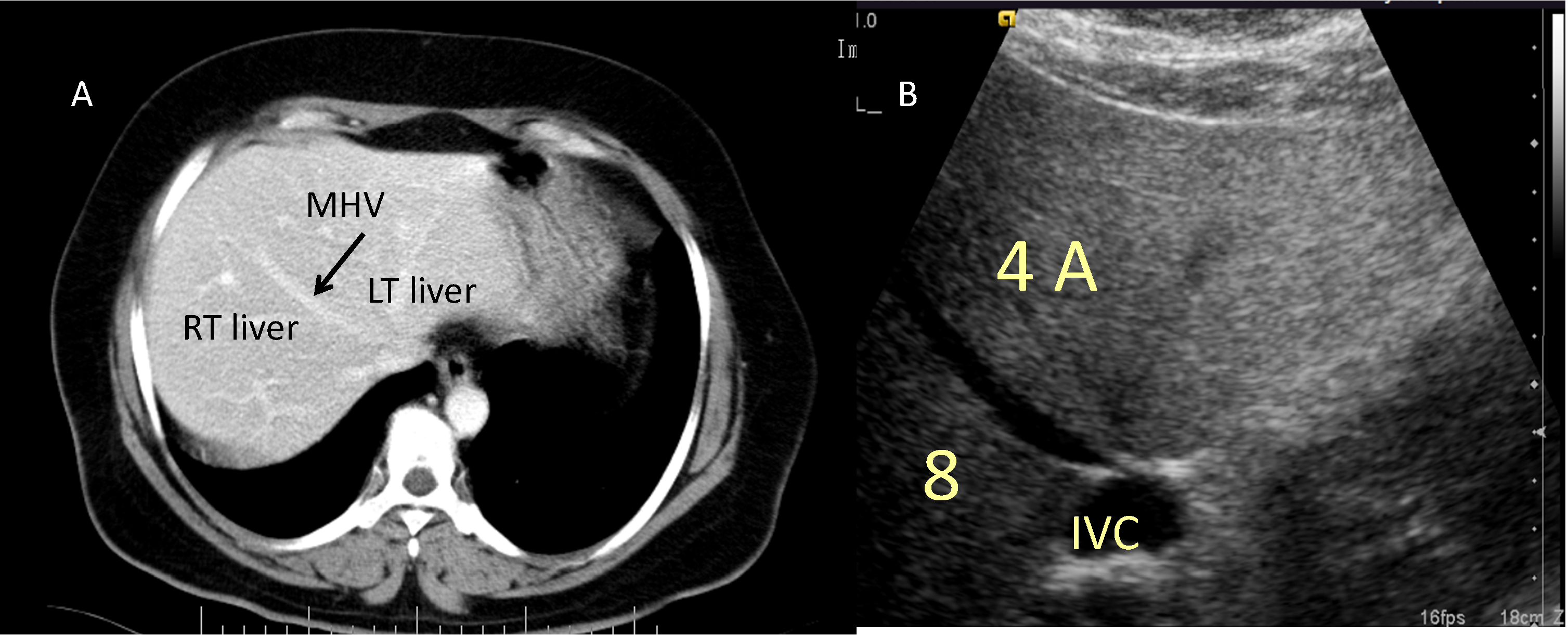
Intimate knowledge of the vascular anatomy of the liver is essential for planning and follow up of liver transplants particularly those involving partial resection of a living donor organ treatment of liver tumors tips and less frequently the management of other liver diseases. The echo reflection pattern of the liver is similar to or slightly higher than that of the renal cortex. A longitudinal sonogram demonstrates a homogeneous liver with midlevel echoes.
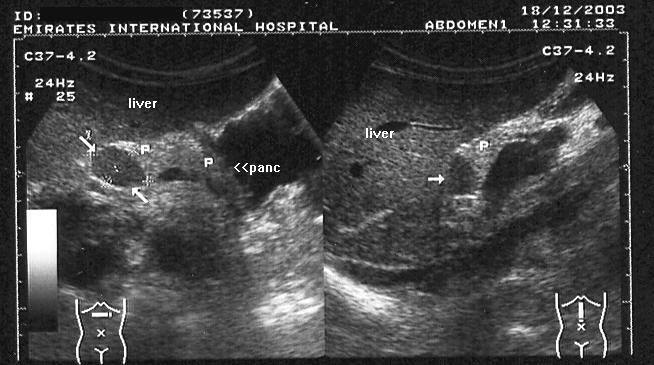
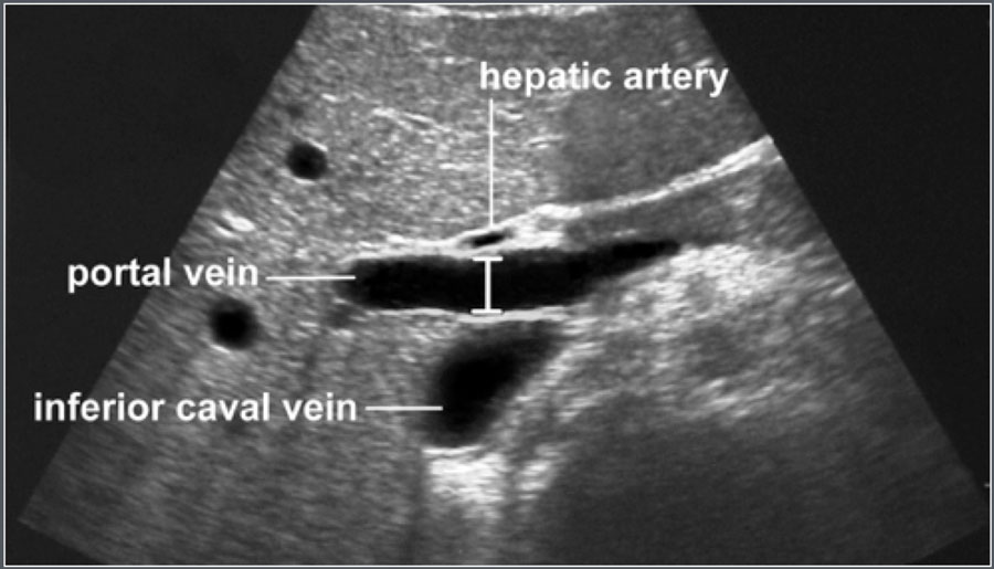
For the most part multislice computed tomography ct and magnetic resonance imaging mri have now replaced angiography in the study of hepatic vascularization. Porta hepatis is seen with an oblique angle 45degree rotation from the sagittal view to the transverse view.
 Liver Ultrasound Anatomy 4 Wmv Youtube
 Liver Anatomy Physiology Review 9 5 12 Test At Labette
 A Gallery Of High Resolution Ultrasound Color Doppler 3d
![]() Us Study Of The Liver Longitudinal And Transverse Scan
Us Study Of The Liver Longitudinal And Transverse Scan
 Stepwise Evaluation Of Liver Sectors And Liver Segments By
 Segmental Oriented Liver Surgery Intechopen
 Adrenal Gland Ultrasound Ultrasound Anatomy
Sagittal Ultrasound Images Of The Liver And Gallbladder Gb
 00283 Updated Complete Liver Protocol
 A Gallery Of High Resolution Ultrasound Color Doppler 3d
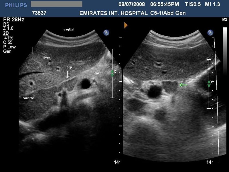
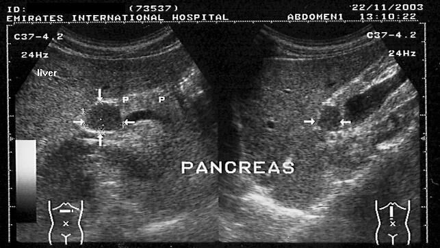
 Basic Sonographic Anatomy Of Pancreas And Kidneys
 Ultrasound Of Liver Segments Anatomy

 Startradiology
 Liver Ultrasound
 A Gallery Of High Resolution Ultrasound Color Doppler 3d


 Presentation1 Abdominal Ultrasound Anatomy

 Startradiology
 Couinaud Liver Segments On Ultrasound Creative Commons
 The Radiology Assistant Normal Values Ultrasound
 Liver Ultrasound Sonography Ultrasound Sonography
 Pin By Katie Ash On Ultrasound Ultrasound Sonography
 Liver Anatomy And Protocol Basics Sonographic Tendencies
 Liver Ultrasound Uzv Ultrasound Vascular Ultrasound
 Segmental Oriented Liver Surgery Intechopen
 Pdf Ultrasound Anatomical Visualization Of The Rabbit Liver

Ultrasound Of The Liver


Posting Komentar
Posting Komentar