The white of your eye. This portion of the conjunctiva covers the anterior part of the sclera the white of the eye.
The front part what you see in the mirror includes.

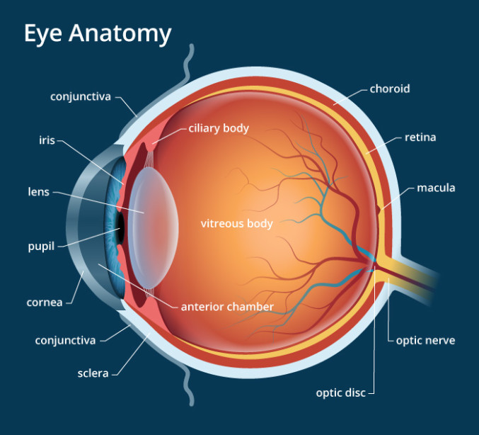
Eye anatomy conjunctiva. It is highly vascularized and home to extensive lymphatic vessels. The conjunctiva of the eye provides protection and lubrication of the eye by the production of mucus and tears. Conjunctiva thin transparent mucous membrane lining the posterior aspect of eye lid anterior aspect of eye ball latin.
It prevents microbial entrance into the eye and plays a role in immune surveillance. The conjunctiva keeps bacteria and foreign material from getting behind the eye. It has two segments.
Conjunctiva the palpebral conjunctiva forms the deepest layer of the eyelid. Conjoin to join it joins the eye ball to the eye lid 3. The conjunctiva is a thin transparent layer of tissues covering the front of the eye including the sclera and the inside of the eyelids.
A thin layer of tissue that covers the entire front of your eye except for the cornea. Anatomy of conjunctiva 1. The conjunctiva is the mucous membrane that lines the eyelid and covers the visible portion of the eyeball except the cornea the transparent part of the eyeball that covers the iris and the pupil.
It is a thin mucous membrane which is reflected onto the sclera of the eyeball bulbar conjunctiva. The eyelids lid a portion of the conjunctiva. The black circular opening in the iris that lets light in.
The clear tissue covering the white part of your eye and the inside of your eyelids. It is composed of unkeratinized stratified squamous epithelium with goblet cells and stratified columnar epithelium. A clear dome over the iris.
The conjunctiva contains visible blood vessels that are visible against the white background of the sclera. The conjunctiva is a tissue that lines the inside of the eyelids and covers the sclera the white of the eye. The conjunctiva is highly vascularised with many microvessels easily accessible for imaging studies.
It lines the inside of the eyelids and provides a covering to the sclera. In eyelid the normal functioning of the conjunctiva and cornea. The conjunctiva is the clear thin membrane that covers part of the front surface of the eye and the inner surface of the eyelids.

 Anatomy Of A Bleb A Photograph Of An Eye With A Fluid
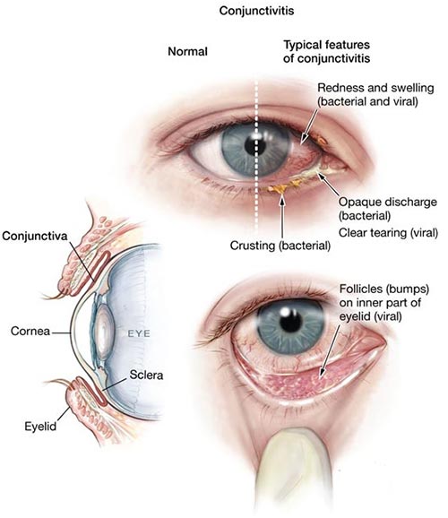 Pink Eye Conjunctivitis Treatment Top Eye Doctor Nyc
 Royalty Free Conjunctiva Stock Images Photos Vectors
 Vision Anatomy And Physiology
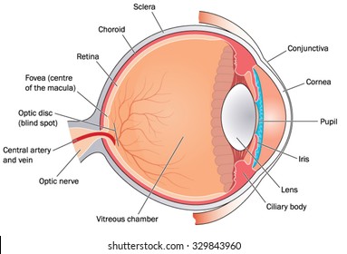
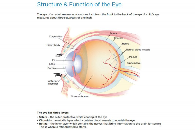 Retinoblastoma Anatomy Of The Eye Memorial Sloan
 Bulbar Conjunctiva Eye Anatomy Medical Anatomy Eyeball

 Conjunctiva Definition And Detailed Illustration
 Anatomy Atlases Anatomy Of First Aid A Case Study Approach
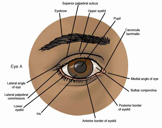
 Eye Structures Front And Side Views Healthlink Bc
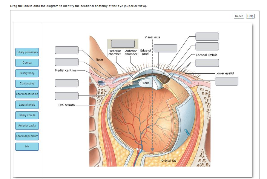 Solved Drag The Labels Onto The Diagram To Identify The S
 How The Human Eye Works Cornea Layers Role Light Rays
 Orbits And Eyes Anatomical Illustrations
 Eye Anatomy Physiology Conjunctiva Sclera By Dr
 Eye Anatomy The Vision Council
 Conjunctivitis In Dogs Elwood Vet
 Anatomy Of The Conjunctiva Eye Anatomy Medical Pictures
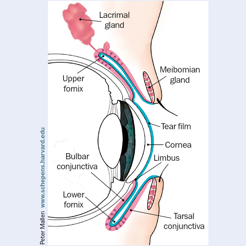
 Anatomy Of The Human Eye 1 Cornea 2 Meibomian Glands 3


 Conjunctiva Anatomy Pi Uptodate


 Eye Anatomy A Closer Look At The Parts Of The Eye
Lecture 13 Anat 102 Anatomy Physiology Ii Studocu


Posting Komentar
Posting Komentar