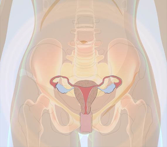
The exception to this compound structure when compared to all other bones is that it has differences that are classified by sex both for functional and general developmental reasons. The cervix is the narrow part that protrudes into the vagina.
 3d Skeletal System The Pelvic Girdle
Explore and learn about the pelvis with our 3d interactive anatomy atlas.
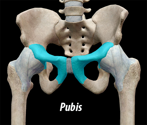
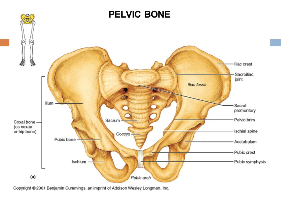
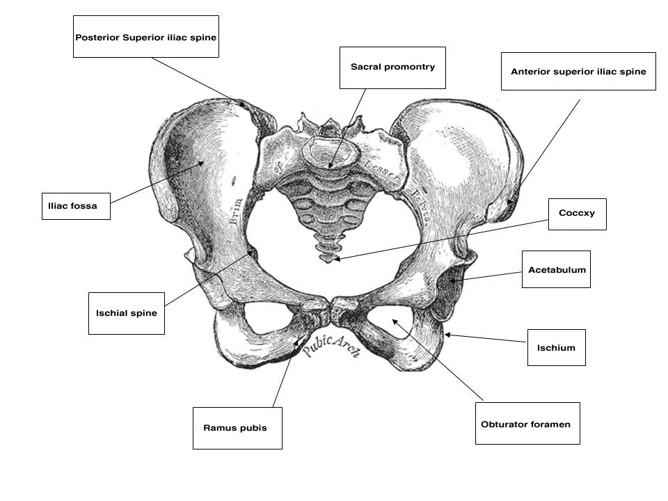
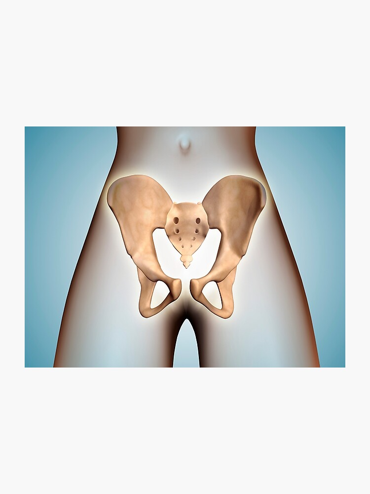
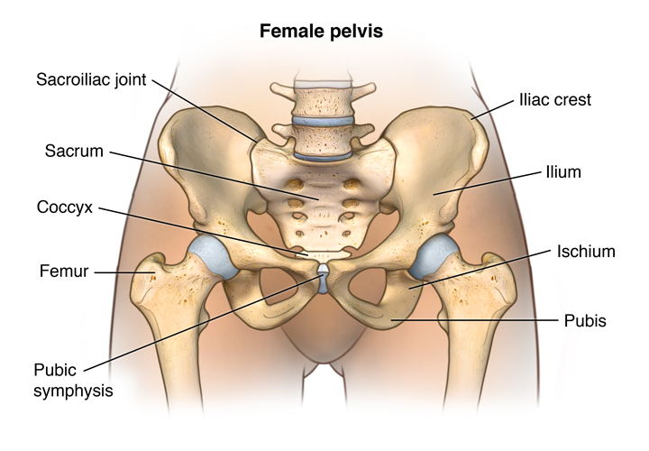
Female pelvic anatomy bones. The body is the part below. The focus in this study session will be on the female pelvis which supports the major load of the pregnant uterus and the fetal skull which has to pass through the womans pelvis when she gives birth. In male pelvis the obturator foramen is round while in female pelvis the obturator foramen is oval.
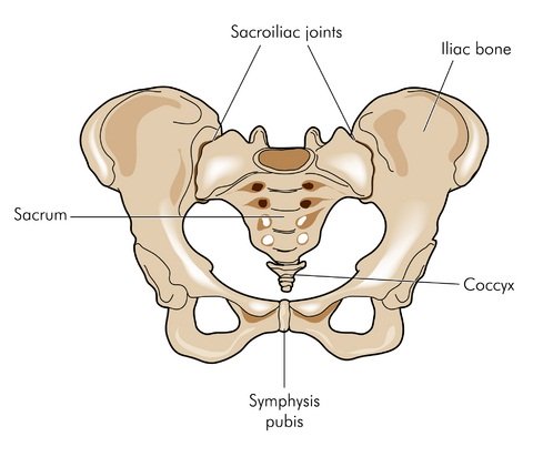
Together they form the part of the pelvis called the pelvic girdle. The pelvis is a bony structure that can be found in both male and female skeletons. There are two hip bones one on the left side of the body and the other on the right.
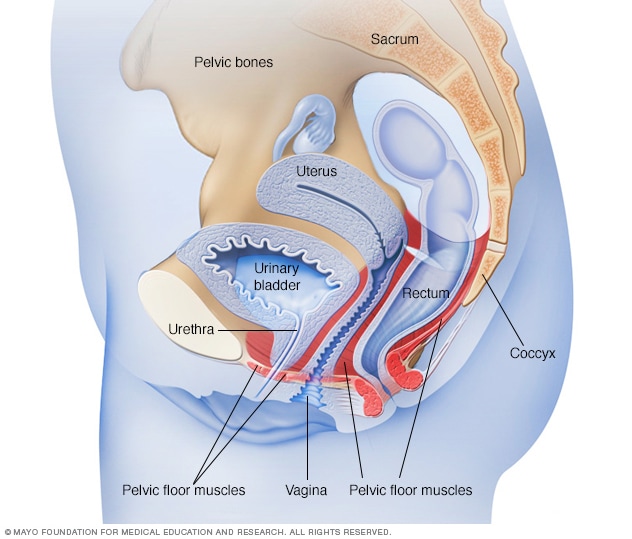
In a nonpregnant female it lies on the urinary bladder. A male pelvic bone is heavier taller and much thicker while a female pelvic bone is thinner and denser. The interior walls are straight the subpubic arch wide the sacrum shows an average to backward inclination and the greater sciatic notch is well rounded.
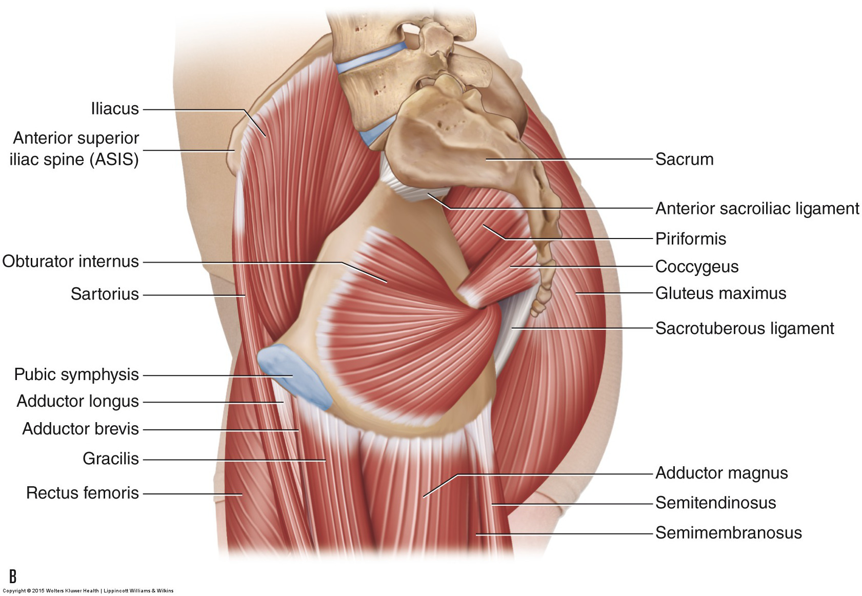
Pelvic wall muscles piriformis exits pelvis through greater sciatic foramen and attaches to greater trochanter of the femur external or lateral hip rotator innervation. It has a cervical canal which opens into the vagina through the external os which is the opening of the uterus. The rest of the human skeleton differs only in size which is genetically determined and is usually slightly larger in males than in females.
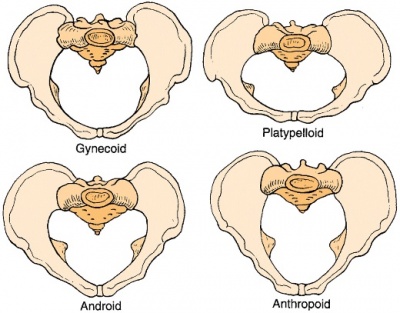
Inferior gluteal lateral sacral and superior gluteal artery injury. The gynaecoid pelvis is the so called normal female pelvis. The fundus lies above the entrance of the uterine tube.
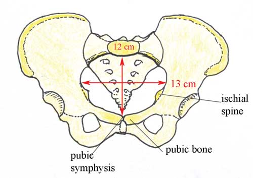
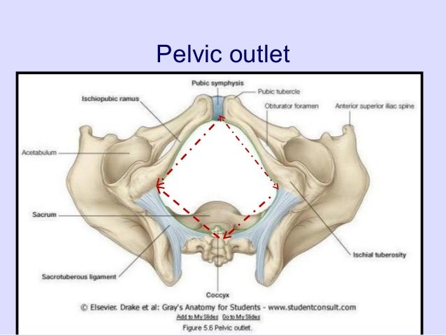
Its inlet is either slightly oval with a greater transverse diameter or round. L5 s1 and s2 blood supply. This is so a baby can pass through the pubic outlet the circular hole in the middle of the pelvic bones during childbirth.
Persistent hip pain obturator internus. The female pelvic bones are typically larger and broader than a males. The pelvic outlet in male pelvis is narrower whereas the pelvis outlet in female pelvis is wider.
The bones of the skeleton have the main function of supporting our body weight and acting as attachment points for our muscles.
 Male And Female Pelvic Floor Anatomy Gu Pelvic Floor
 Pelvis Images Stock Photos Vectors Shutterstock
 Pelvic Floor Anatomy Physiopedia

Female Pelvic Anatomy
 Anatomy Of Female Pelvic Ppt Video Online Download
 Pelvis Hip Anatomy
 Royalty Free Female Pelvis Stock Images Photos Vectors
 Pelvic Floor Wikipedia
 Female Pelvis Bone Anatomy Canvas Print
 Female Pelvic Anatomy 2 Wichita Urology
![]() Pelvis And Perineum Anatomy Vessels Nerves Kenhub
Pelvis And Perineum Anatomy Vessels Nerves Kenhub
 The Pelvis Anatomy Images Pelvic Floor Connective Tissues
 Female Pelvic Floor Muscles Mayo Clinic
 Antenatal Care Module 6 Anatomy Of The Female Pelvis And
 Anatomy Of Pelvic Bone On Female Body Photographic Print
 What Is Urogynecology Star Clinic
 Fertility Testing Anatomical Evaluation Sonograms Dallas Ivf
 8 3 The Pelvic Girdle And Pelvis Anatomy And Physiology
 Rear View Female Pelvis Anatomy Hip Anatomy Psoas Muscle
 S61f Premier Academic Series Skeleton Female Pelvis Unpainted Hanging Mount
 Female Pelvic Anatomy Ppt Video Online Download
![]() Pelvis Anatomy Bones Joints Ligaments And Foramina Kenhub
Pelvis Anatomy Bones Joints Ligaments And Foramina Kenhub
 Male Vs Female Pelvis Differences Anatomy Of Skeleton
 Anatomy Of The Male And Female Pelvis Comprehensive
 Female Bony Pelvis And Fetal Skull For Undergraduate
 Bony Pelvis Anatomy Bone And Spine
Pelvis Wikipedia


Posting Komentar
Posting Komentar