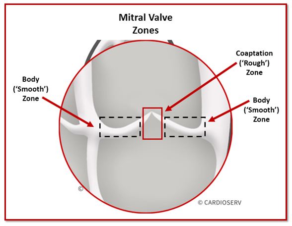
Anatomy the mitral valve consists of two valve leaflets the anterior leaflet amvl and the posterior leaflet pmvl which together have a surface of 4 6 cm 2. Table 3 mitral valve apparatus components in normal and diseased states.
 Example Of Typical Stress Strain Curve Observed In Mitral
Describe the detailed anatomy of the mitral valve mv using two dimensional 2d transesophageal echocardiography tee based on the american society of echocardiographysociety of cardiovascular anesthesiology guidelines.
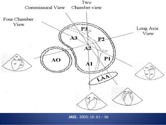
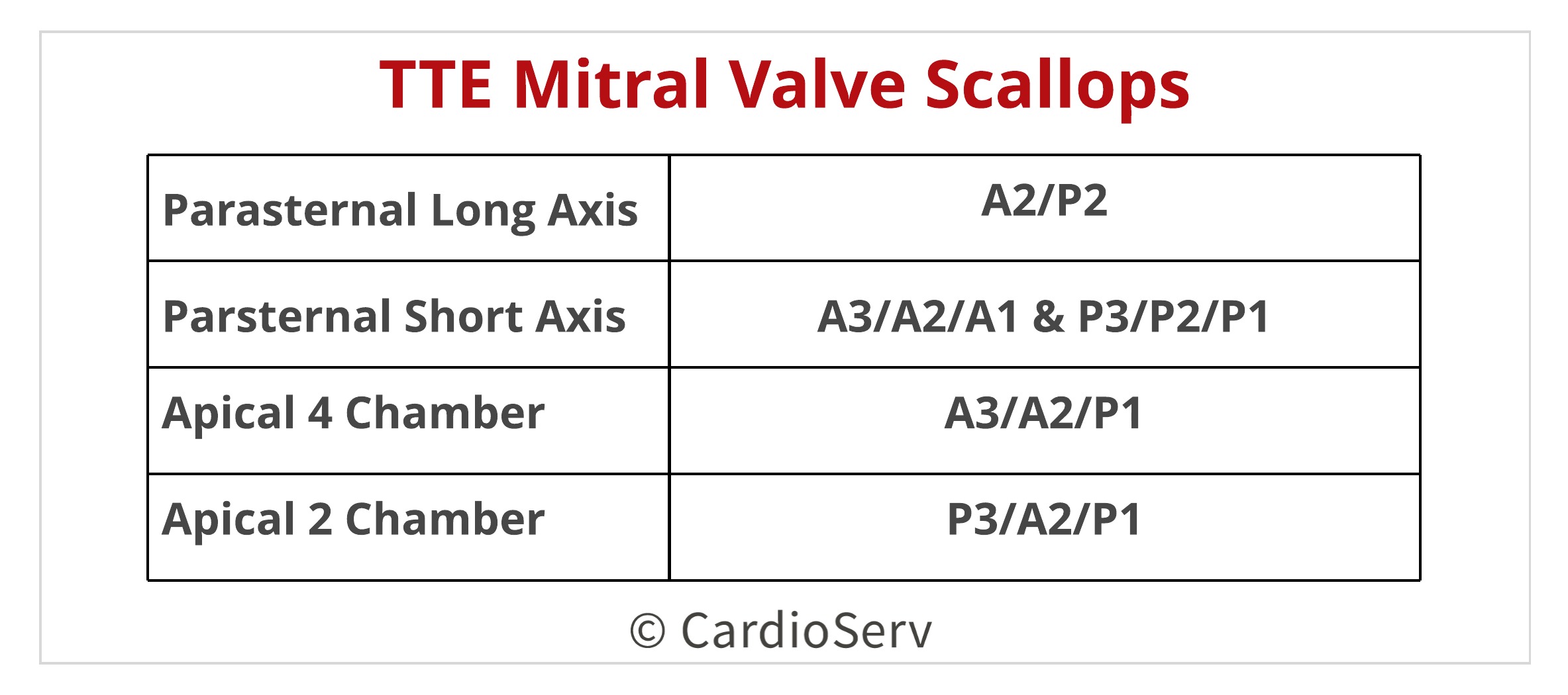
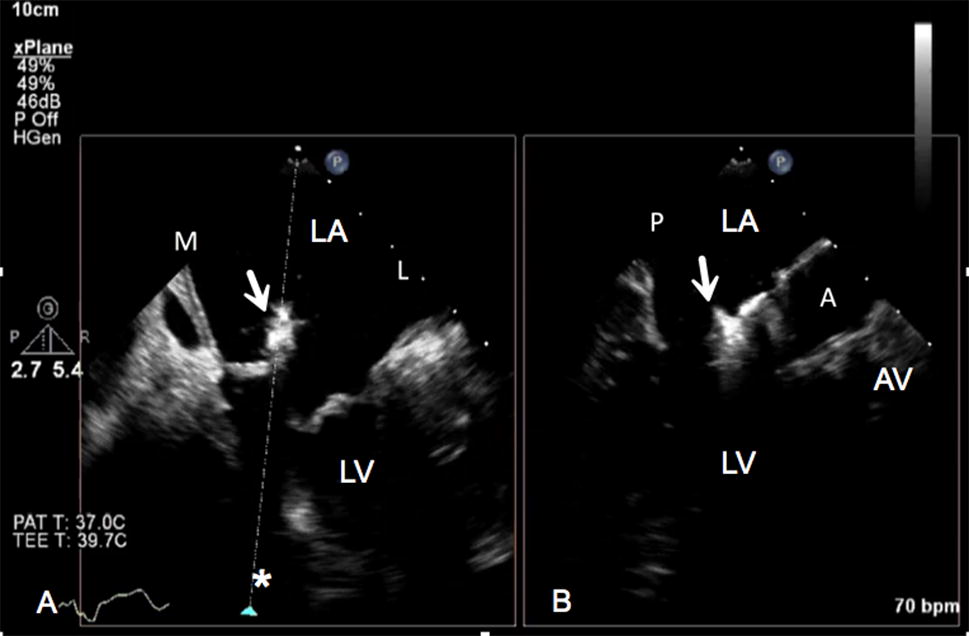
Mitral valve anatomy tee. The comprehensive tee examination of the mitral valve consists of a series of eight cross sectional views. Ography tee has enabled accurate noninvasive imag ing of the complex mitral valve anatomy in real time and from unique orientations previously available only to surgeons. You can now confidently identify 5 components of the mitral valve apparatus.
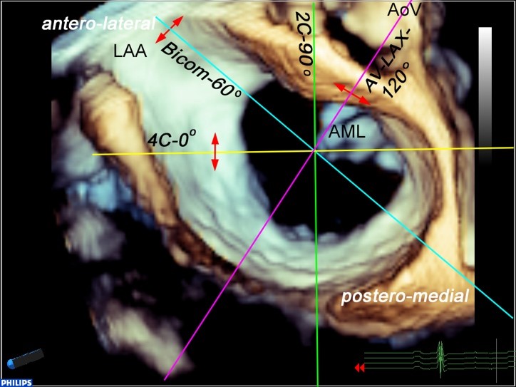
The standard modalities of real time 3d tee have recently been described 32. Anatomy of mitral valve mitral valve apparatus mitral valve annulus. Perturbations of the normal anatomic relations can result in mitral valve dysfunction table 3.
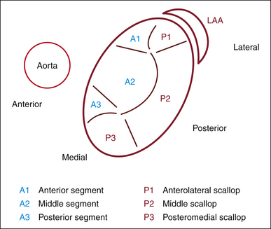
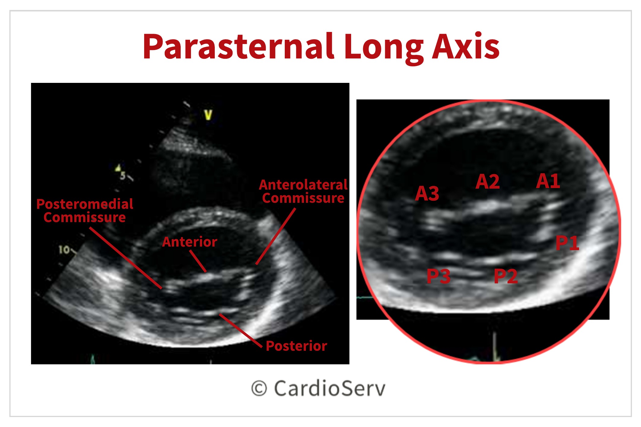
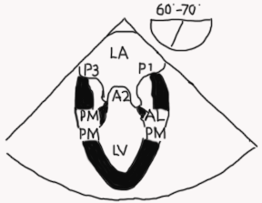
Mitral leaflets with commissures. The two leaflets of the mv are noticeably different in structure and are referred to as the anterior and posterior leaflets by clinicians. Assessment of mitral valve dr.
Normal mitral valve anatomy leaflets. Transesophageal echocardiography tee is performed intraoperatively in all patients undergoing valve surgery and is critical to assess and localize valvular dysfunction. Feasibility of mitral repair 1.
Via chordae tendineae small tendons which ensure that the leaflets do not prolapse the valve leaflets are attached to two major papillary muscles anterolateral en posteromedial in the left ventricle. This week we reviewed mitral valve anatomy to lay the foundation for our in depth review of quantification of mitral valve regurgitation. Assessment of mitral valve by tee 1.
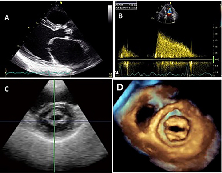
Assessment of mitral valve anatomy by real time 3 dimensional 3d transesophageal echocardiography tee has proven to be superior compared to 2 dimensional tee 121. The mv comprises two leaflets annular attachment at the atrioventricular junction tendinous chords and the papillary muscles pms. Surgeons skill and experience 2.
Accurate identification the anatomic lesions of the mitral valve echocardiography is pivotal in defining the functional anatomy of the mitral valve surgeon and echocardiographer speaking a common language mutual respect and honesty knowing when to send the. Mitral valve anatomy is designed to promote and maintain normal mitral valve apparatus function. Join us next week as we start our discussion on correct scanning techniques for the mitral valve.
Altogether called as mitral.
 Quantitative Assessment Of Mitral Regurgitation
 Understanding The Role Of Echocardiography In The Assessment
 Finally Mitral Valve Orientation Explained
 Role Of Transesophageal Echocardiography In Mitral Valve
 Anatomy Of The Mitral Valve Complex According To Fluoroscopy
 Finally Mitral Valve Orientation Explained


 Echocardiographic Guidance For Transcatheter Mitral Valve


Mitral Valve Tee2013 Dr Dharmesh
 Mitral Valve Anatomy Name 5 Components

 Multimodality Imaging In The Context Of Transcatheter Mitral
 Anatomy Of The Tricuspid Valve
 How To Image The Mitral Valve With The Help Of Tee
 Myxomatous Mitral Valve Disease Comparison Of Different

 Mitral Valve Level Tee Level Mitral Valve Cardiac
 Echocardiographic Guidance For Transcatheter Mitral Valve

 Apical 3 Chamber View Tee Cardiac Sonography Cardiac


Posting Komentar
Posting Komentar