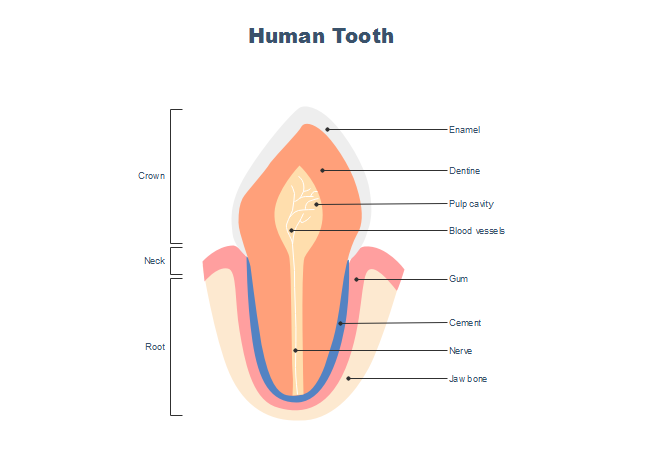
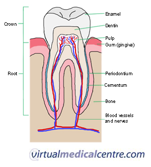
Dentine forms the major component of each tooth and extends almost the entire length of the tooth. These teeth erupt at around age 18 but are often surgically removed to prevent displacement of other teeth.
 Dental Anatomy Quick Study Academic Inc Barcharts
In contrast to the brittle nature of enamel dentine is elastic and compressible.
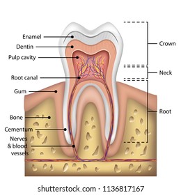
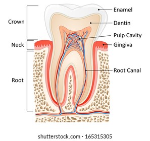
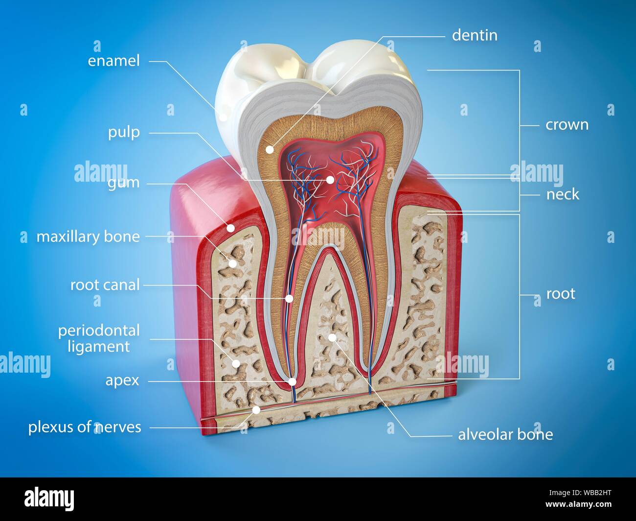
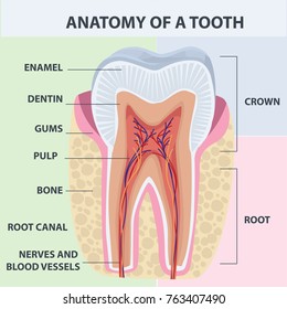
Teeth anatomy diagram. The shape of the crown determines the tooths function. It makes up approximately two thirds of the tooth. The root canal is a passageway that contains pulp.
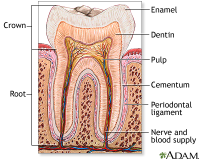
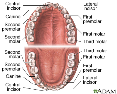
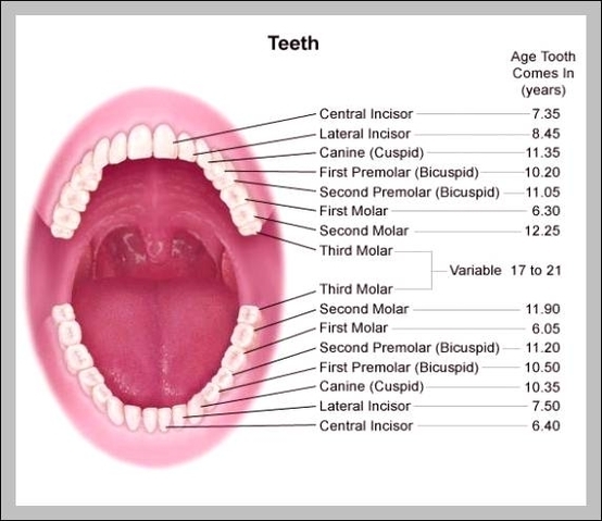
The enamel dentin pulp cementum and the periodontal ligaments are important parts of the human tooth anatomy. The root is the part of the tooth that extends into the bone and holds the tooth in place. Molars 8 total.
This process of enamel erosion by acids is. Teeth anatomy of the tooth. The enamel that covers the crown in each tooth can be broken down by acids produced by bacteria that live in the mouth and assist in digestion of small bits of food.
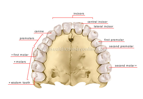
For example front teeth are sharp and chisel shaped for cutting while molars have flat surfaces for grinding. Gumline where the tooth and the gums meet. Teeth dental cavities.
Without proper brushing and flossing plaque and tartar can build up at the gumline leading to gingivitis and gum disease. Also called cement this bone like material covers the tooths root. Flat teeth in the rear of the mouth best at grinding food.
Tooth decay and cavities are important health concerns related to the teeth. What are the different parts of a tooth. While enamel dentin and cementum are the hard tissues present in a human tooth the pulp is a soft living tissue.
Wisdom teeth or third molars 4 total. Crown the top part of the tooth and the only part you can normally see. It is a living tissue softer than enamel with a structure similar to bone.
It is sensitive and is protected by enamel on the crown portion and cementum on the roots. Structure and anatomy of a human tooth. Premolars 8 total.
Its made up of several parts. Teeth between the canines and molars.
 Disorders Of The Teeth And Jaw Anatomical Chart
 The Mouth Pharynx And Esophagus Anatomy And Physiology Ii
 4 Simplified Cross Section Of A Tooth Incisor And Jaw
 Human Tooth Anatomy Chart Diagram Teeth Illustration Buy
 1 General Tooth Anatomy Indicating The Four Primary Types
 Cancer Of The Tongue Mouth Cheeks And Lips
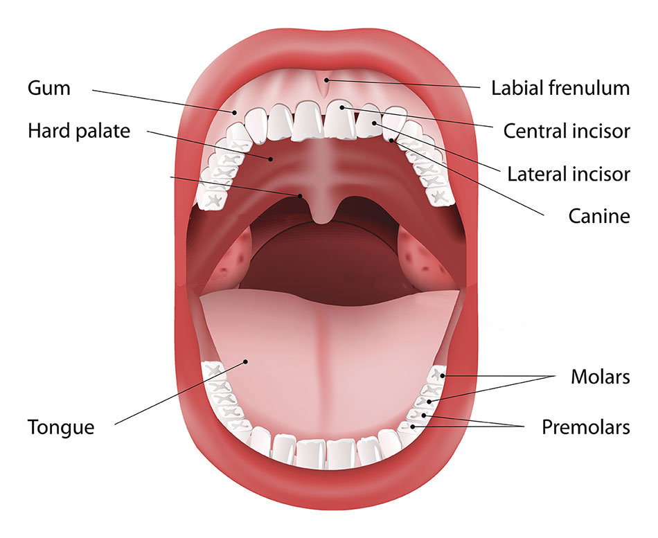
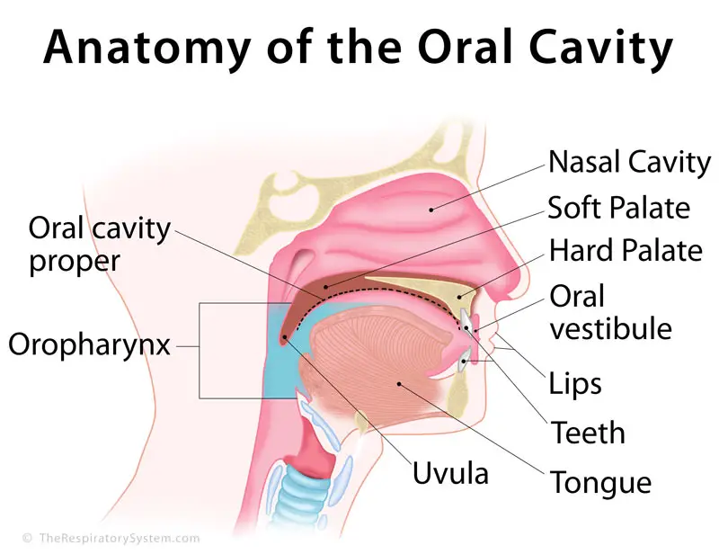 Oral Cavity Definition Anatomy Functions Diagram
 Free Incisor Tooth Anatomy Templates
 Tooth Anatomy Chart Orthodontist Human Teeth Loss Diagram
 Tooth Types Dental Health Foundation
 Tooth Anatomy Images Stock Photos Vectors Shutterstock
 Tooth Anatomy Medlineplus Medical Encyclopedia Image
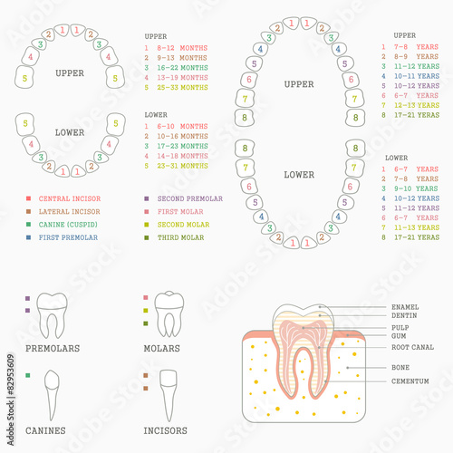
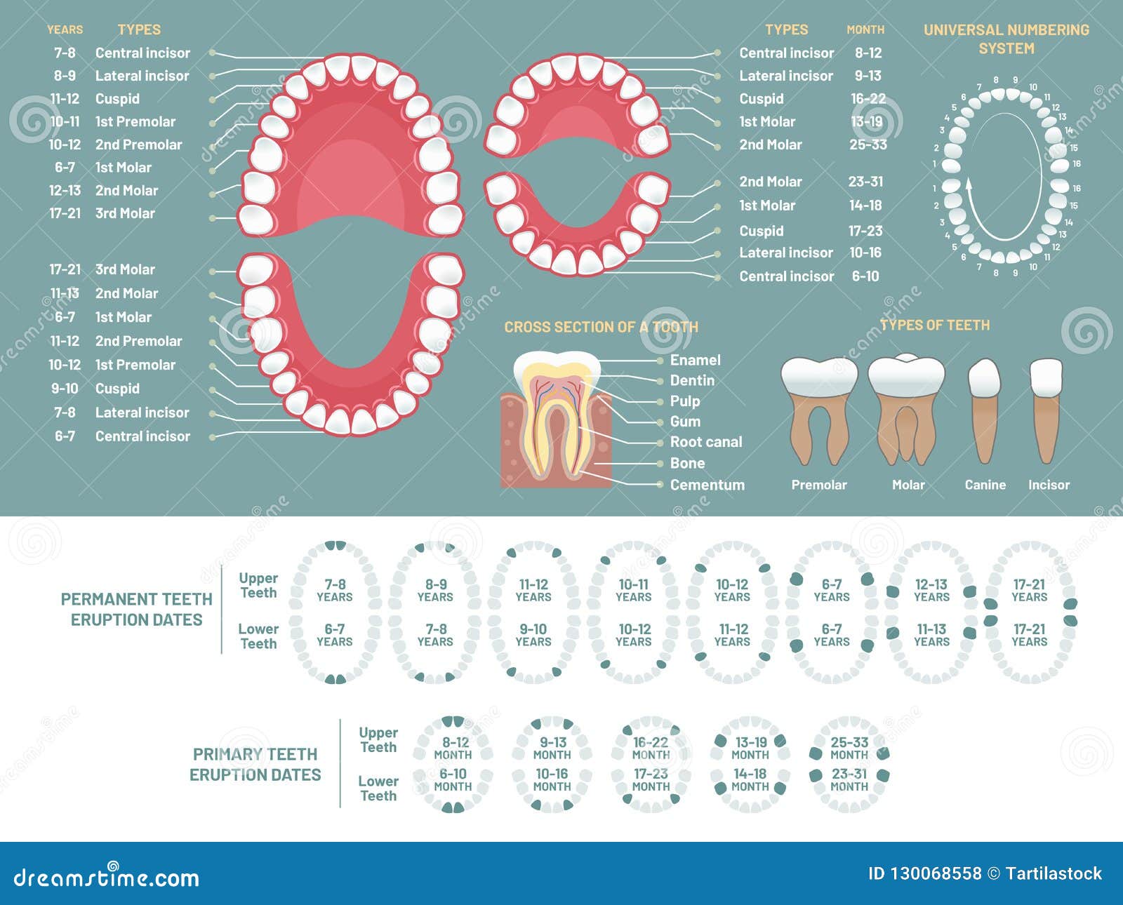
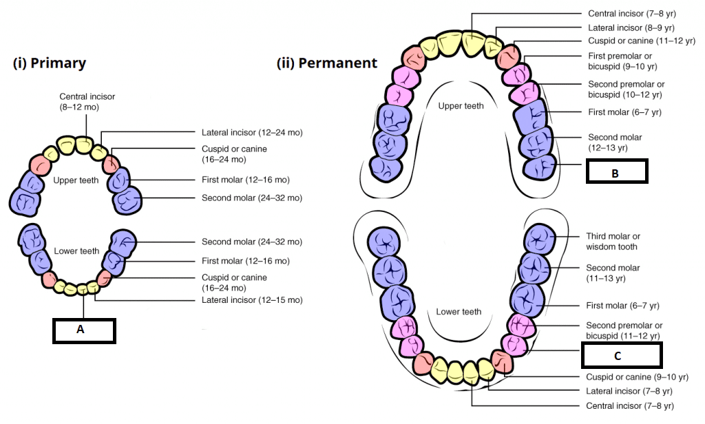 Child And Adult Dentition Teeth Structure Primary
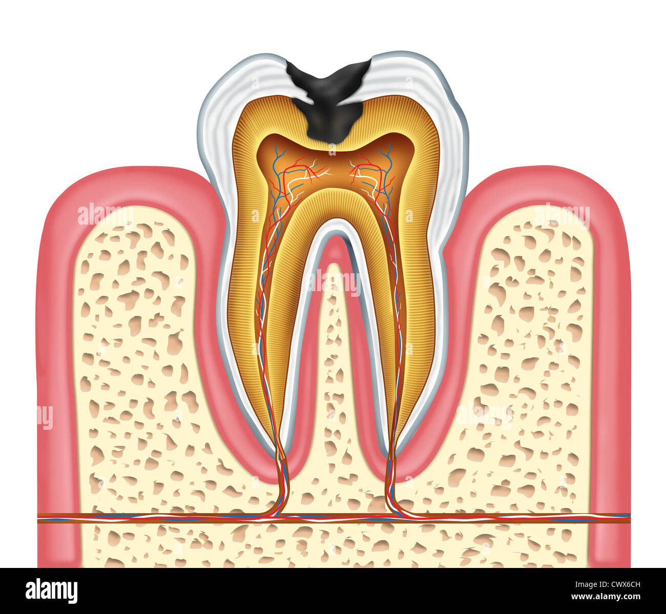 Tooth Inner Anatomy Diagram As A Dentist Surgeon Teeth
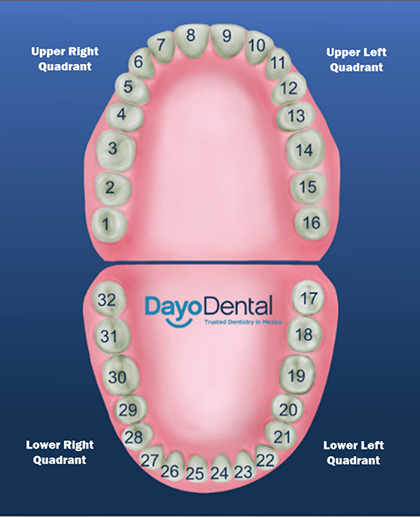 Diagram Of Human Teeth By Number Types Of Electrical

 Pin On Basic
 Vector Clipart Orthodontist Human Tooth Anatomy Vector
 Teeth Anatomy Adult Teeth Permanent Dentition
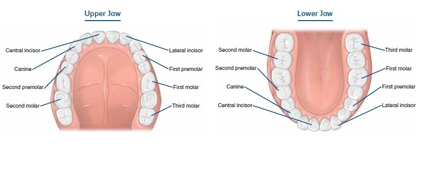
 Teeth Names And Locations In Human Mouth And Their Functions
 Teeth Vector Anatomy Stock Vector Illustration Of Caries
 Great Collection Of Tooth Anatomy Diagrams Beautiful Bright
 Anatomy Of The Teeth Anatomical Chart
Human Teeth Images Stock Photos Vectors Shutterstock
 Dental Anatomy Medlineplus Medical Encyclopedia Image
 Molar Tooth Cross Section Stock Photos Molar Tooth Cross

 Human Tooth Anatomy Cross Section Dental Diagram 20x14 Inch
 Oral Anatomy Diagram Anatomy1 Dental Hygiene School
 Teeth Names And Numbers Diagram Names Number And
 Tooth Anatomy Images Stock Photos Vectors Shutterstock


Posting Komentar
Posting Komentar