It is a finger like projection from the main body of the la. The left atrial appendage laa is a derivative of the atrial primordium that has anatomical and physiologic variations from the left atrium la which is an extension of the embryological pulmonary vein pv bud.
 Relational Anatomy Of The Septum And Left Atrial Appendage
Laa appears as a windsock in appearance.

Left atrial appendage anatomy. The junction is fairly well defined by a narrowing at the orifice of the appendage. It acts as a reservoir for the left atrium of the heart. A major endocrine organ it mainly produces anp atrial natriuretic peptide supply inside the heart.
Left atrial appendage anatomy and function. Short term response to sustained atrial fibrillation m weigner s katz p douglas and w manning department of medicine cardiovascular division and the harvard thorndike laboratory of beth israel deaconess medical center 330 brookline avenue boston ma 02215 usa. Left atrial appendage anatomy.
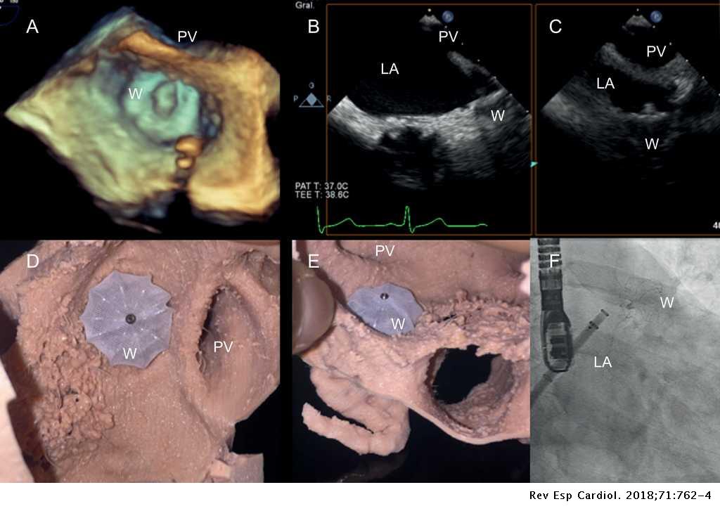
Background transesophageal echocardiography tee is the diagnostic modality of choice for visualizing the left atrial appendage laa. It is proximal to the unfastened wall of the cardiac chamber known as the left ventricle. Percutaneous left atrial appendage laa closure represents a complementary option and effective treatment for patients at risk of thromboembolism especially in patients for whom it may be difficult to achieve satisfactory anticoagulation control or where anticoagulation treatment is not possible or desirable.
This study defined the morphology of the laa in normal autopsy specimen hearts and considered the implications of these findings for tee studies. Left atrial appendage function. The laa derives from the primordial left atrium la which is formed mainly by the adsorption of the primordial pulmonary veins and their branches 6.
The left atrial appendage laa is a pouch like projection from the main body of the left atrium lies in the atrioventricular sulcus in close proximity to the left circumflex artery the left phrenic nerve and the left pulmonary veins.

 Left Atrial Anatomy Revisited Circulation Arrhythmia And
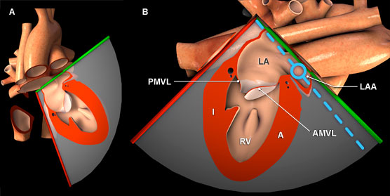
 Left Atrial Appendage Anatomy And Imaging Landmarks
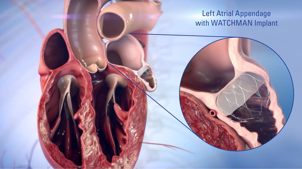
 Treating A Fib With The Watchman Left Atrial Appendage Closure Device
 Akmal Arshad On Twitter Watchman Device Close Off The Left
![]() Heart Right And Left Atrium Anatomy And Function Kenhub
Heart Right And Left Atrium Anatomy And Function Kenhub

 The Left Atrial Appendage Anatomy Function And


 An Anatomical Review Of The Left Atrium Sciencedirect
 Left Atrial Appendage Closure Device Watchman Device

 Watchman Left Atrial Appendage Occlusion
 A Low Dose Dual Phase Cardiovascular Ct Protocol To Assess
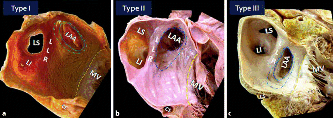
 Anatomy Of The Normal Left Atrial Appendage Circulation
 Left Atrial Appendage Anatomy And Clinicals
Left Atrial Appendage
 Anatomy Of The Atria Springerlink

 Left Atrial Appendage Anatomy And Imaging Landmarks
 Anatomy Of The Atria Springerlink
 Percutaneous Left Atrial Appendage Occlusion For Stroke
 Cardiac Interventions Today Assessing Anatomy For Left
 Left Atrial Appendage Anatomy And Imaging Landmarks
 Left Atrial Appendage Closure Heart Care Intermountain

 Three Dimensional Volume Rendering Images Showed The Number
 Left Atrial Appendage Closure Devices Arrhythmia
 Patient Specific 3d Printed Cardiac Model For Percutaneous
The Radiology Assistant Cardiac Anatomy


Posting Komentar
Posting Komentar