Nerve signals that contain visual information are transmitted through the optic nerve to the brain. The eye is surrounded by the orbital bones and is cushioned by pads of fat within the orbital socket.
Anatomy Physiology Pathology Of The Human Eye
Anatomy physiology pathology of the human eye included are descriptions functions and problems of the major structures of the human eye.
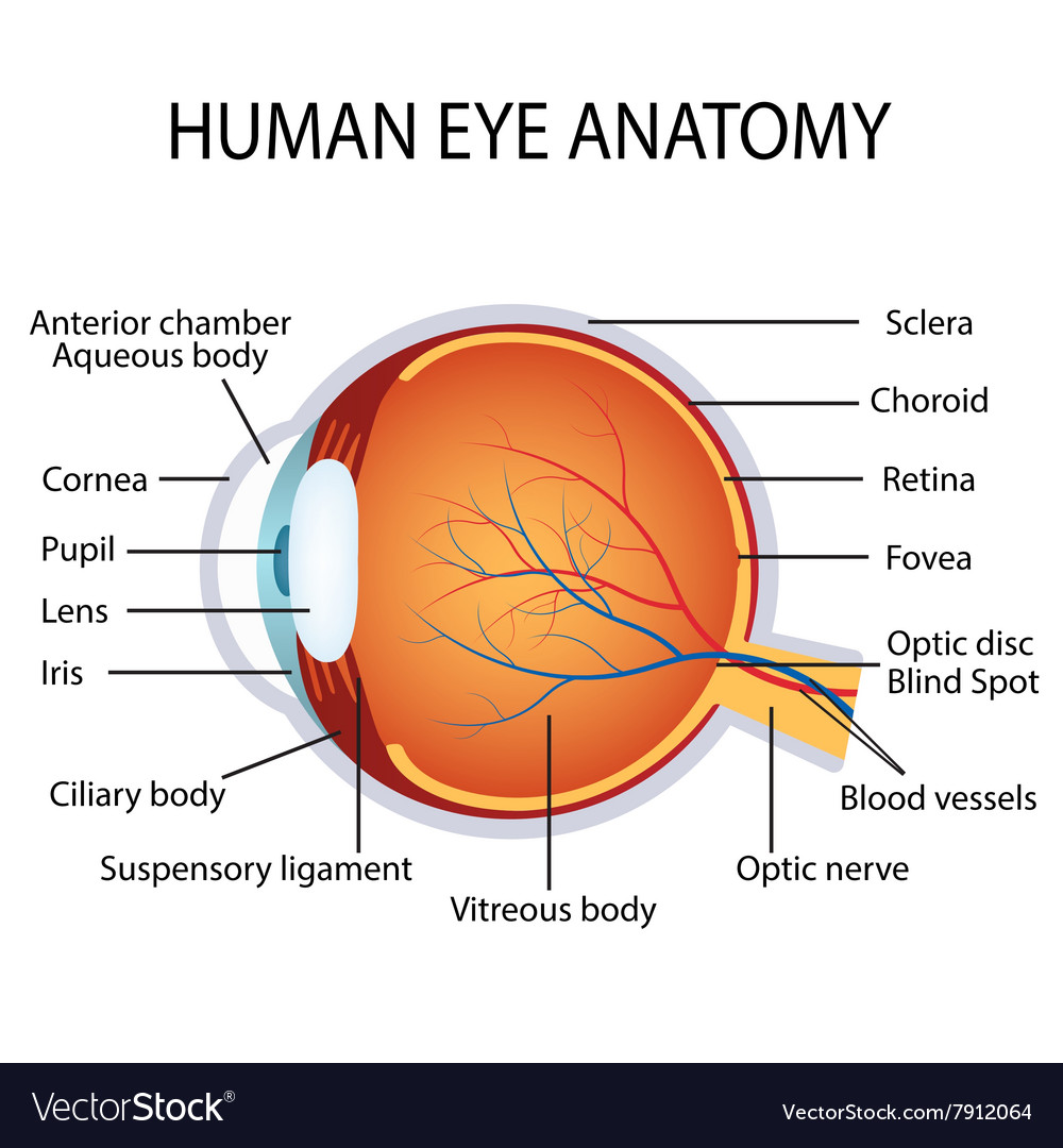
Anatomy of the human eye. Clear front window of the eye that transmits and focuses light into the eye. In biology human biology physics and practical courses in medicine nursing and therapies. Picture of eye anatomy detail.
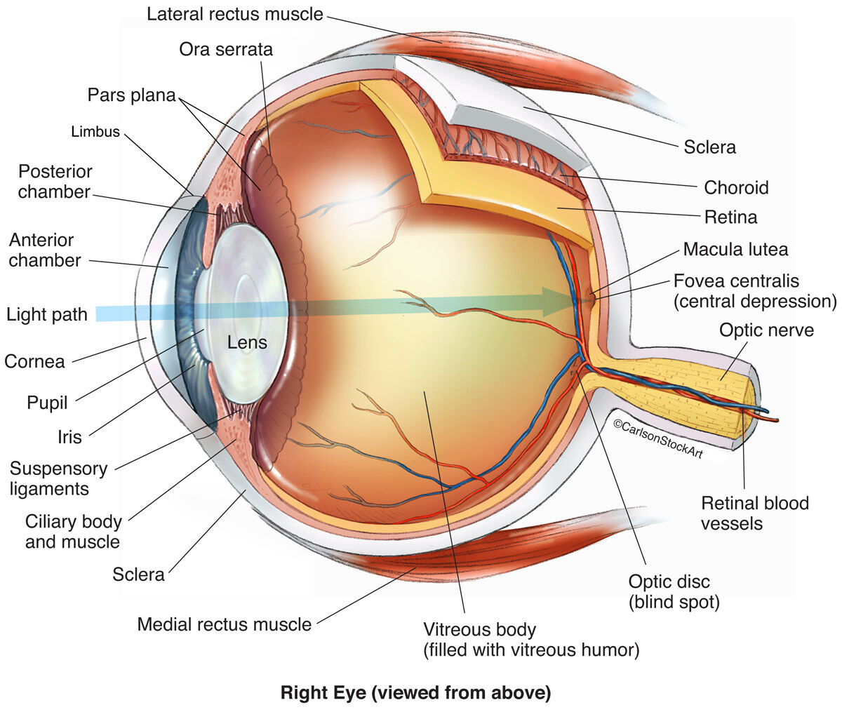
The anatomy of the eye includes auxillary structures such as the bony eye socket and extraocular muscles as well as the structures of the eye itself such as the lens and the retina. Vision is our window to the outside world. Conjunctiva cornea iris lens macula retina optic nerve vitreous and extraocular muscles.
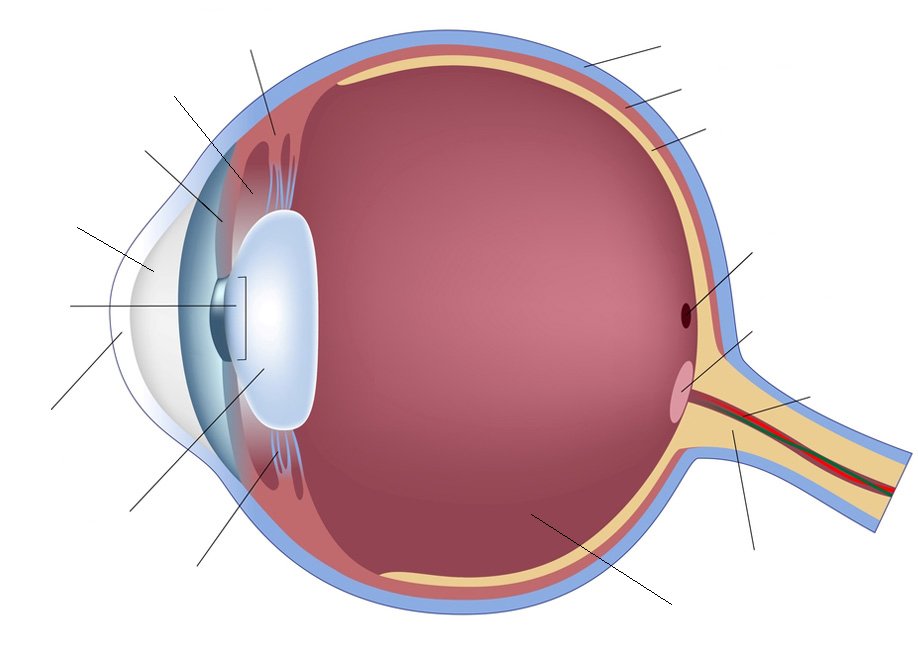
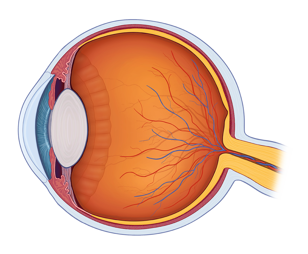
A closer look at the parts of the eye by liz segre when surveyed about the five senses sight hearing taste smell and touch people consistently report that their eyesight is the mode of perception they value and fear losing most. The diagrams below show cross sections of the human eyeball. The inside lining of the eye is covered by special light sensing cells that are collectively called the retina.
The human eye can differentiate between about 10 million colors and is possibly capable of detecting a single photon. It converts light into electrical impulses. The eye is the organ responsible for vision.
Anatomy of the eye the anatomy and physiology of the human eye is an important part of many courses eg. The human eye is an organ that reacts to light and has several purposesthe eye is a complex structure with layers lens muscles receptors that is surrounded by many boneswatch various parts. The eye is our organ of sight.
This article explores the anatomy of the eye looking at the different structures of the human eye and their function. Anatomy of the eye. The eye has a number of components which include but are not limited to the cornea iris pupil lens retina macula optic nerve choroid and vitreous.
The human eye is an organ that reacts to light and allows vision. This simple introduction the subjects of the eye and visual optics includes a simple diagram of the eye together with definitions of the parts of the eye labelled in the illustration. Behind the eye your optic nerve carries.
Extraocular muscles help move the eye in different directions. Anatomy of the eye. Rod and cone cells in the retina allow conscious light perception and vision including color differentiation and the perception of depth.
Human eye specialized sense organ in humans that is capable of receiving visual images which are relayed to the brain.
 The Poor Design Of The Human Eye The Human Evolution Blog
 Human Eye Anatomy La Pine Eyecare Clinic
Human Eye Snowy Owl Vs Human Eyes
 Eye Anatomy Detail Picture Image On Medicinenet Com
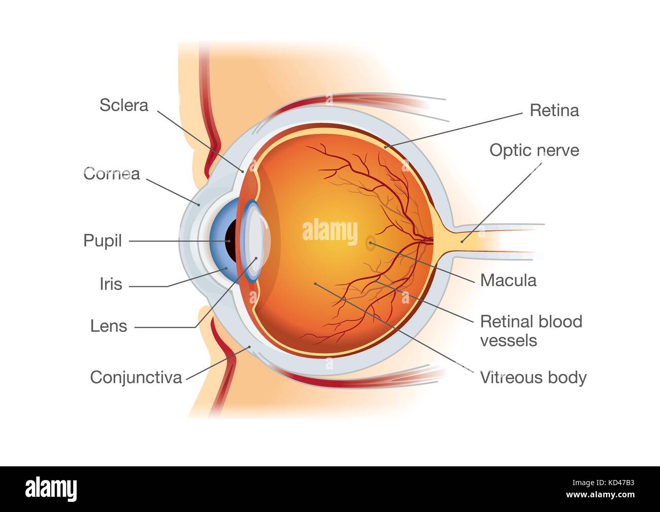 Human Eye Anatomy In Side View Stock Vector Art
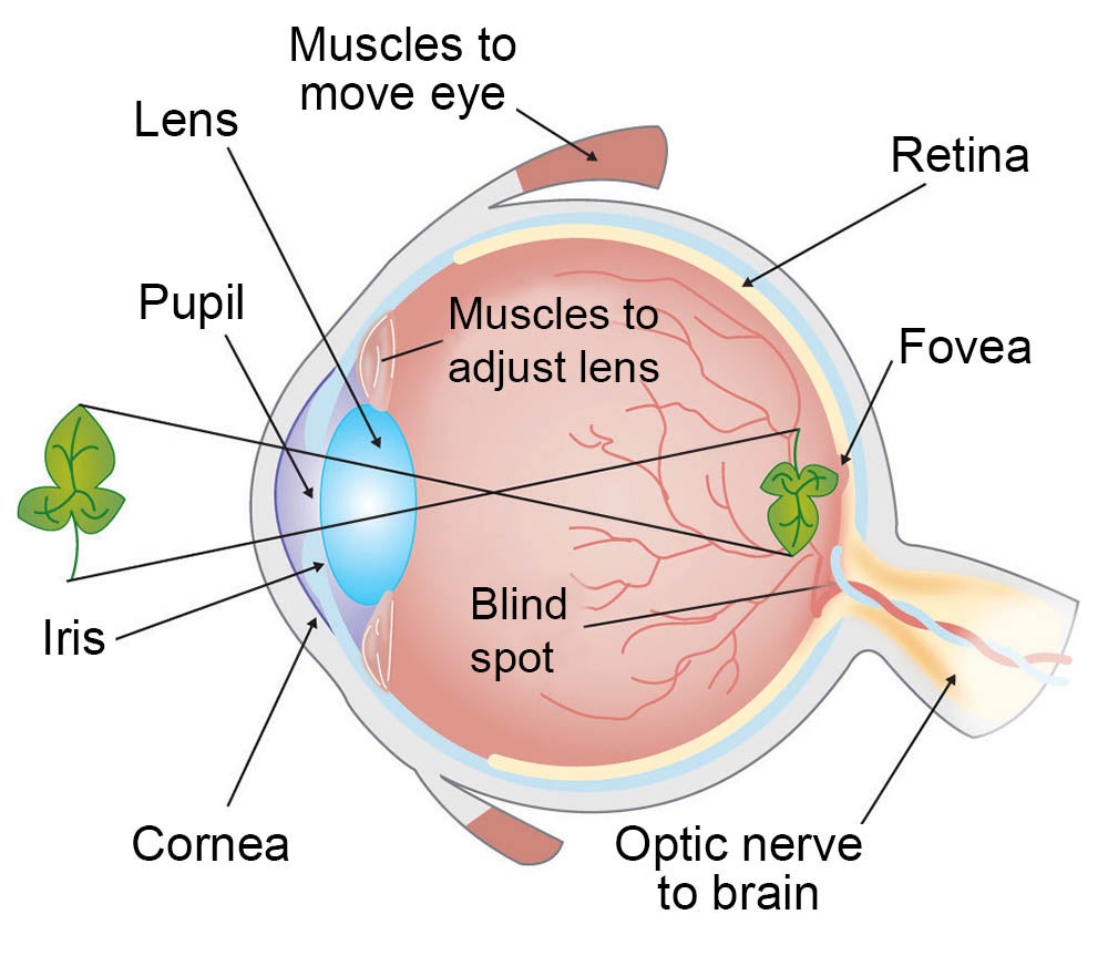 How Do We See Light Ask A Biologist
 Anatomy Of The Eye Editable Powerpoint Presentation Main Parts Of Human Eye Free Ppt
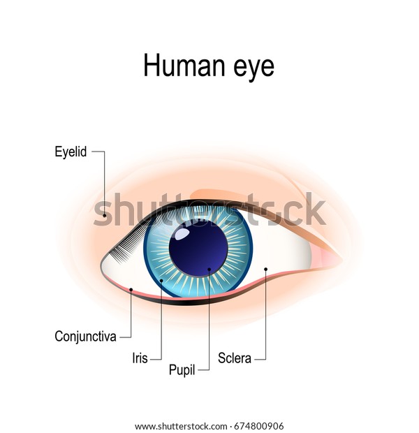 Anatomy Human Eye Front View External Stock Vector Royalty
Eye Anatomy And Physiology How The Eye And Vision Work

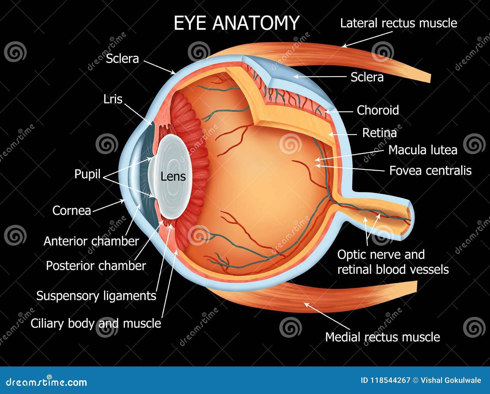 Human Eye Anatomy Full Details Stock Illustration
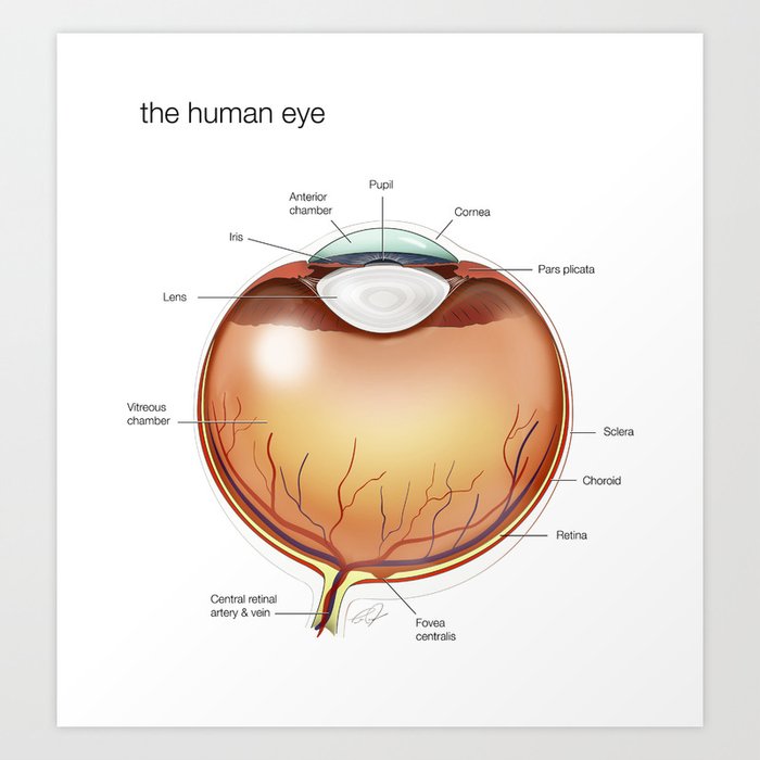 Human Eye Anatomy Illustration Art Print By Cjonesscivi
 2 Cross Section Of A Human Eye Anatomy Download
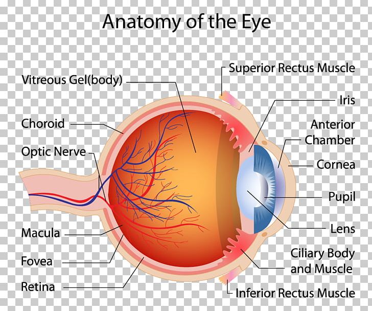 Human Eye Macula Of Retina Muscle Anatomy Png Clipart
 How To Draw Human Eye Anatomy Diagram Easily
1 Basic Anatomy Of The Human Eye As Seen Through A Cross
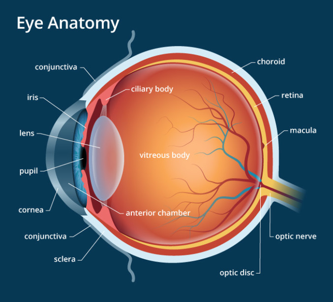 Eye Anatomy A Closer Look At The Parts Of The Eye
 Eye Diagram Unlabelled Human Eye Diagram Unlabelled Human
 Eye Anatomy 1 Illustration Carlson Stock Art

Gross Anatomy Of The Eye By Helga Kolb Webvision
 Anatomy Of The Human Eye Visual Acuity Light Perception
 Human Eye Anatomy Vector Image 1815155 Stockunlimited
 The Human Eye In Anatomical Transparencies Books Health




Posting Komentar
Posting Komentar