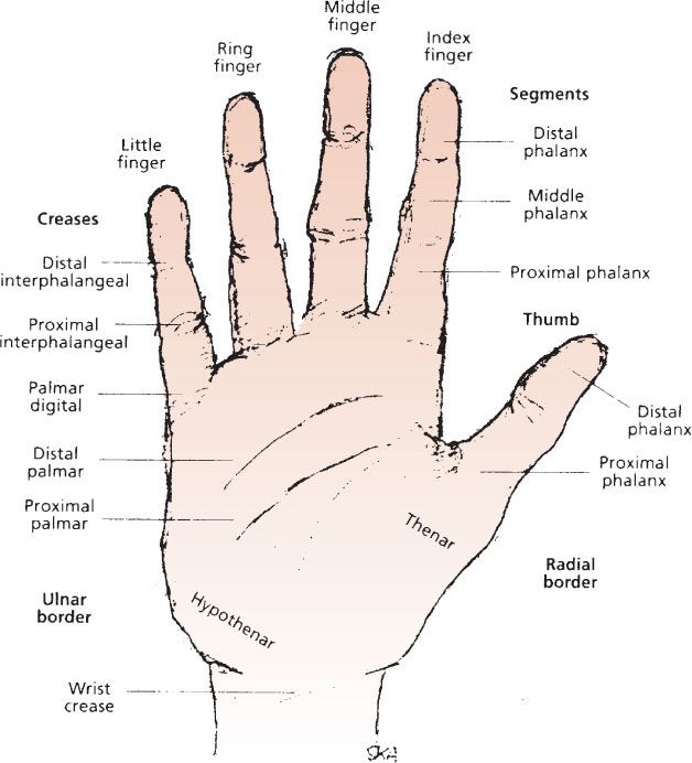
Hand surgeons often describe an injury or problem as being on the ulnar side or radial side of the hand. Here are some landmarks of the wrist and hand surface anatomy.
 Surface Anatomy Advanced Anatomy 2nd Ed
Interosseous muscles of the hand.

Surface anatomy of hand. Three unipennate muscles between the metacarpals and are attached to the index ring and little fingers. The intrinsic muscles of the hand are located within the hand itself. The tendons of the flexor carpi radialis and the palmaris longus are palpable.
Surface anatomy of the hand the hand is twice as long as it is broad. Hand surface anatomy 1. Naming the digits thumb index.
Thenar and hypothenar are two terms that describe the fleshy mass of skin fat. Surface anatomy distal phalanx. Abductor digiti minimi flexor digiti minimi opponens digiti minimi and palmaris brevis.
Naming the digits thumb index middle. Naming the digits thumb. They are responsible for the fine motor functions of the hand.
Hook of the hamate. This region is called the hypothenar eminence and consists of the four hypothenar muscles. These adduct the fingers toward the midline all interosseous muscles are innervated by the deep branch of the ulnar nerve.
Naming the digits the birmingham hand centre assessment and early management. These include the adductor pollicis palmaris brevis interossei lumbricals thenar and hypothenar muscles. Naming the digits thumb index.
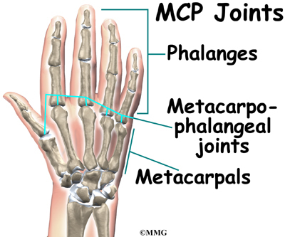
In this article we shall be looking at the anatomy of the intrinsic muscles of the hand. Section 2 surface pertinent anatomy. The length of the middle finger from the metacarpophalangeal joint to its extremity is equal to the distance from the metacarpophalangeal joint to the radiocarpal joint.
Also on the palmar surface of the hand the thenar eminence has a corresponding fleshy region on the ulnar side of the hand. Surface anatomy involves the specific systematic implementation of topographic and anatomical knowledge through targeted palpation of the living human body. The tendons of extensor digitorum are visible on the back of the hand.
The head of the. If you have symptoms isolated on one side or the other this can be valuable information that leads to a correct diagnosis. It is easily palpated and visible at the base of the little finger.
The pisiform bone is palpated just below the hypothenar eminence. Index long ring and little metacarpals. Four bipennate muscles in the dorsum of the hand that abduct the index middle and ring fingers away from midline palmar interossei.
207 this chapter on surface anatomy intends to provide practitioners with a systematic method for quickly and reliably finding all of the structures that are important for treating the hand. Scaphoid tuberosity and scaphoid waist within the anatomic snuffbox.
 Hand Surface Anatomy India Future Society
 Illustrations Of The Human Hand Showing The Anatomical Basis
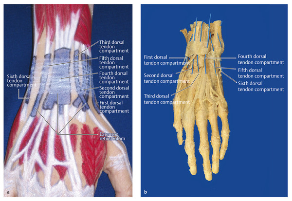 Surface Anatomy Of The Forearm Wrist And Hand Structures
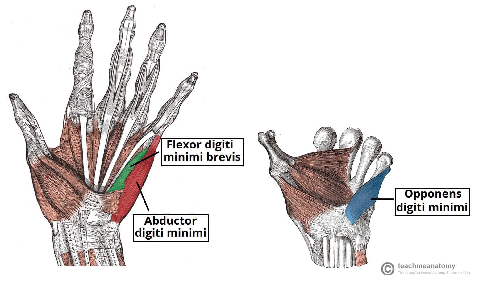 The Ulnar Nerve Course Motor Sensory Teachmeanatomy
 Flexor Retinaculum Of The Hand Wikipedia
 I Examination Of The Wrist Surface Anatomy Of The Carpal
 Figure 9 From Human Hand Modeling From Surface Anatomy
 Surface Anatomy Anatomy Human Anatomy Surface
Hand Surface Anatomy Language Of Hand And Arm Surgery Series
 Manus And Carpus Anatomy Of The Dog On Ct
Hand Anatomy Midwest Bone Joint Institute Elgin Illinois
 The Hand Three Figures Left To Right Bones Muscles And
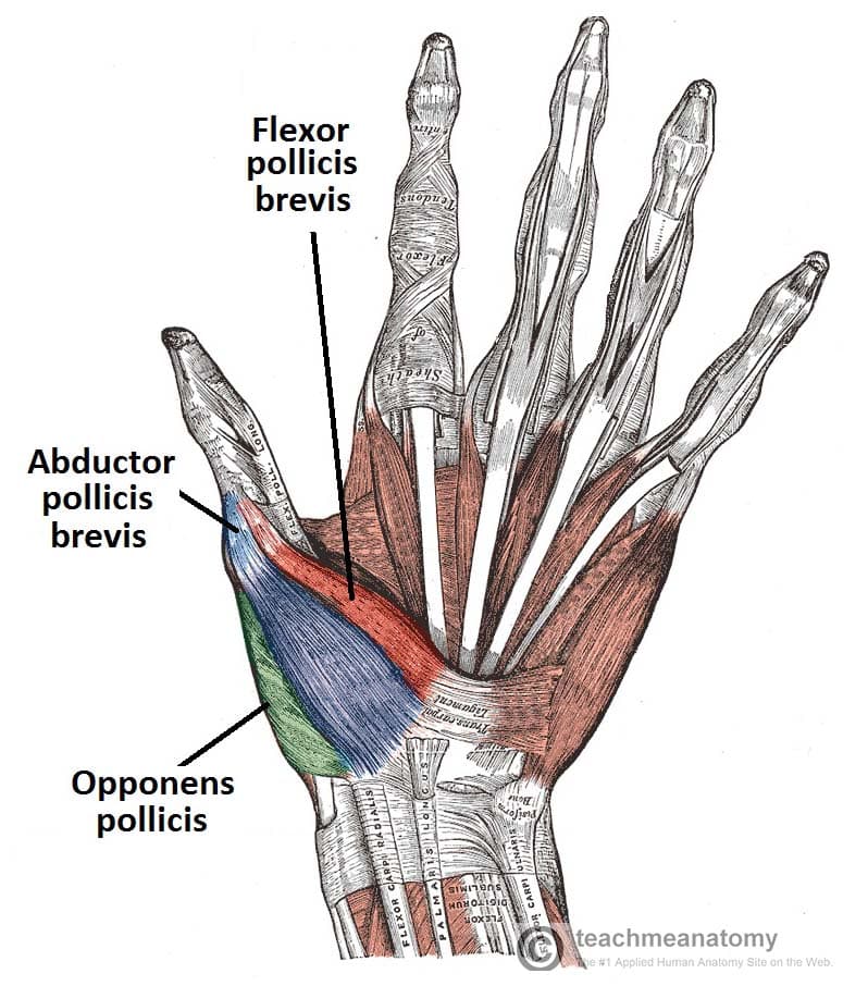 The Muscles Of The Hand Thenar Hypothenar Teachmeanatomy
 Hand And Wrist Anatomy Musculoskeletal Key
 Surface Anatomy Normal Wrist Medlineplus Medical
 Surface Anatomy Hand Palmar Aspect Model
 Surface Anatomy Of Upper Limb Bones
 Forearm Shaft Approach Forearm Surface Anatomy Ao
 The Bones Of The Forearm Stock Image F001 8064 Science
 Surface Anatomy Hand Surgery Source
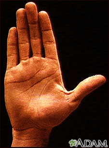 Surface Anatomy Normal Palm Medlineplus Medical


Posting Komentar
Posting Komentar