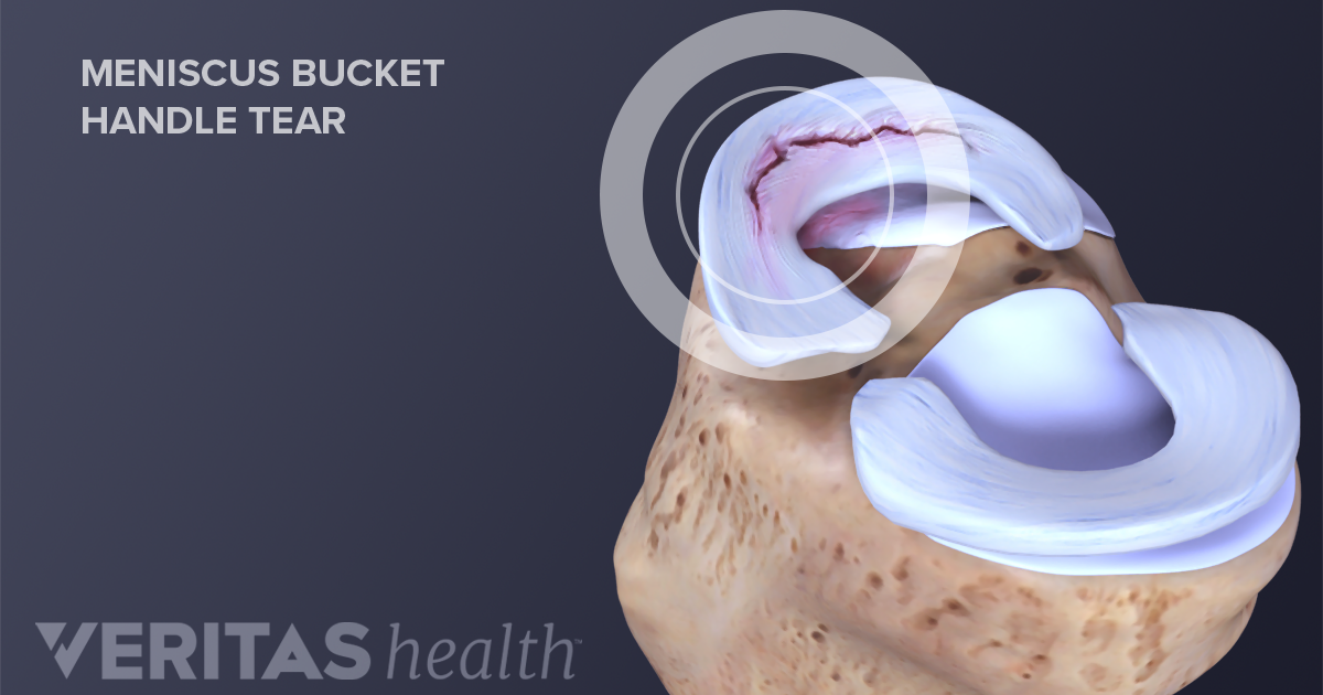
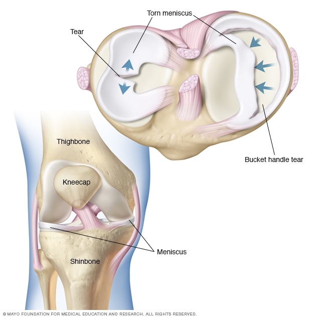
However anyone at any age can tear a meniscus. When people talk about torn cartilage in the knee they are usually referring to a torn meniscus.

This provides a.

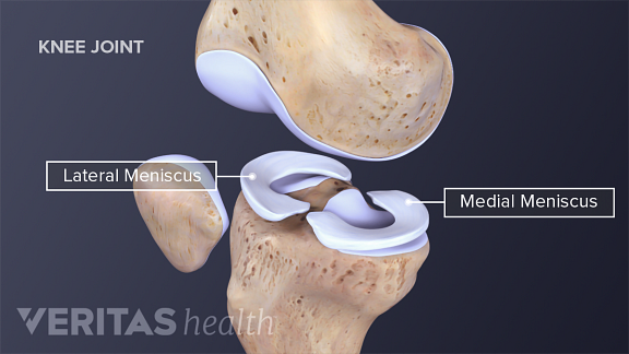
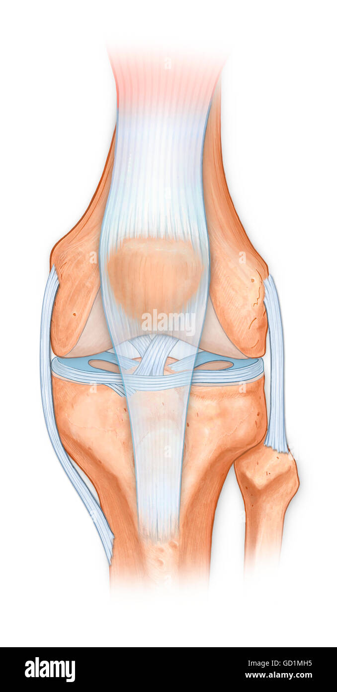
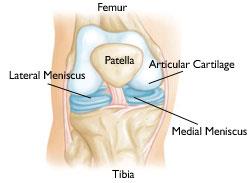
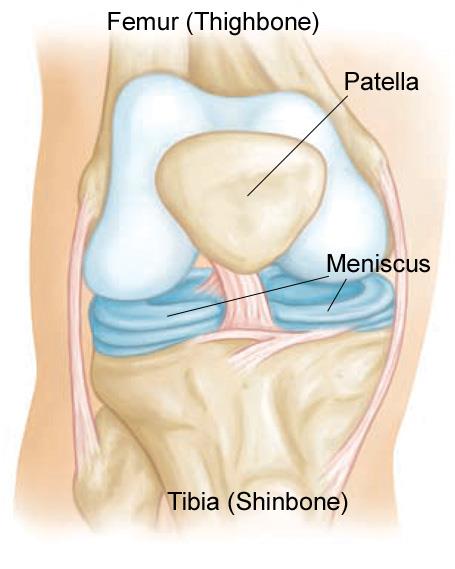
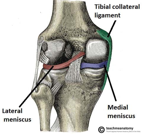
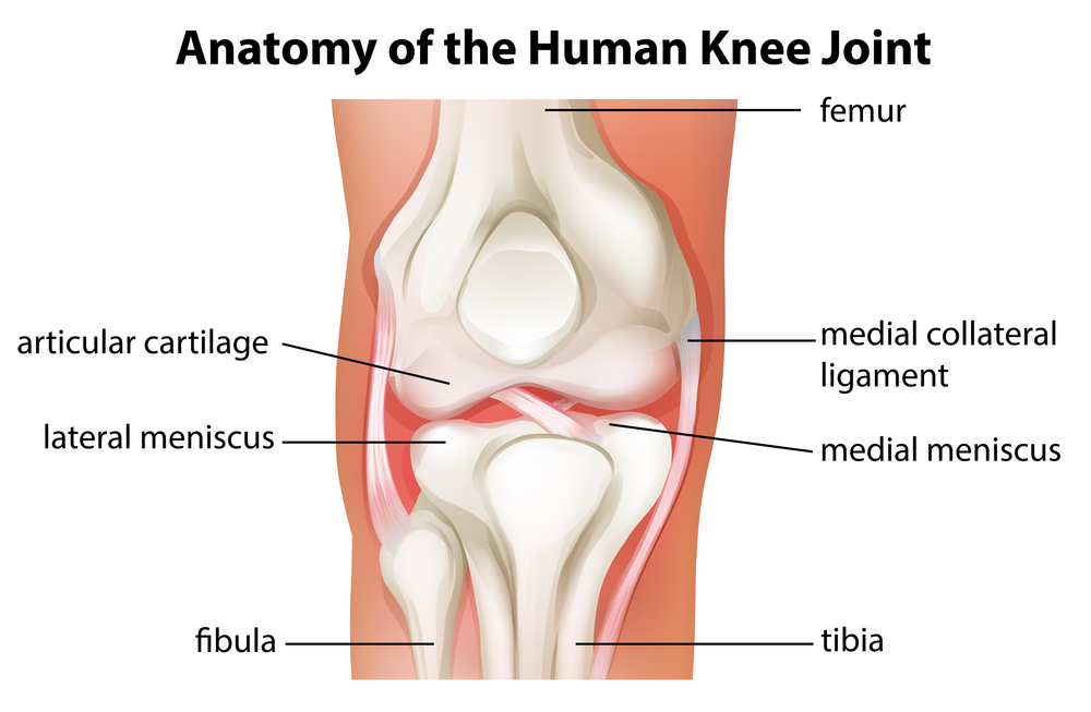
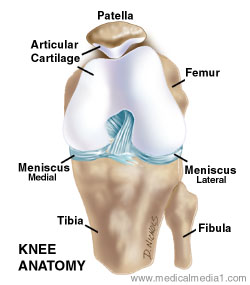
Anatomy of the knee meniscus. Medial meniscus and lateral meniscus. The medial meniscus is the central band of cartilage attached to the tibia or shinbone. The job of the meniscus is to cushion the knee joint and transfer forces between the tibia and femur the thigh and shin bones.
In humans they are present in the knee wrist acromioclavicular sternoclavicular and temporomandibular joints. Large tears may cause the. A meniscus is a crescent shaped fibrocartilaginous anatomical structure that in contrast to an articular disc only partly divides a joint cavity.
The band goes around the knee joint in a crescent shaped path and is located between the medial condyles of. The meniscus is a cushion structure made of cartilage which fits within the knee joint between the tibia and femur. Any form of arthritis or injury may cause a knee effusion.
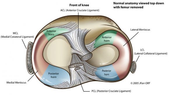
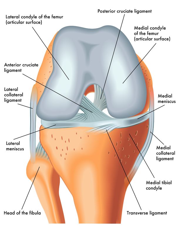
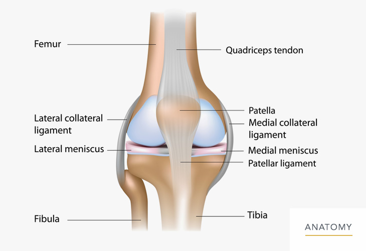
In the healthy knee there are two menisci. The knees menisci are two half moon wedge shaped pieces of cartilage the lateral and medial meniscus acting as lubricant and elastic buffer distributing forces evenly between the femur upper leg and tibia lower leg in the knee joint. Athletes particularly those who play contact sports are at risk for meniscus tears.
The medial meniscus is on the inner part of the knee. Meniscus tears are among the most common knee injuries. The knee meniscus is a special layer of extra cartilage that lines the knee joint.
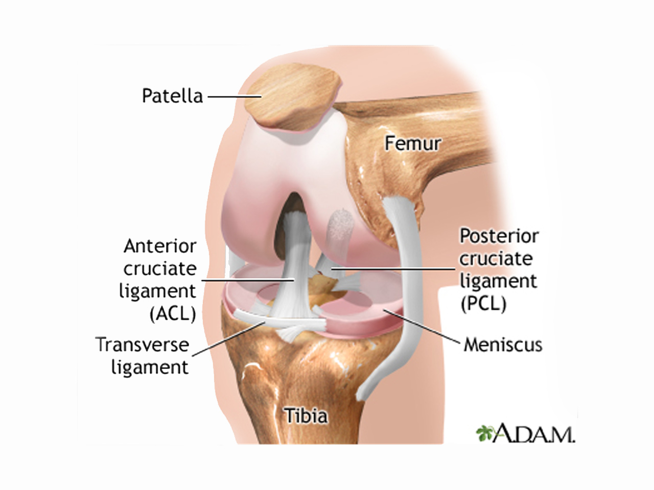
There are two menisci inside the knee joint. Integral to the knees functionality is the meniscus. Damage to a meniscus the cartilage that cushions the knee often occurs with twisting the knee.
2 in other animals they may be present in other joints. The meniscus is a c shaped piece of tough rubbery cartilage that acts as a shock absorber between your shinbone and thighbone. It can be torn if you suddenly twist your knee while bearing weight on it.
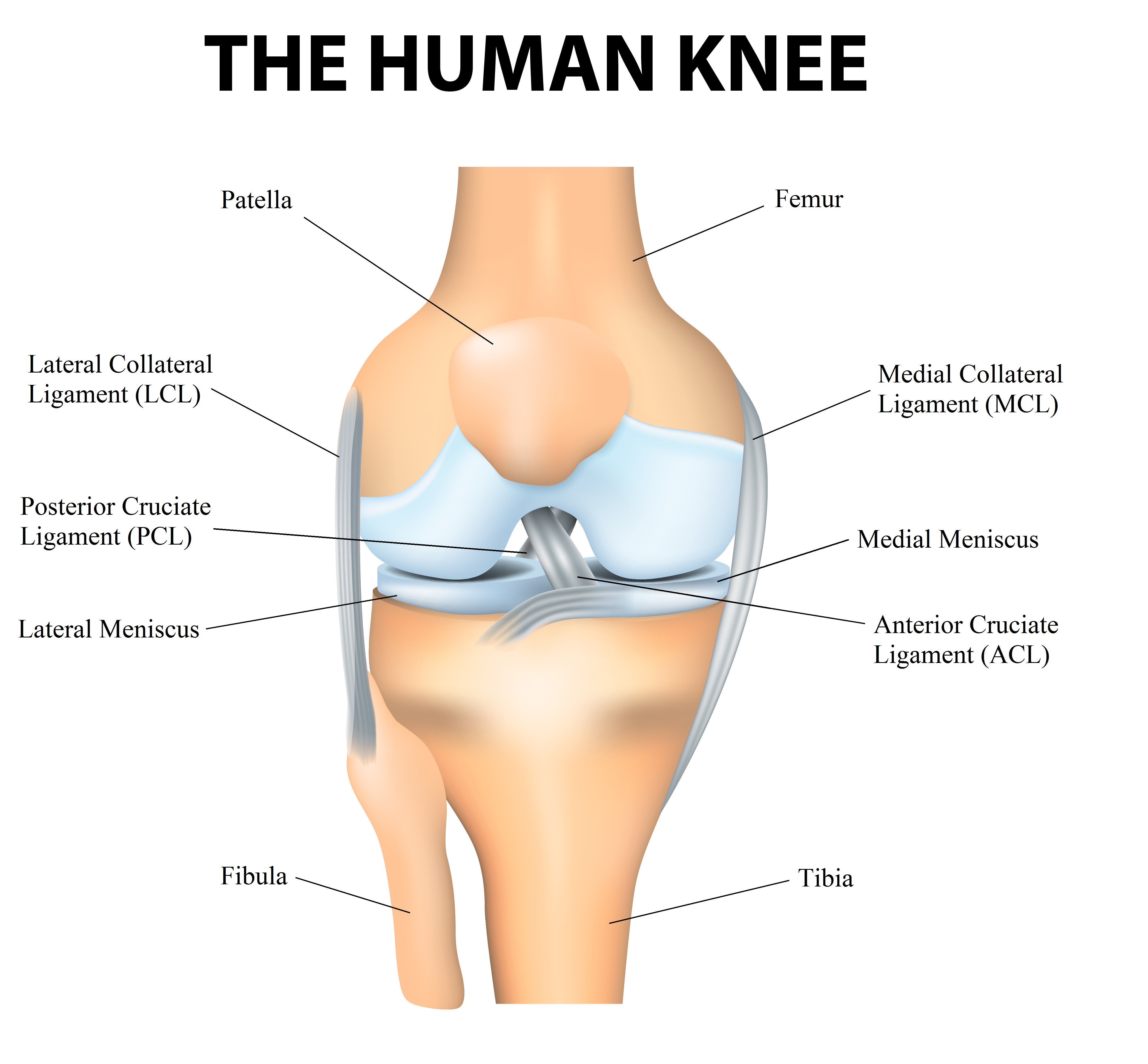
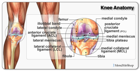
The knee joint contains the meniscus structure comprised of both a medial and a lateral component situated between the corresponding femoral condyle and tibial plateau figure 1 each is a glossy white complex tissue comprised of cells specialized extracellular matrix ecm molecules and region specific innervation and vascularization. The meniscus is a rubber like c shaped disc that sits within the knee between the end of the thighbone femur and the top of the shinbone tibia. Most of the joints in our body are lined with a thin layer of articular cartilage made of collagen and chondroitin.
 Understanding Meniscus Tears
 The Injury Zone Basic Anatomy And Function Of The Meniscus
 Anatomy And Function Of The Knee Skagit Northwest Orthopedics
 Acl Solutions Acl Knee Anatomy And Diagram Images
 Understanding Meniscus Tears
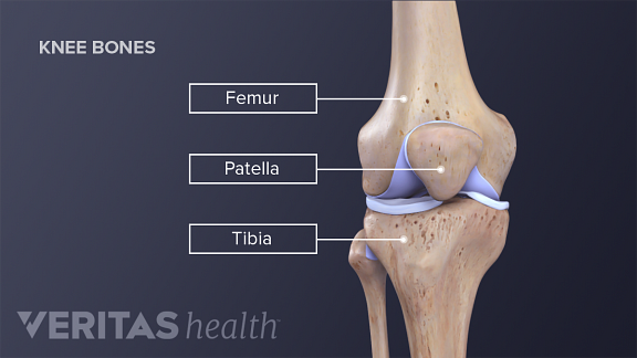
 Normal Anatomy Of The Knee Joint Femur Tibia Fibula
 Meniscal Transplant Surgery Orthoinfo Aaos
 Lateral Meniscus Wikipedia
 Torn Meniscus Symptoms And Causes Mayo Clinic
 Matthew Boyle Orthopaedic Surgeon Knee Anatomy Knee
 Redding Hospital Knee Anatomy
 Torn Meniscus Anatomy And Causes Video Jeffrey H Berg
 Knee Anatomy
The Injury Zone Basic Anatomy And Function Of The Meniscus

 Life With A Degenerative Meniscus Tear Buffalo Rehab Group
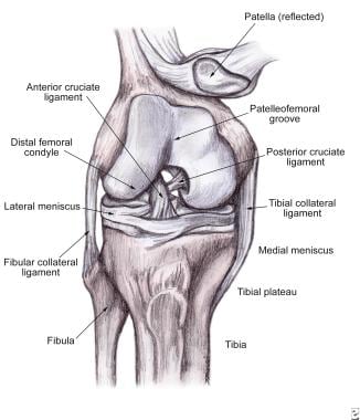
 Clinical Anatomy Knee Mensicus And Knee Joint
 Lateral Meniscus An Overview Sciencedirect Topics
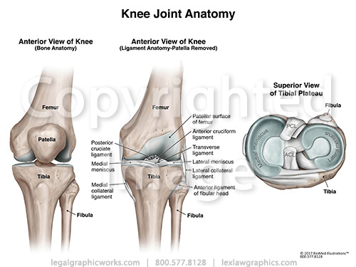 Anterior Tibial View Of Knee Joint Anatomy
 Soft Tissue Knee Injury Practice Essentials Background
 The Knee Meniscal Injuries
 Meniscus Tears Orthoinfo Aaos
 The Knee Joint Articulations Movements Injuries
 Torn Meniscus Treatments Physical Therapy Just As Good As
 Knee Calf Orthopedic Specialist Of Northern California
 Anatomy Of The Knee Baxter Regional Medical Center

 Can Stem Cells Treat An Acl Tear Or Torn Meniscus
 Meniscus Tear
 The Knee Anatomy Injuries Treatment And Rehabilitation
 Meniscal Tears Knee Cartilage Deterioration And Treatment
 Anatomy Of The Knee Joint Paley Orthopedic Spine Institute


Posting Komentar
Posting Komentar