Unsubscribe from scc pathology. Radiology residency umjmh 6447 views.
The pelviss frame is made up of the bones of the pelvis which connect the axial skeleton to the femurs and therefore acts in weight bearing of the upper body.
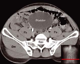
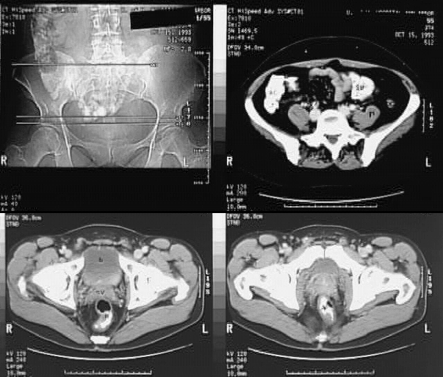
Pelvic anatomy ct. The pelvis ct anatomymp4 scc pathology. This mri male pelvis axial cross sectional anatomy tool is absolutely free to use. Anatomy ct axial abdomen and pelvis male male abdomen and pelvis ct scan form no 1.
This photo gallery presents the anatomy of the abdomen by means of ct axial coronal and sagittal reconstructions. Atlas of ct anatomy of the abdomen. Newer high resolution ct scanners combined with mechanical intravenous contrast medium injectors and thinner sections have substantially improved the imaging of female genital tract anatomy.
The pelvic cavity is inferior part of the abdominopelvic cavity and is in direct connection with the abdominal cavity. Ct anatomy of the pelvis. Talos i f jakab m kikinis r.
It is divided into. Learn the diagnosis of ct and methods of computed tomography. Use the mouse scroll wheel to move the images up and down alternatively use the tiny arrows on both side of the image to move the images on both side of the image to move the images.
The pelvis is the lower portion of the trunk located between the abdomen and the lower limbs. 15 liver 16 oesophagus 17 stomach 41 descending abdominal aorta 43 inferior vena cava 55 thoracic vertebra. Approach to the abdominal pelvic ct duration.
The pelvic cavity is bounded by the bony pelvis and the pelvic musculature and primarily contains reproductive organs and the rectum. Anatomy of the abdomen and male pelvis using cross sectional imaging ct interactive atlas of human anatomy we have created an anatomical atlas of abdominal and pelvic ct which is an interactive tool for studying the conventional anatomy of the normal structures based on a multidetector computed tomography. The ct appearance of the normal ligamentous vascular and visceral anatomy of the female pelvis can be confusing.
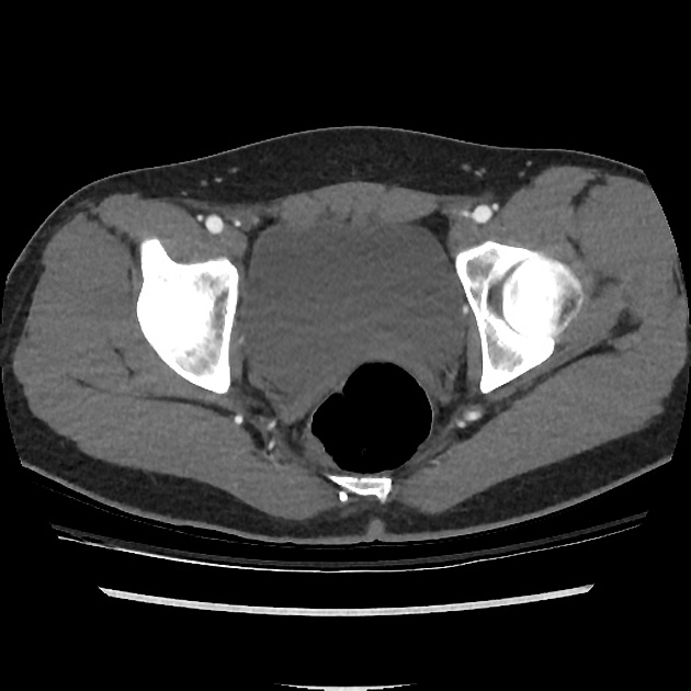 Normal Ct Angiogram Of Pelvis Radiology Case Radiopaedia Org

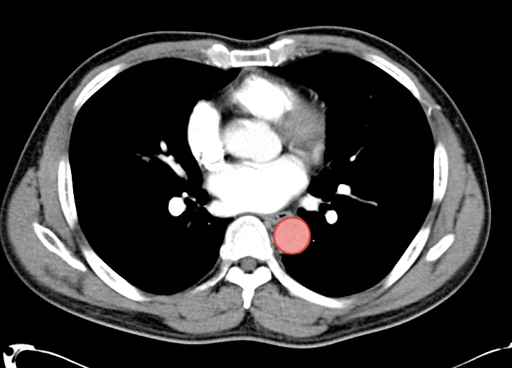 A Systematic Approach To The Interpretation Of Ct Abdomen Pelvis
 Ct Scan Tips Protocols Ct Anatomy Of The Female Pelvis
 Ct Abdomen And Pelvis With Contrast Anatomy Labelled
Atlas Of Ct Anatomy Of The Abdomen
Uams Gross Anatomy X Ray Atlas
 Pelvis An Overview Sciencedirect Topics
 Pelvis Computed Tomograph Axial Ct Muscle Anatomy
 Sagittal Ct Of Pelvis And Lumbar Spine
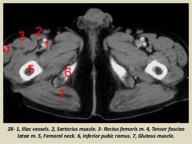 Presentation1 Pptx Ct Normal Anatomy Of The Abdomen And Pelvis
 The Computerized Tomography Ct Scan Of Abdomen And Pelvis
 Ct Of The Abdomen And Pelvis Chapter 33 Clinical
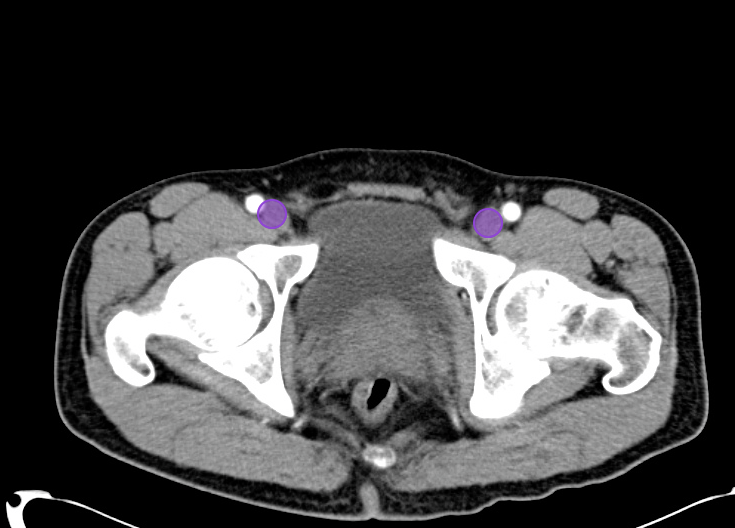 A Systematic Approach To The Interpretation Of Ct Abdomen Pelvis
 Figure 3 From Ct Anatomy Of The Female Pelvis A Second Look
 Abdominal Ct Anatomy Radiology Key
 Gross Anatomy Cross Sections Of The Abdomen And Pelvis Part 3 Axial Cuts
 Ct Scan Of Abdomen Stock Photo Image Of Anatomy Verticle
 Mri Pelvis Anatomy Free Male Pelvis Axial Anatomy
 Mri Pelvis Anatomy Free Male Pelvis Axial Anatomy
Image Guided Percutaneous Procedures In Deep Pelvic Sites
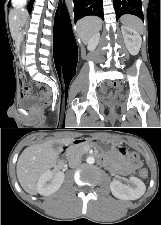 Computed Tomography Of The Abdomen And Pelvis Wikipedia
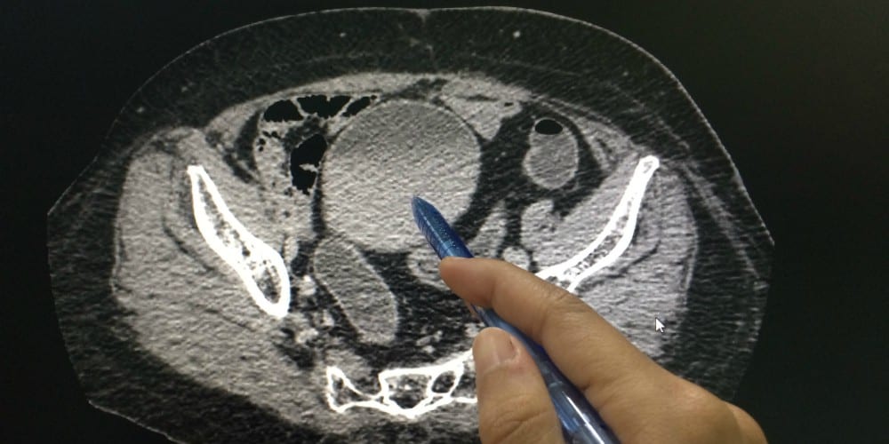 Which Cancers Can A Pelvic Ct Scan Detect American Health
 Normal Ct Pelvis Adult Male Radiology Case Radiopaedia Org


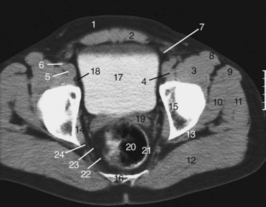

Posting Komentar
Posting Komentar