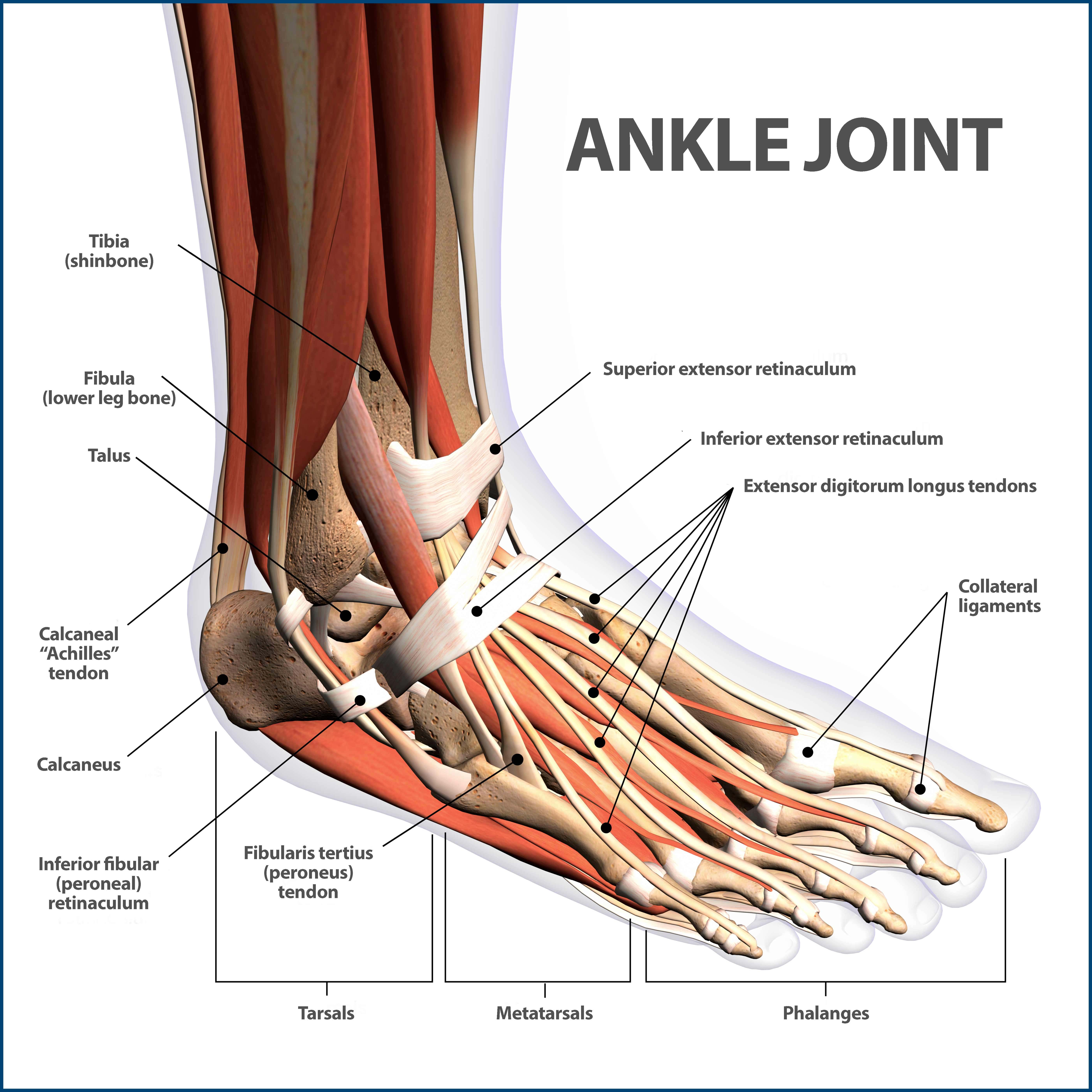
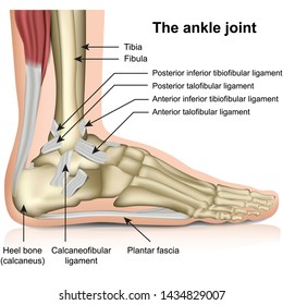
The ankle joint also known as the talocrural joint allows dorsiflexion and plantar flexion of the foot. The last two together are called the lower ankle joint.
 28 Best Ankle Anatomy Images In 2019 Ankle Anatomy
Footeducation is committed to helping educate patients about foot and ankle conditions by providing high quality accurate and easy to understand information.
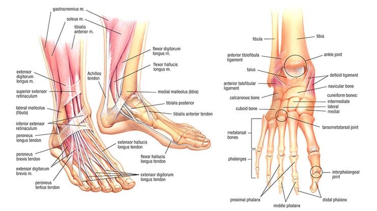
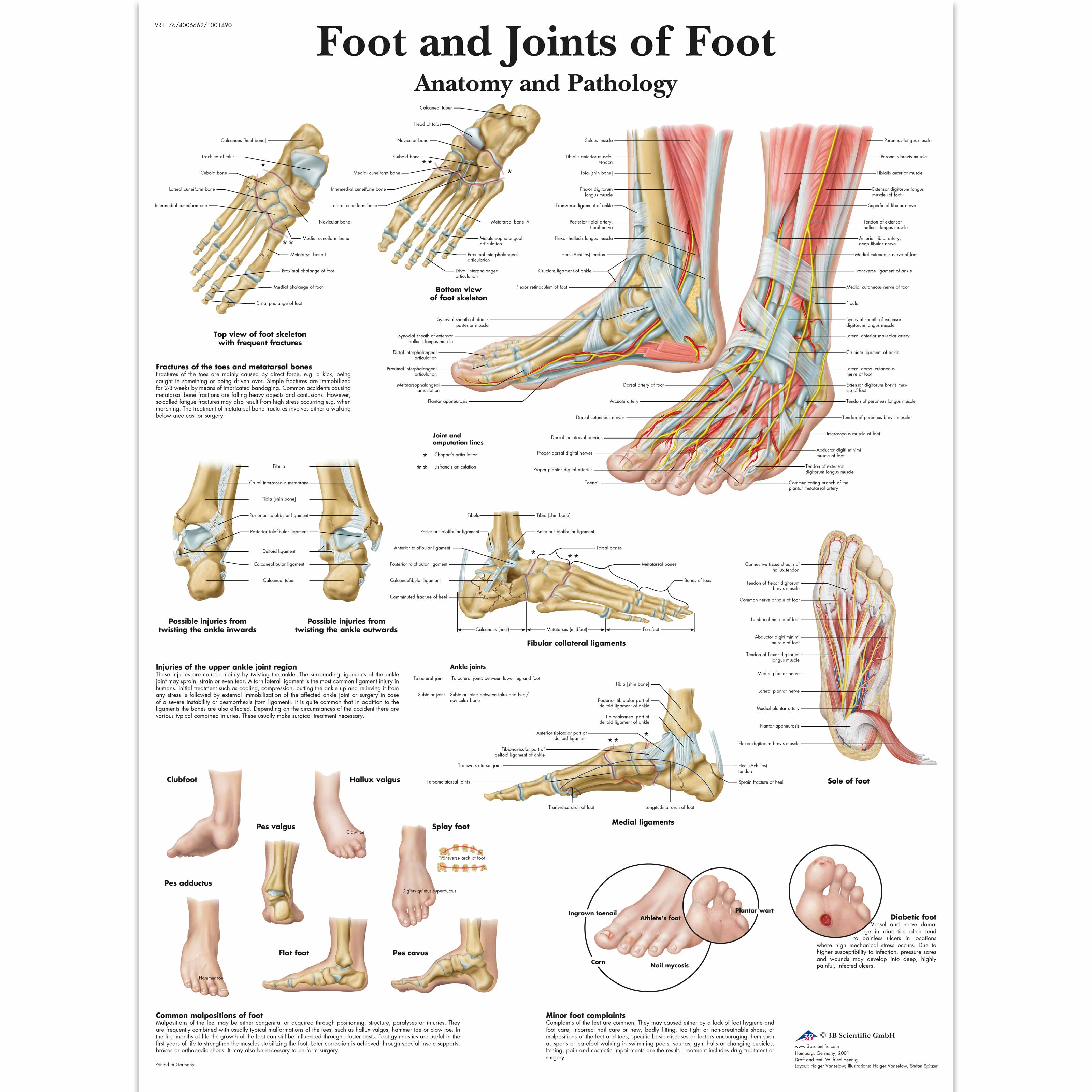
The anatomy of the foot and ankle. The subtalar joint sits below the ankle joint and allows side to side motion of the foot. The largest and strongest tendon of the foot is the achilles tendon which extends from the calf muscle to the heel. The ankle is the joint between the foot and leg composed of three separate bones.
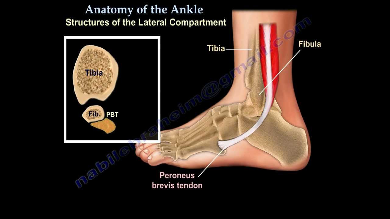
Ebraheims educational animated video describes anatomical structures of the foot and ankle the bony anatomy the joints ligaments and the compartments in a simple and easy way. Its strength and joint function facilitate running jumping walking up stairs and raising the body onto the toes. Foot ankle anatomy muscles tendons and ligaments.
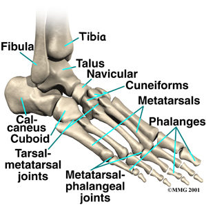
These all work together to bear weight allow movement and provide a stable base for us to stand and move on. The bones of the foot and ankle begin with the ankle joint itself. The outer bone is the fibula or calf bone.
Upper ankle joint tibiotarsal talocalcaneonavicular and subtalar joints. It is made up of three joints. Ligaments hold the tendons in place and stabilize the joints.
The talus bone supports the leg bones tibia and fibula forming the ankle. The ankle joint allows up and down movement of the foot. The ankle joint talocrural joint is formed where the distal end of the leg meets the foot.
Medically reviewed by healthline medical team on april 8 2015. Foot and ankle anatomy is quite complex. The inner bone is the tibia or shinbone which supports most of a persons weight when standing.
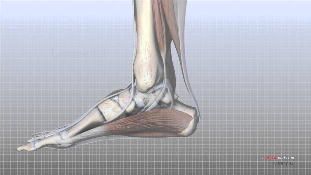
Lateral side of the ankle joint capsule. Use our anatomy tools to learn about bones joints ligaments and muscles of the foot and ankle. It is formed by the bones of the leg tibia and fibula and the foot talus.
It is formed by the bones of the leg tibia and fibula and the foot talus. The ankle joint is formed where the talus the uppermost bone in the foot and the tibia shin meet. The ankle joint or talocrural joint is a synovial joint located in the lower limb.
The calcaneus heel bone is the largest bone in the foot. The hindfoot forms the heel and ankle. Numerous ligaments made of tough moveable.
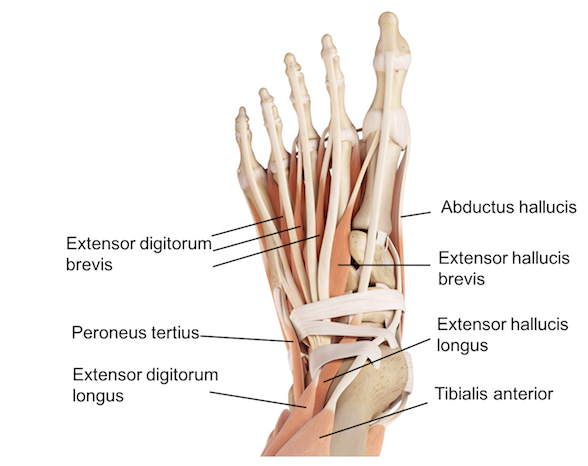
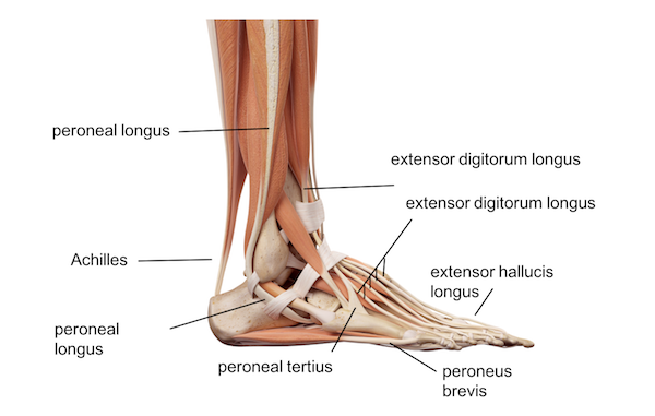
The foot consists of thirty three bones twenty six joints and over a hundred muscles ligaments and tendons.
 Ankle Fractures Broken Ankle Florida Orthopaedic Institute
 Ankle Foot Anatomy
 Foot Anatomy Laminated Poster
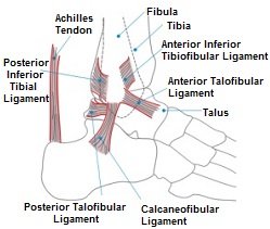
 Ankle Joint Anatomy Overview Lateral Ligament Anatomy And
Foot Exercises For Foot Pain Tight Ankles Calves Gmb
 Ankle Foot Anatomy
 3b Scientific A31 1l Human Left Loose Foot And Ankle Skeleton
 Anatomy Of The Foot Ankle Everything You Need To Know Dr Nabil Ebraheim
 Bones The Of Foot Stock Vector Illustration Of Orthopedic
 Get To Know The Ankle Joint Yoga Journal
 Foot Anatomy Animated Tutorial
 Foot And Ankle Patient Education
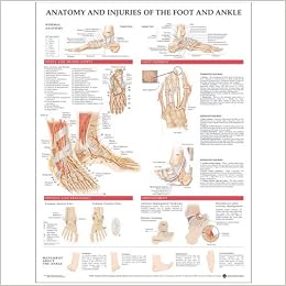 Anatomy And Injuries Of The Foot And Ankle 9781587798375

 Foot And Ankle Skeleton Model
 Foot And Ankle Anatomy Bones Muscles Ligaments Tendons
 Ankle Images Stock Photos Vectors Shutterstock
 Foot Anatomy Spokane Valley Wa Foot Doctor
 Foot And Joints Of Foot Chart Anatomy And Pathology
![]() Ankle And Foot Anatomy Bones Joints Muscles Kenhub
Ankle And Foot Anatomy Bones Joints Muscles Kenhub
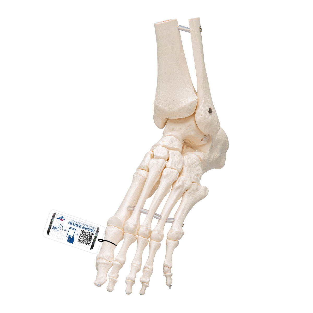 Foot Ankle Skeleton Elastic Mounted 3b Smart Anatomy
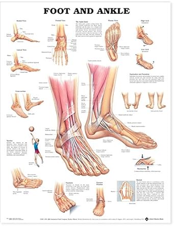 Foot And Ankle Anatomical Chart
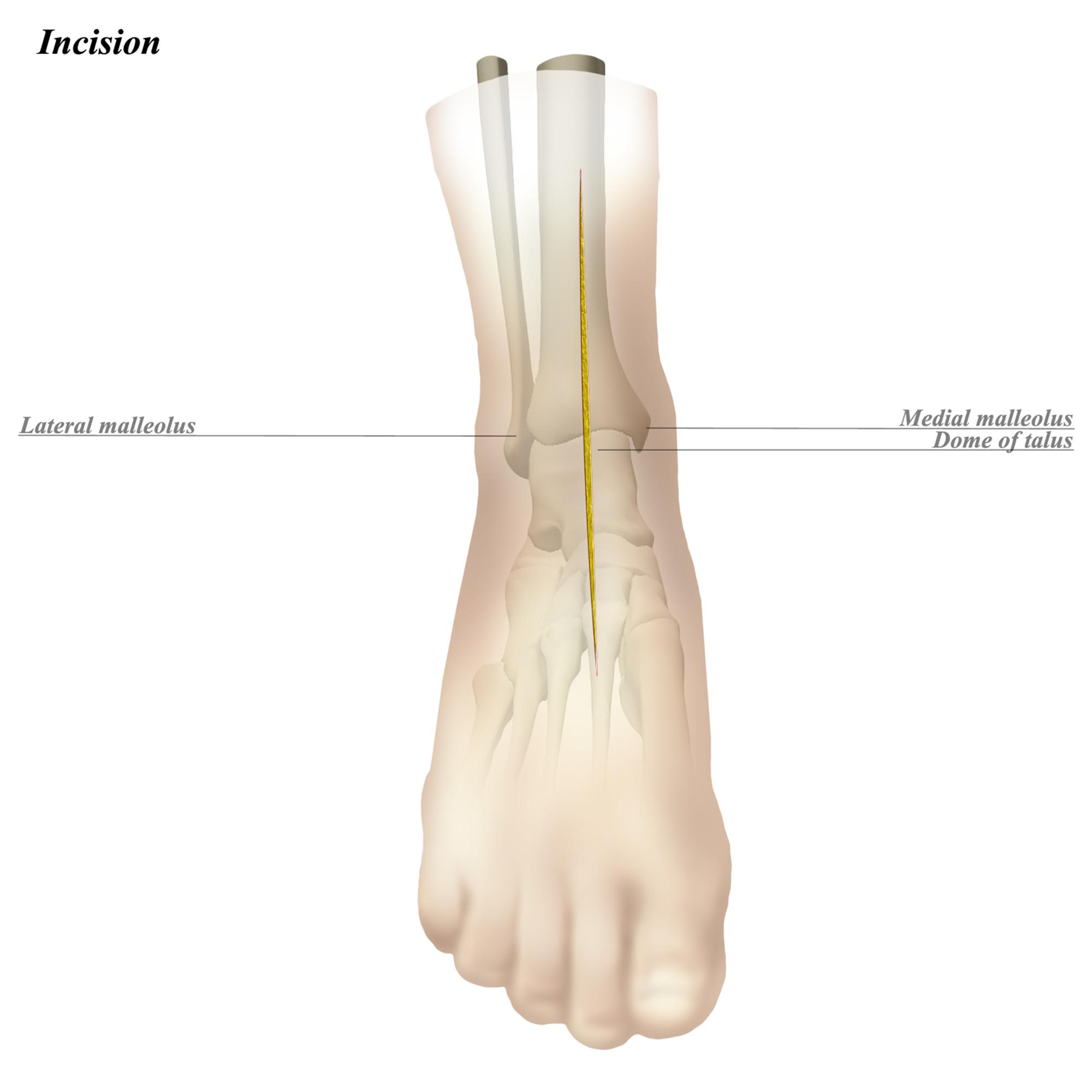 Ankle Anterior Approach Approaches Orthobullets
 Foot And Ankle Orthopedics Seaview Orthopaedic Medical


Posting Komentar
Posting Komentar