The word cornea has come from kerato. It covers the pupil the opening at the center of the eye iris the colored part of the eye and anterior chamber the fluid filled inside of the eye.
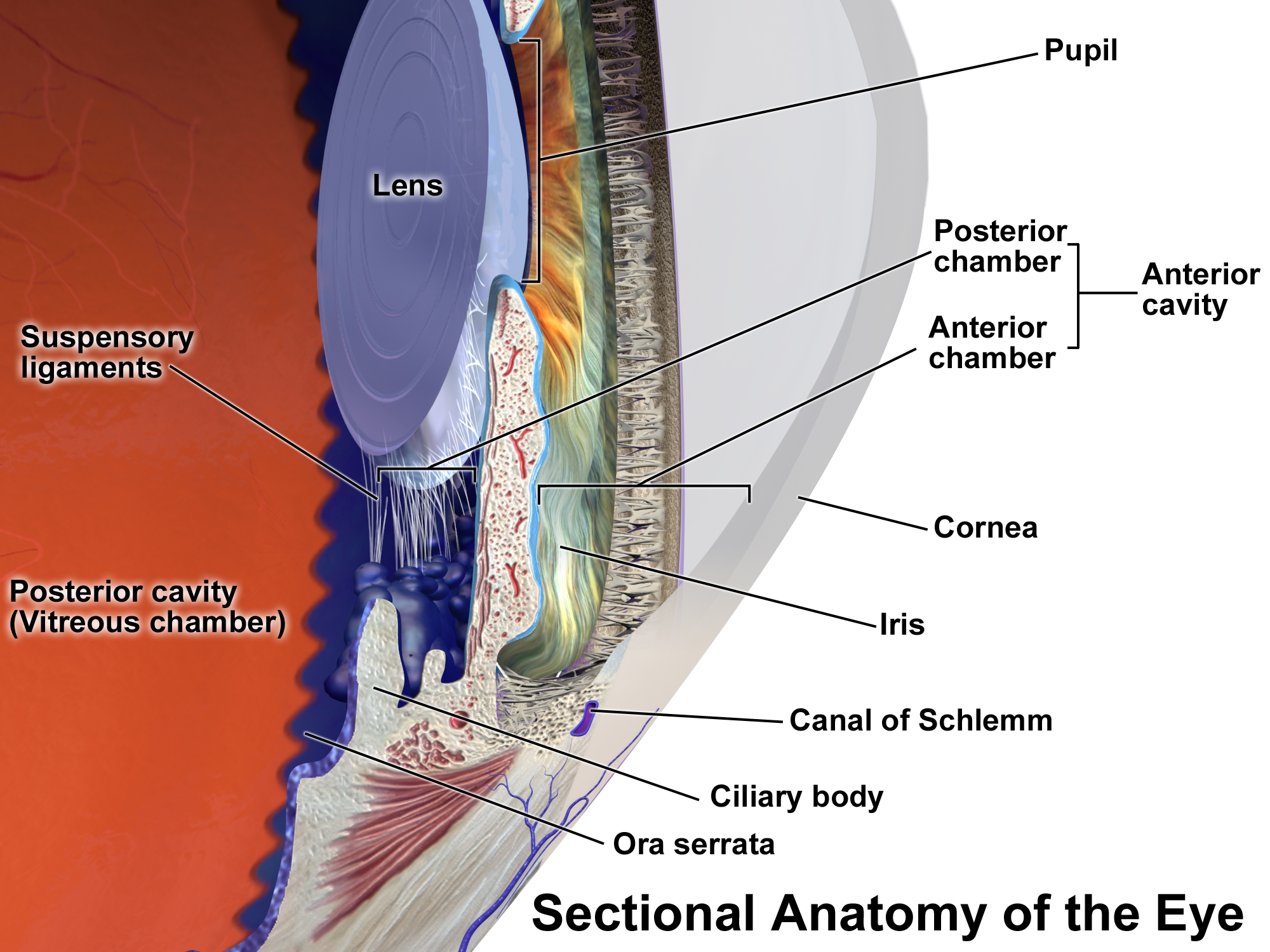 Anterior Chamber Of Eyeball Wikipedia
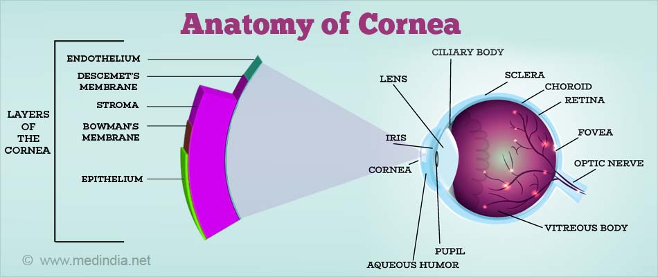
Anatomy of cornea 1.
Anatomy of cornea. Parthopratim dutta majumder. The complexity of structure and function necessary to maintain such elegant simplicity is the wonder. And the endothelium or inner lining.
The cornea is a transparent structure that together with the lens provides the refractive power of the eye. Dimensions anterior surface. The epithelium or outer covering.
It is approximately 05 mm thick at the center and gradually increases in thickness toward the periphery. The stroma or supporting structure. The cornea composes the outermost layer of the eye.
The term kerato in greek means horn or shield like. This outer layer of the cornea is five to seven cells thick. The cornea with the anterior chamber and lens refracts light with the cornea accounting for approximately two thirds of the eyes total optical power.
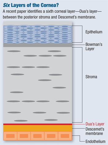
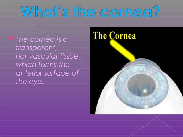
The cornea is the transparent front part of the eye that covers the iris pupil and anterior chamber. This middle layer of the cornea is approximately 500 microns thick. It contains five distinguishable layers.
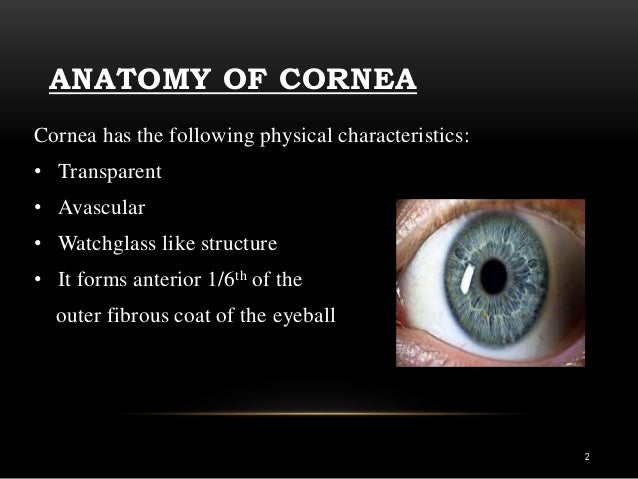
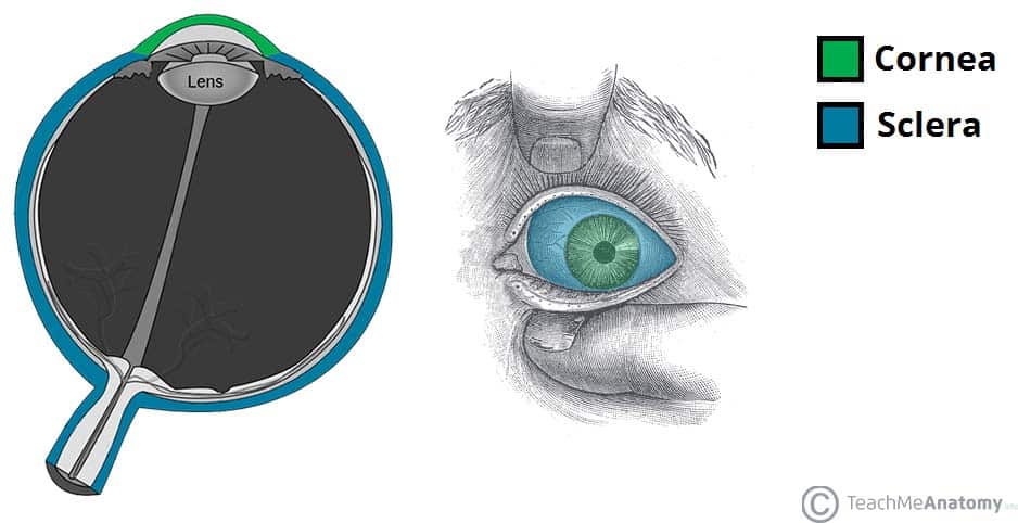
In the average adult the horizontal diameter of the cornea is 115 to 120 mm1 and about 10 mm larger than the vertical diameter. Is made up of the cornea and the sclera. The cornea is the transparent tissue that covers the front of the eyeit forms anterior 16th of the outer fibrous coat of eyeball.
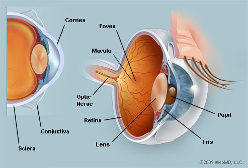
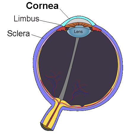
This magnified image of a section of the eye demonstrates the structure of the cornea and the limbus. The cornea is the transparent window of the eye. Corneal anatomy the cornea is the transparent front part of the eye that covers the iris pupil and anterior chamber.
Anatomy of cornea dr nithin keshav. Vertical 117 mm horizontal 106 mm posterior surface. From front to back these layers are.
Yet without its clarity the eye would not be able to perform its necessary functions. Together with the lens the cornea refracts light accounting for approximately two thirds of the eyes total optical power. The anatomy and structure of the adult human cornea.
Anatomy of the cornea. Anatomy and physiology of the cornea. This is a very thin 8 to 14 microns and dense fibrous sheet.
Radius of curvature anterior 78 mm. Introduction cornea medieval latin co rne a te la horny web latin cornu horn. The corneas main function is to refract or bend light.
The cornea is the transparent part of the eye that covers the front portion of the eye. Anatomy of cornea text graphicsdr. The cornea lacks the neurobiological sophistication of the retina and the dynamic movement of the lens.
 Corneal Anatomy Symptoms And Examination American
Cornea Anatomy Functions Linkocare
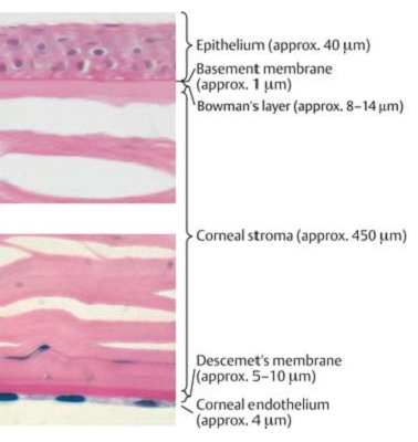 Anatomy Of The Cornea Cornea The Outer Layer Anatomy
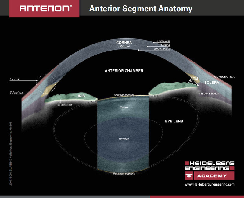 New Anterior Segment Anatomy Handout Heidelberg
Eye Anatomy And How The Eye Works
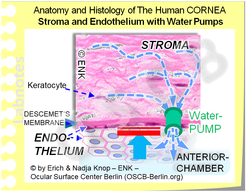 The Cornea Ocular Surface Center Berlin
Rutnin Gimbel Lasik Centre Pioneer In Laser Eye Surgery
The Anatomy Of The Eye Anterior Segment Precision Family
Anatomy Of Cornea Of Dr Sohel Mahmud Authorstream
 Corneal Anatomy And Physiology 2
 Anatomy Of A Normal Human Eye Amdf
Cornea Transplant Cleveland Clinic
 Amazon Com Ambesonne Educational License Plate Human Eye
 Cornea Definition And Detailed Illustration
 Vision And The Eye S Anatomy Healthengine Blog
 More Details On Dua S Layer Of The Cornea
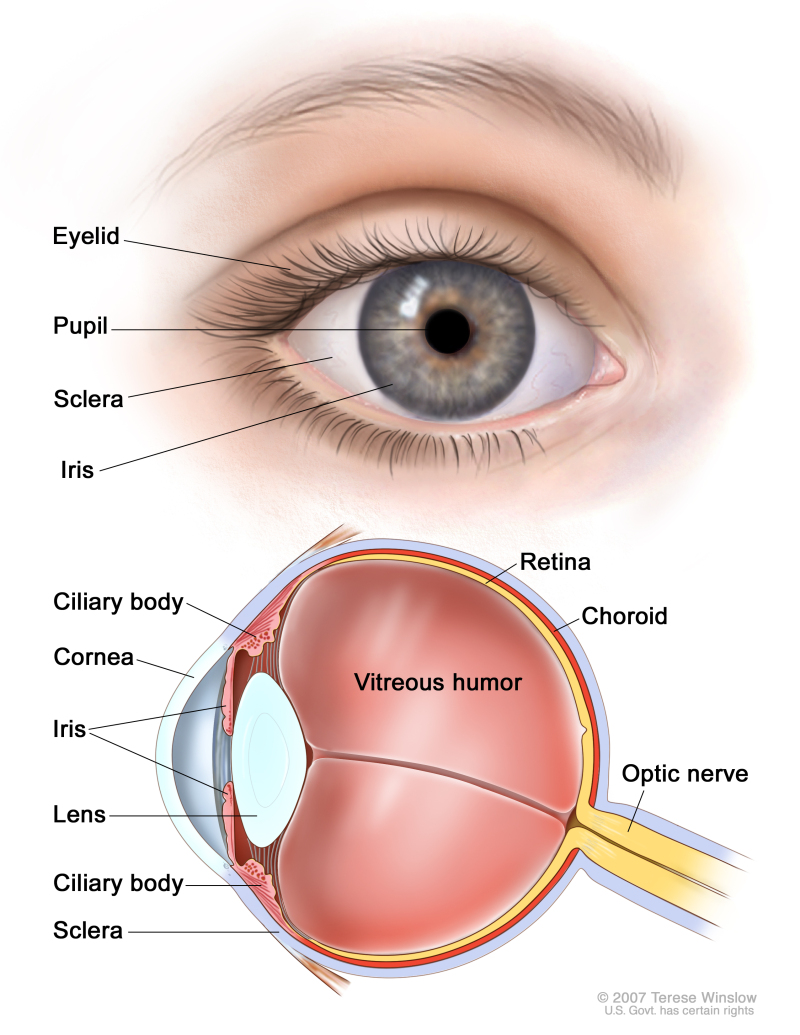 Figure Anatomy Of The Eye Showing Pdq Cancer
 The Eyes Human Anatomy Diagram Optic Nerve Iris Cornea
 Eye Anatomy Glaucoma Research Foundation
Optimed Eye And Laser Clinic Anatomy The Cornea The
Parts Of The Eye American Academy Of Ophthalmology
 Anatomy Of The Cornea A Section Of The Anterior Part Of
 Eye Anatomy Detail Picture Image On Medicinenet Com
 The Eyeball Structure Vasculature Teachmeanatomy
Anatomy Of Cornea By Dr Parthopratim Dutta Majumder
 The Anatomy Of The Domestic Animals Veterinary Anatomy
Major Ocular Structures Laramy K Independent Optical Lab



Posting Komentar
Posting Komentar