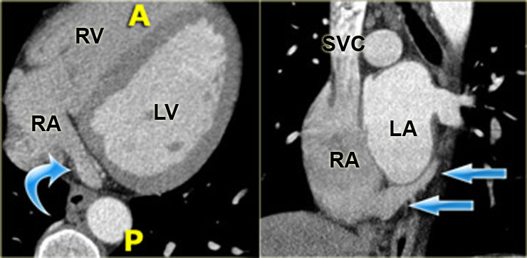
A linear low attenuation structure extending anteriorly from the crista terminalis is visible. Atlas of ct anatomy of the abdomen.
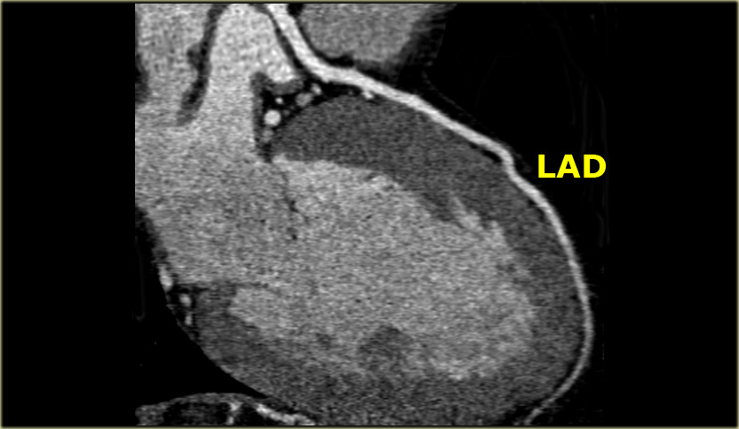
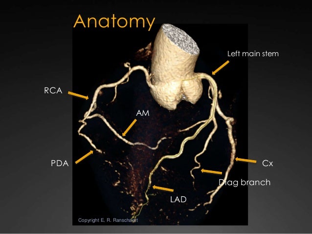
 A Coronary Angiogram Superimposed On Computed Tomography
Usually coronary ct angiography ccta is performed as it contains data about coronary and cardiac anatomy.

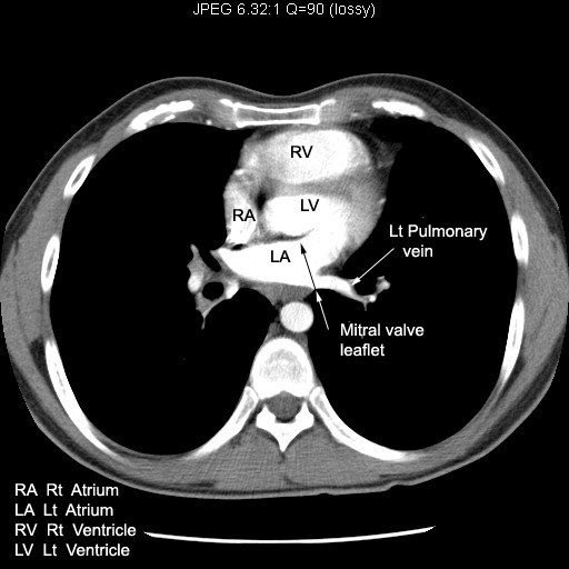
Ct heart anatomy. Cardiac ct is a heart imaging test that uses ct technology with or without intravenous iv contrast dye to visualize the heart anatomy coronary circulation and great vessels which includes the aorta pulmonary veins and arteries. With these scanners the heart and coronary arteries are routinely imaged as a motion free volume of data. This structure represents the septum spurium.
Atlas of ct anatomy of the abdomen. The advent of multidetector computed tomography ct particularly with scanners having 64 or more detectors has continued to improve temporal resolution and allows the acquisition of isotropic voxels. The crista terminalis is a vertical fibromuscular ridge that separates the smooth portion of the right atrium which receives the superior and inferior vena cavae and coronary sinus from the right atrial appendage and the remainder of the right atrium containing pectinate muscles.
Due to recent innovations during the last two decades new ccta protocols allow for significant dose reductions with reported mean sub millisievert doses. Anatomy of the heart quiz ct click on the image description. This photo gallery presents the anatomy of the abdomen by means of ct axial coronal and sagittal reconstructions.
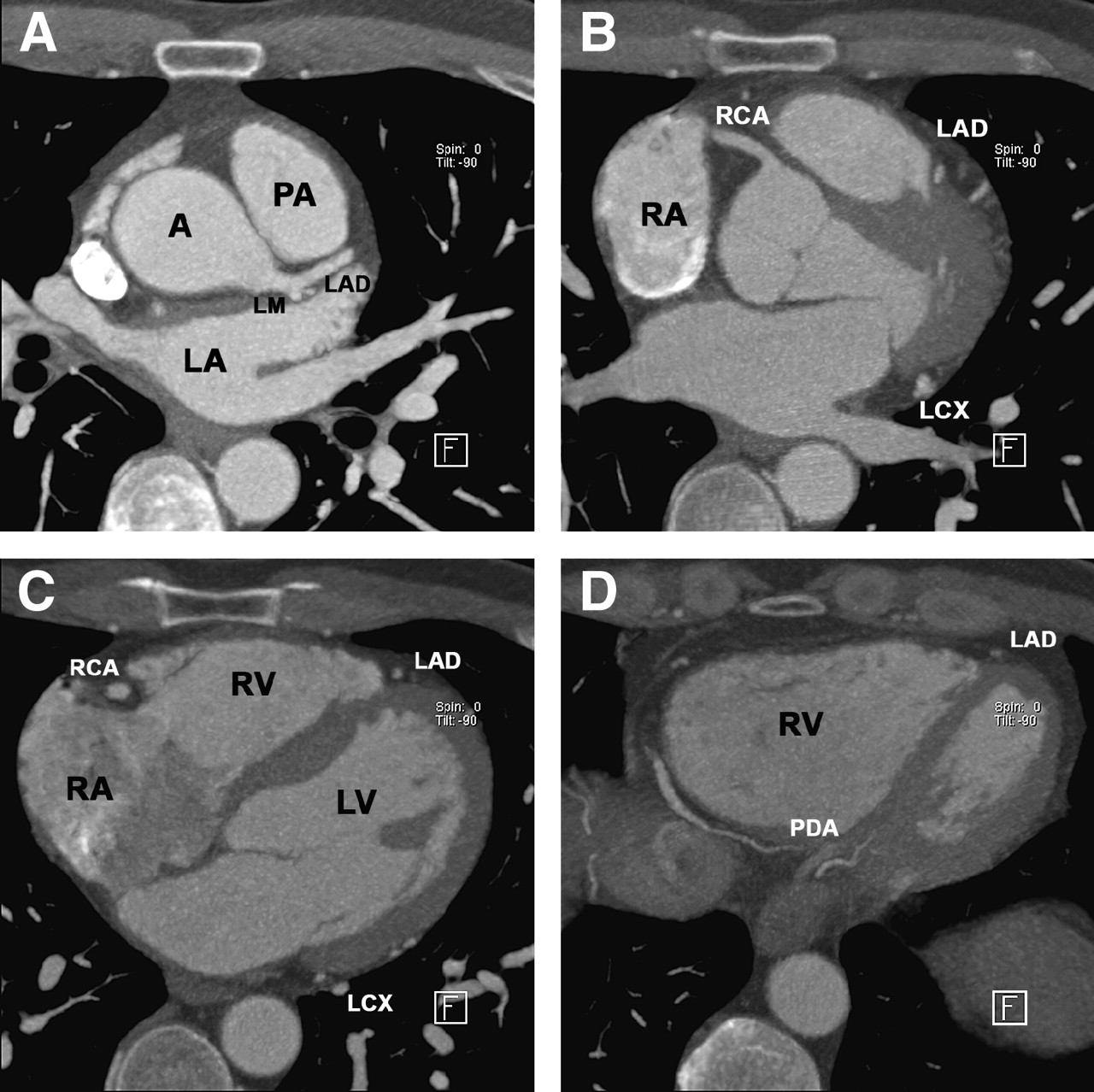
Click on different parts of the heart and coronary vessels on this axial ct and answer corresponding questions. Anatomy of the heart coronary ct interactive atlas of the human body using cross sectional imaging in this interactive anatomy atlas of the human heart the anatomical structures are visible on a contrast materialenhanced computed tomography ct of the heart and coronary arteries. The thoracic duct sits to the left of the esophagus in the superior mediastinum.
The esophagus enters the thorax by penetrating the diaphragm at the esophageal hiatus at the level of t10 the upper half is formed by striated muscles fibers where as the lower half is formed by smooth muscle. The septum spurium is the most prominent of the anterior pectinate muscles arising from the crista terminalis.
 Ffr Ct Is Ready For Prime Time Evaluation Of Coronary
Cardiac Ct Cross Sectional Anatomy Cellular And Molecular
 Anatomy Of The Coronary Arteries And Veins In Ct Imaging
 The Radiology Assistant Coronary Anatomy And Anomalies
 Ct Anatomy Of The Heart Semantic Scholar

 Anatomy Of The Heart And Coronary Arteries Coronary Ct
 E Anatomy Radiologic Anatomy Atlas Of The Human Body
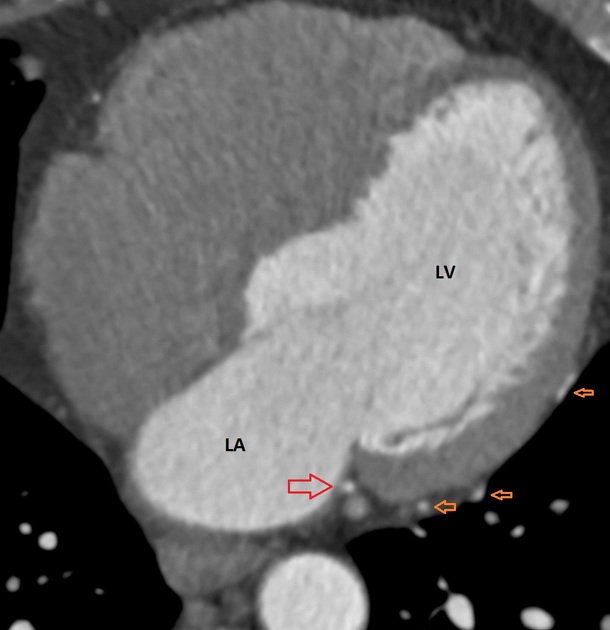
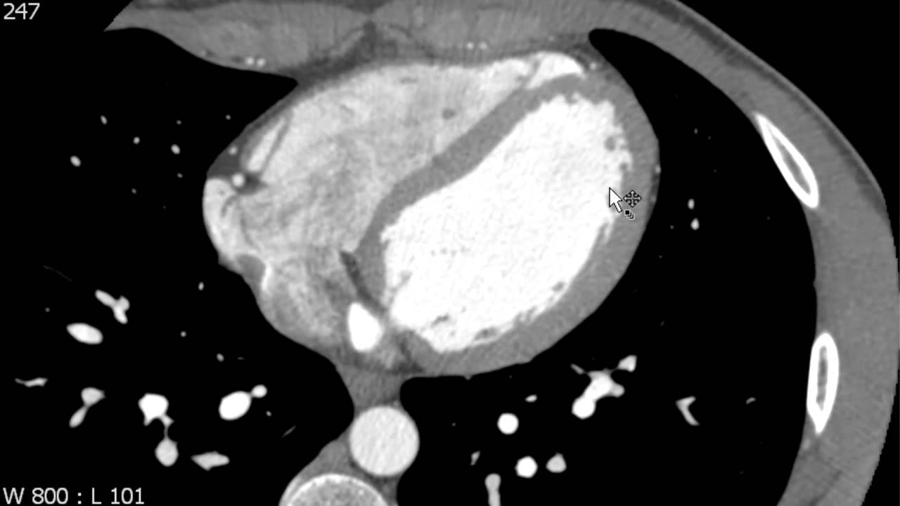 Left Ventricle Anatomy On Axial Cardiac Ct
 Ct Cross Sectional Anatomy Nuclear Medicine Human Anatomy
 State Of The Art Cardiac Ct Of The Coronary Arteries
 Left Dominant Coronary Circulation Radiology Case
 Cardiac Ct Cross Sectional Anatomy Cellular And Molecular

 Ct Scan Wikipedia
 Ct Anatomy Of The Heart Semantic Scholar
 Using 4d Ct To Understand Anatomy Device Interaction Across
 E Anatomy Radiologic Anatomy Atlas Of The Human Body
 Ct Anatomy Of The Heart Semantic Scholar
 The Radiology Assistant Cardiac Anatomy
![]() Abnormal Left Coronal Artery Case And Images Kenhub
Abnormal Left Coronal Artery Case And Images Kenhub
 The Radiology Assistant Cardiac Anatomy
 Cardiac Findings On Non Gated Chest Computed Tomography A
 Science Source Internal Heart Anatomy 3d Ct Scan


Posting Komentar
Posting Komentar