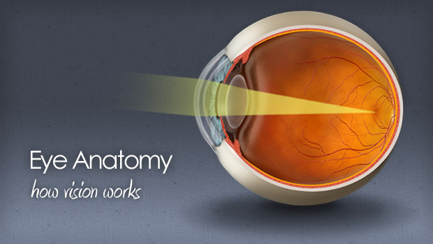
Last week we discussed vision from a historical perspective in order to understand how johannes kepler rené descartes and bishop berkeley discovered the importance of the mind in effecting vision. Watch an animation on how vision works.
 A P 2 Lab Test Eye Ear Anatomy Physiology 123 With
Learn about the anatomy of the eye with this free multiple choice quiz with links to over 200 other anatomy physiology and pathology quizzes.

Physiology and anatomy of eye. The eye is contained within the bony orbit of the head. Anatomy parts and structure. A tapetum is a mirror type membrane that is reflective on the back of the eye activating the rod gives better night vision vitreous humor posterior portion of eye filled with jelly like substance.
The bony orbit is a cavity comprising parts of the lacrimal bone includes fossa for nasolacrimal duct and the maxilla includes caudal foramen of infraorbital canal. Anatomy of the eye. Anatomy physiology pathology of the human eye included are descriptions functions and problems of the major structures of the human eye.
Extraocular muscles help move the eye in different directions. It is situated on an orbit of skull and is supplied by optic nerve. Physiology of vision 3.
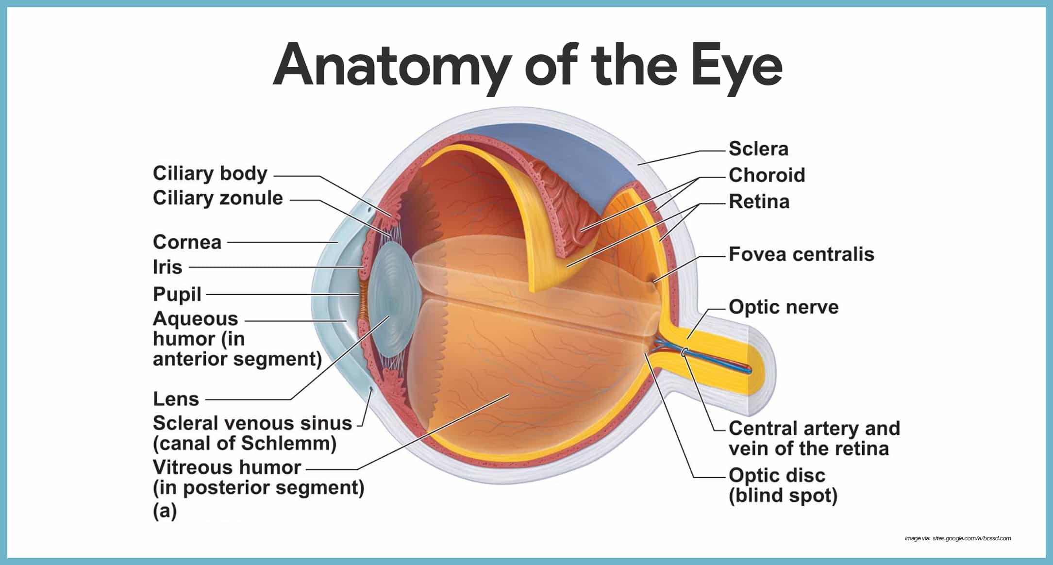
Development anatomy and physiology of the eye the word perspective comes from the latin per through and specere look at. Anatomy of the eye. Sight begins when light rays from an object enter the eye through the cornea the clear front window of the eyeball.
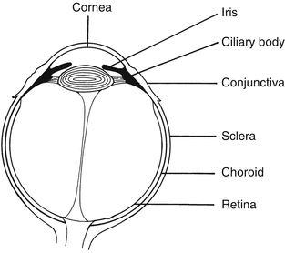
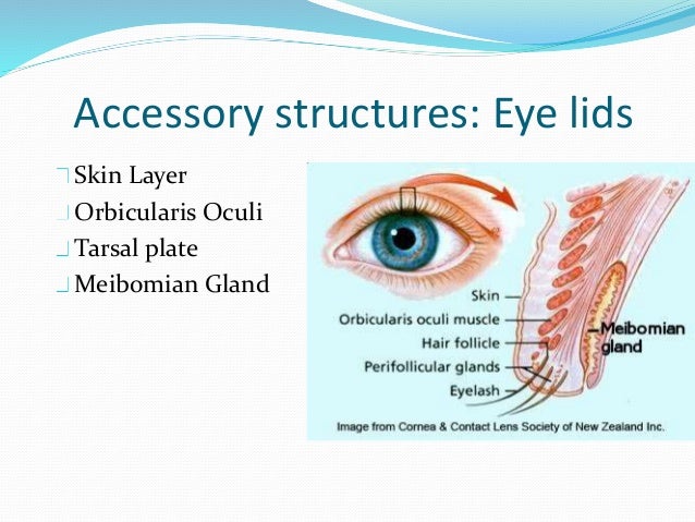
The eye is surrounded by the orbital bones and is cushioned by pads of fat within the orbital socket. They have more rods. Conjunctiva cornea iris lens macula retina optic nerve vitreous and extraocular muscles.
Physiology of the eye 1. There are 6 sets of muscles attached to outer surface of eye ball which helps to rotate it in different direction. Eye anatomy how the eye works.
Eye ball structures 1 fibrous tunic 2 vascular tunic 3 nervous tunic 3. The anterior cavity and the posterior cavity. The eye is the photo receptor organ.
The eye is also divided into two cavities. The innermost layer of the eye is the neural tunic or retina which contains the nervous tissue responsible for photoreception. It is continuous with the temporal bone and the pterygopalatine fossa caudally.
Nerve signals that contain visual information are transmitted through the optic nerve to the brain. Human eye is spherical about 25 cm in diameter. Interior of the ball 1 anterior cavity 2 vitreous chamber 3 lens b.
The eye a guide to the many parts of the human eye how and why vision works and functions. The anterior cavity is the space between the cornea and lens including the iris and ciliary body.
 Health Problem Solutions Eye Physiology And Anatomy
 Sense Organs Anatomy And Physiology
 Pdf Anatomy And Physiology Of The Eye Effect Of Drugs On
 Special Senses Anatomy And Physiology Nurseslabs
 Basic Anatomy And Physiology Of The Eye Springerlink
 Special Senses Vision Anatomy And Physiology I
 Anatomy And Physiology Special Senses The Eyes Nursing Crib
28 The Electric Signals Originating In The Eye
What Is The Anatomy Of The Eye Quora
Archive Cats Comparative Physiology Of Vision

 Human Anatomy Physiology Human Eye Organ Lens Eye Png
 Human Eye Ball Anatomy Physiology Diagram
Eye Anatomy And Vision Course Hero
Understanding Eye Structure Fiteyes Com
 Anatomy And Physiology Of The Eye
 Human Eye Ball Anatomy Physiology Diagram
Iknow Interactive Anatomy And Physiology Of The Human Eye
 Special Senses Eye Anatomy And Physiology Biology
 Eye Anatomy And Physiology How The Eye And Vision Work
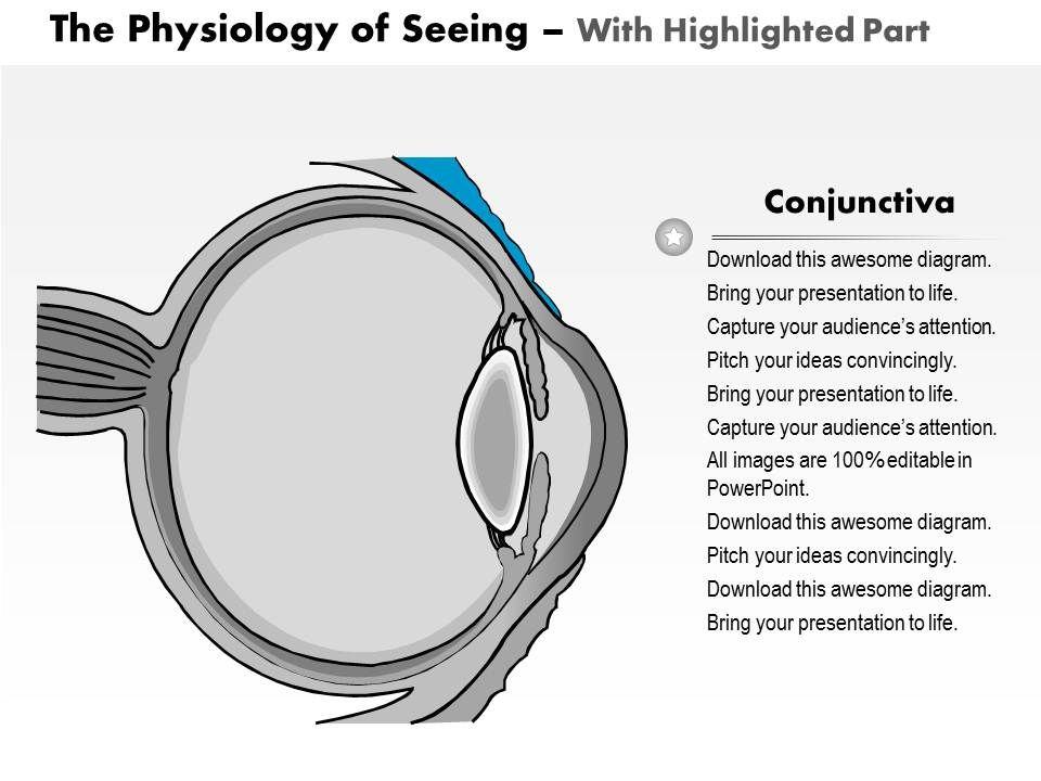 0514 Physiology Of Seeing Eye Anatomy Medical Images For
 Exam 1 Anatomy Physiology Of The Eye Dr Chamcheu
 Medical Surgical Eye Disorders Brilliant Nurse
 Simple Eye Anatomy And Physiology Liquid Conscience
 The Special Senses Ross And Wilson Anatomy And Physiology
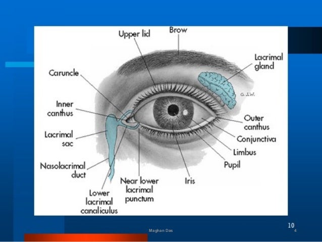 Anatomy And Physiology Of Eye By Maghan Das
Posting Komentar
Posting Komentar