The epidural space situated between the wall of the vertebral canal and the spinal dura mater contains a variable amount of fat. The spinal cord measures approximately 42 45 cm in length 1 cm in diameter and 35 g in weight.
Brain Dissections Neuroanatomy Video Lab
Chapter 43 spinal cord and spinal nerves.
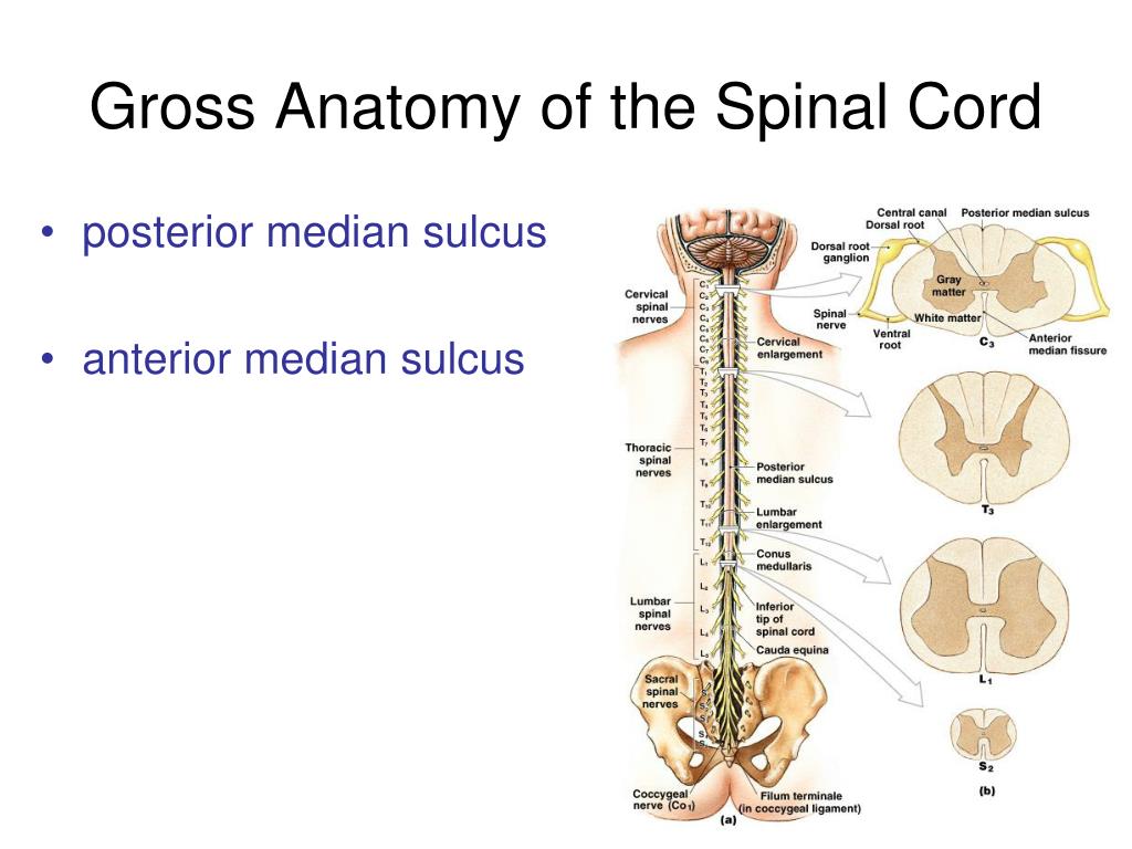
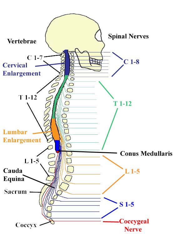
Spinal cord gross anatomy. Spinal cord gross anatomy. The vertebral column grows faster than the spinal cord therefore the lower end of the spinal cord gradually shifts to a higher level. This chapter deals with the gross anatomy of the structures which lie within the vertebral canal and its extensions through the intervertebral foramina the spinal nerve or radicular root canals.
Gross anatomy spinal cord and spinal nerves. 12 coccygeal 1 each connects. Cross section anatomy spinal cord duration.
Terms in this set 85 what are neurons responsible for. Delicate and closely associated with the spinal cord surface. Spinal cord gross anatomy.
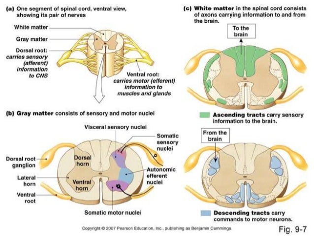
Like the brain it is composed of grey and white matter however opposite to the brain the grey matter is on the internal aspect of the cord and the white matter tracts are external. Spinal cord gross anatomy internal structure meninges. The spinal cord is part of the central nervous system cns which extends caudally and is protected by the bony structures of the vertebral column.
Samuel chen 169796 views. Inferiorly the spinal cord tapers off into the conus medullaris. Reception integration and transmission of nerve impulses.
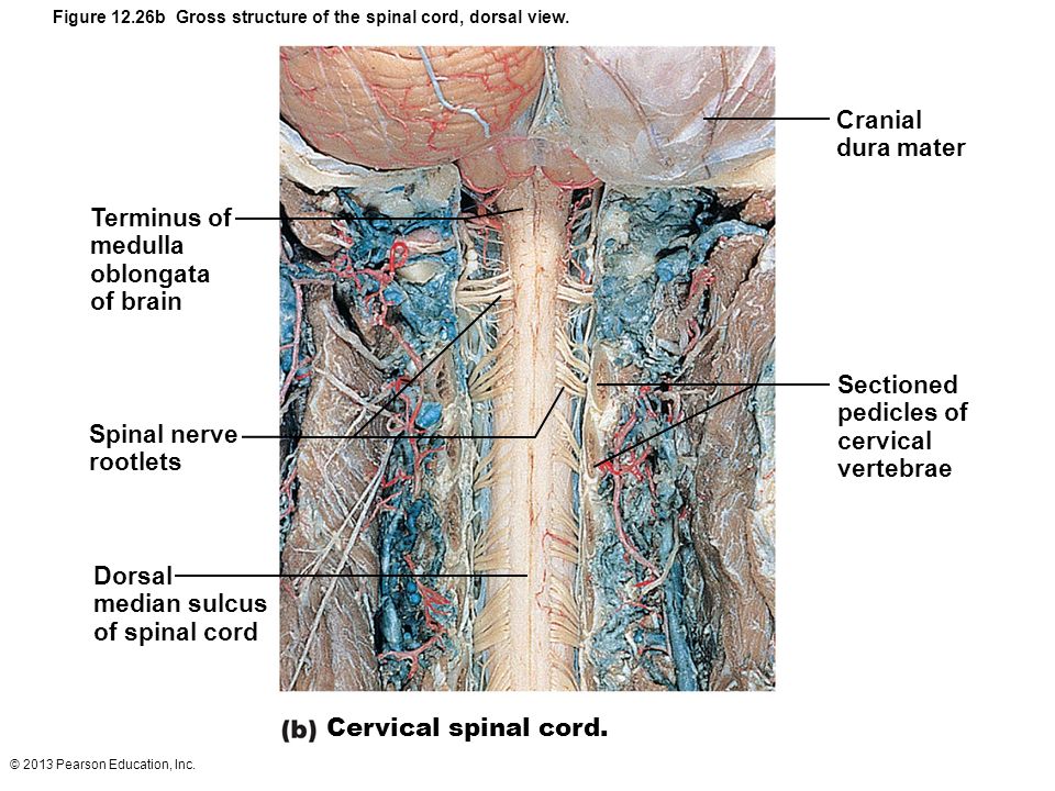
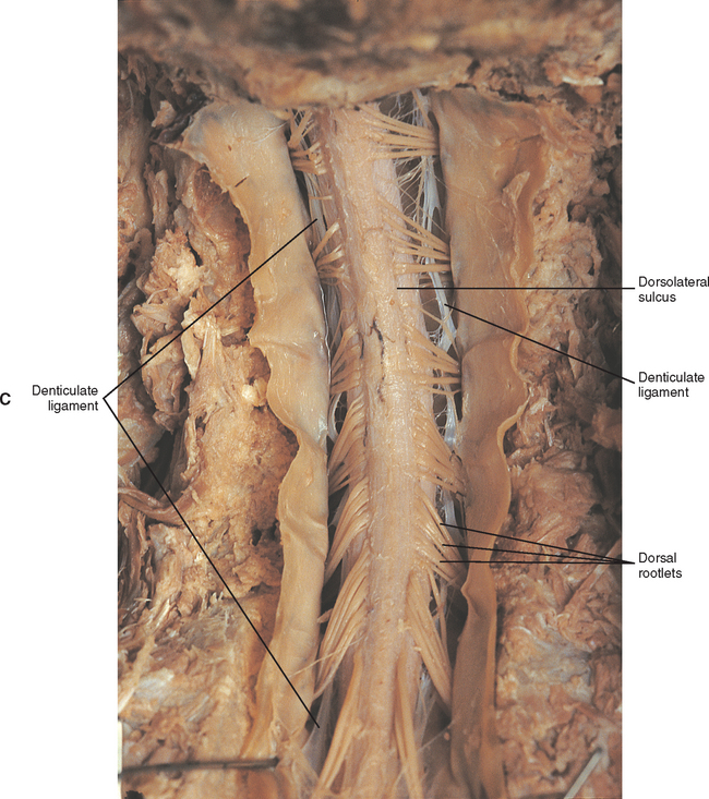
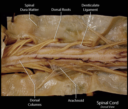
Q it is surrounded by the dura mater the arachnoid mater and the pia mater. Within dura mater the spinal cord is suspended by bilateral denticulate ligaments and surrounded by subarachnoid space filled with cerebrospinal fluid. Spinal cord is the lower cylindrical part of central nervous system that is located in the vertebral canal.
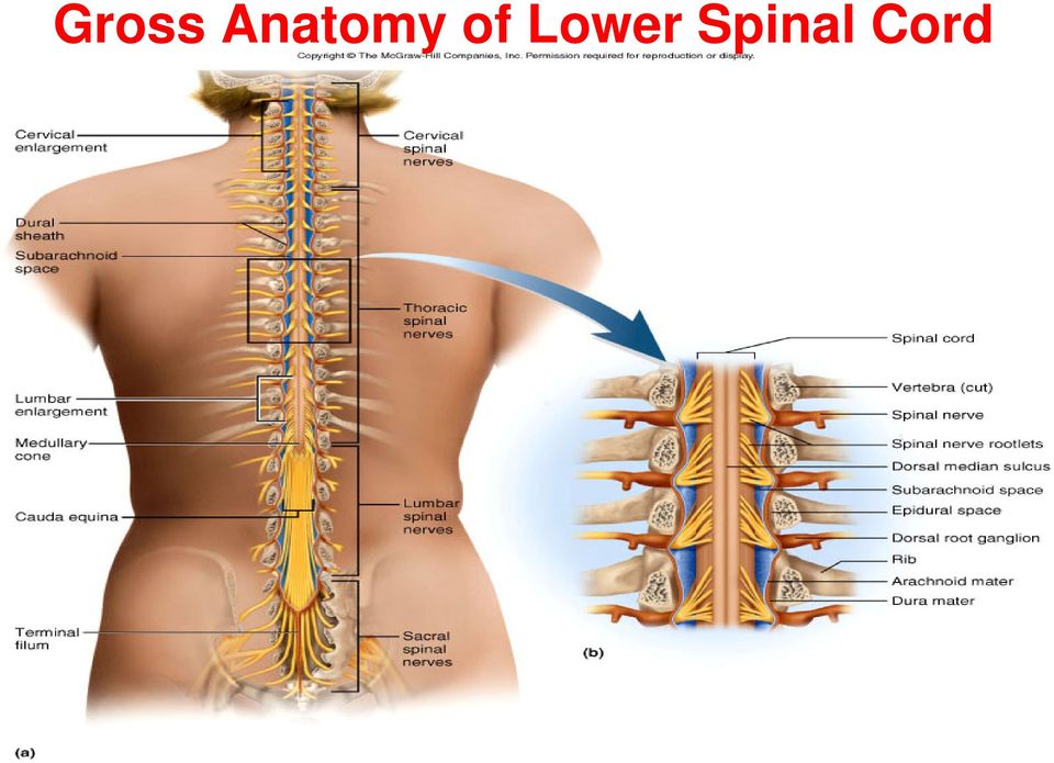
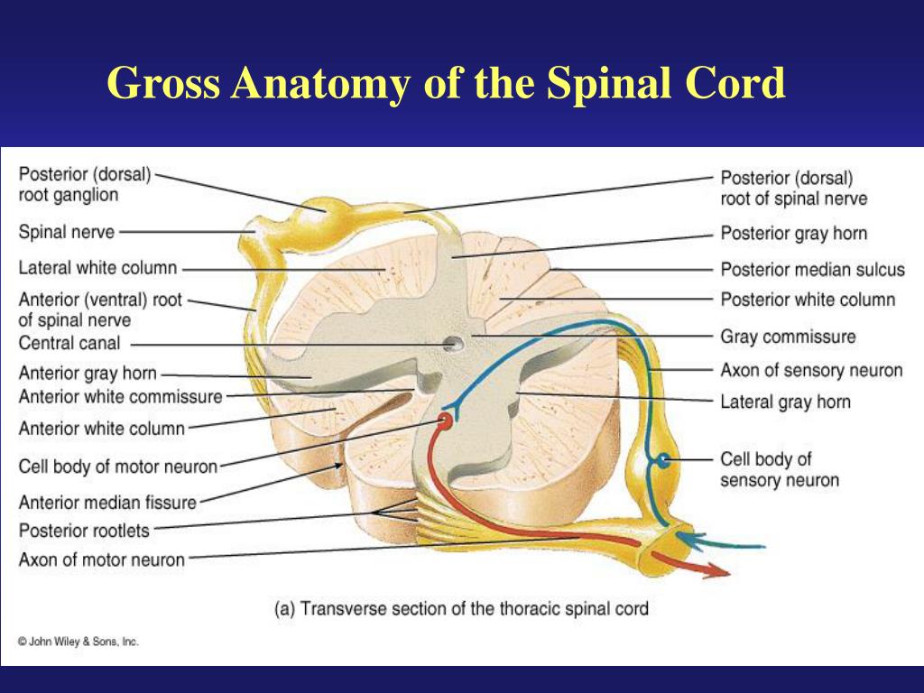
It is covered by the three membranes of the cns ie the dura mater arachnoid and the innermost pia mater. Structure long and cylindrical 42cm in length and 18cm thick has two enlarged portions o cervical enlargement c4 and t1 receives sensory input from the upper limbs sends motor output to the upper limbs. The spinal cord its blood vessels and nerve roots lie within a meningeal sheath the theca.
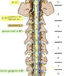
Spinal cord and spinal nerves. Throughout its length paired dorsal and ventral nerve roots enter its dorsolateral and ventrolateral surface respectively.
 Gross Anatomy Of Spinal Cord And Plexuses Diagram Quizlet
 Anatomy Of The Brain And Spinal Cord Seattle Cancer Care
 Ppt The Spinal Cord Nerves And Reflexes Powerpoint
 Nervous System Anatomy Gross Anatomy Of Spinal Cord Youtube
 The Central Nervous System Central Nervous System Spinal

![]() Spinal Cord Anatomy Structure Tracts And Function Kenhub
Spinal Cord Anatomy Structure Tracts And Function Kenhub
 Nii Gross Anatomy Of Adult Spinal Cord Fontal And Lateral
 Human Anatomy Physiology Spinal Cord Spinal Nerves And
 Md Spinal Cord Gross Anatomy See Pinterest Com Pin
![]() Spinal Cord Anatomy Structure Tracts And Function Kenhub
Spinal Cord Anatomy Structure Tracts And Function Kenhub
 Solved Examining The Gross Anatomy Of The Spinal Cord And
 Spinal Cord Gross Anatomy And Protection Ppt Video Online
 General Anatomy Of The Spinal Cord Basicmedical Key
 Figure 16 1 Gross Anatomy Of The Spinal Cord Purposegames
 The Gross Anatomy Of Spinal Cord
 Table 1 And 2 Gross Anatomy And Cross Section Of The
 Lumbar Spine Anatomy Overview Gross Anatomy Natural Variants
 Ppt Gross Anatomy Of The Spinal Cord Powerpoint
 Module Spinal Cord And Spinal Nerve 4 Of 14
 Spinal Cord And Ventral Rami In Situ
 Game Statistics Gross Anatomy Spinal Cord Nerves
 The Gross Anatomy Of Spinal Cord
![]() Spinal Cord Anatomy Structure Tracts And Function Kenhub
Spinal Cord Anatomy Structure Tracts And Function Kenhub


Posting Komentar
Posting Komentar