The ratio of fatty tissue to glandular tissue varies among individuals. Breast tests physical exam.
 The Female Breasts Anatomy Of The Female Breasts The
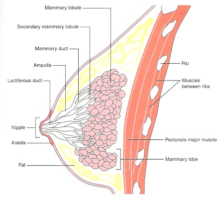
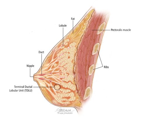
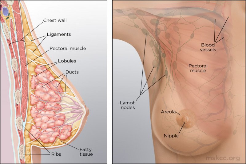
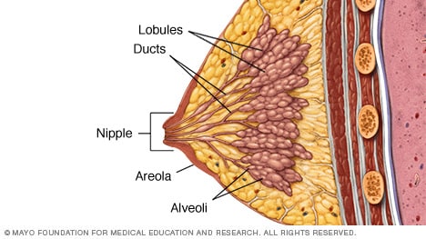
The epithelial component of the tissue consists of lobules where milk is made which connect to ducts that lead out to the nipple.

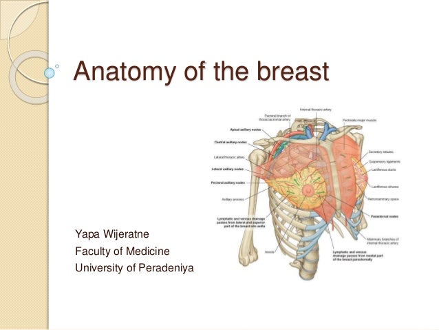
The breast anatomy. A mammography machine compresses each breast and takes low dose x rays. Each breast also contains blood vessels and vessels that transport lymph. These nerves contain both sensory and autonomic nerve fibres the autonomic fibres regulate smooth muscle and blood vessel tone.
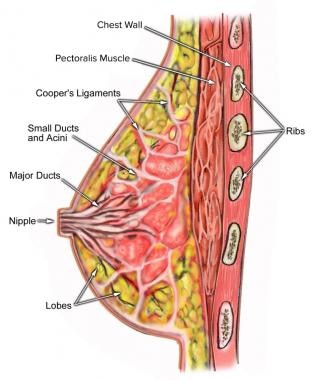
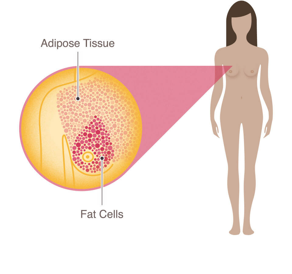
In addition with the onset of menopause ie decrease in estrogen levels the relative amount of fatty tissue increases as the glandular tissue diminishes. Breast anatomy the structure of the breast breasts are made up of fat and breast tissue along with nerves veins arteries and connective tissue that helps hold everything in place. The breast is made up of fatty tissue and glandular milk producing tissues see the image below.
Anatomy physiology of the breast the breast is an organ whose structure reflects its special function. The breast is innervated by the anterior and lateral cutaneous branches of the 4th to 6th intercostal nerves. Ducts are thin tubes that carry milk to the nipple.
Lobes lobules and milk ducts a healthy female breast is made up of 1220 sections called lobes. The areola contains small glands that lubricate the nipple during breastfeeding. The who what where when and sometimes why.
The nipple is located in the middle of the areola which is the darker area surrounding the nipple. See a picture of breast anatomy and learn more about the health topic. There are no muscles in the breasts but muscles lie under each breast to cover the ribs.
Figure 11 shows the different parts of the breast. The nipple is in the center of a dark area of skin called the areola. The female breast is mostly made up of a collection of fat cells called adipose tissue.
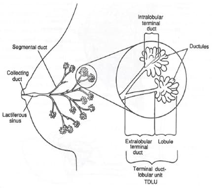
A mammogram that stores the electronic images of each breast in a digital. Lobules are arranged in clusters like bunches of grapes. Breast anatomy picture the breast refers to the front of the chest or more specifically to the mammary gland.
Breast cancers can form in the ducts and the lobes. By examining the breast and nearby underarm tissue for lumps skin changes. The production of milk for lactation breast feeding.
This tissue extends from the collarbone down to the underarm and across to the middle of the ribcage. Learn about the normal anatomy of the breast.

 Anatomy Of The Breasts Children S Wisconsin
 Normal Breast Anatomy Sagittal View Medical Art Works
 Anatomy Of A Healthy Breast Rome Daily Sentinel
 Overview Of The Breast Breast Pathology Johns Hopkins
 Eps Illustration Female Breast Anatomy Vector Clipart
 General Anatomy Of The Breast In Frontal And Sagittal Views
 Breast Anatomy
 Breast Anatomy Overview Vascular Anatomy And Innervation
 Breast Anatomy National Breast Cancer Foundation
 Breast Anatomy Picture Image On Medicinenet Com
Anatomy Of The Breasts Children S Wisconsin
 Anatomy Of The Breast Frontal View Lecture 24 Purposegames
 Anatomy Of The Breast

 Anatomy Of The Breast Memorial Sloan Kettering Cancer Center
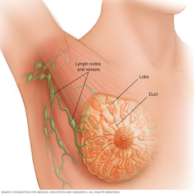 Breast Anatomy Mayo Clinic
 Anatomy Of The Breast Healthlink Bc

 The Anatomy And Physiology Of The Breast
 Woman Breasts Anatomy Cross Section Close Up View Of Female
 Breast Anatomy
 Figure Anatomy Of The Male Breast Pdq Cancer
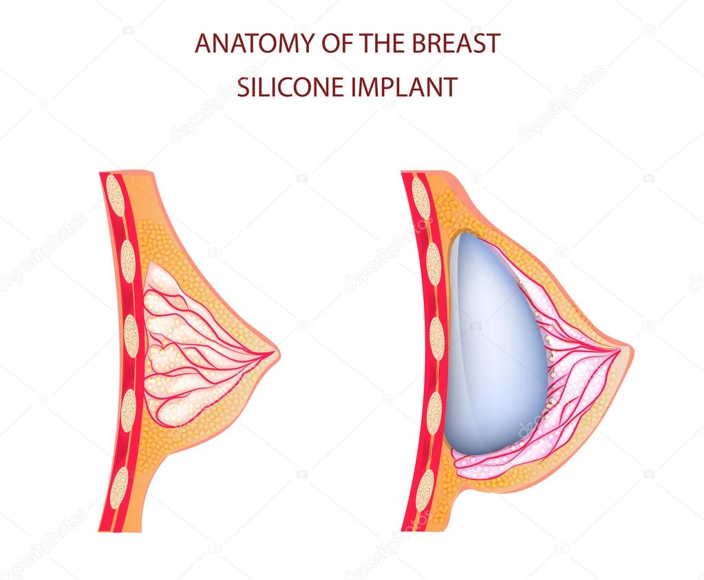 Anatomy Of Breast Silicone Implant Stock Vector C Artemida
 Slide Show Female Breast Anatomy Mayo Clinic
 Figure Anatomy Of The Female Breast Pdq Cancer


Posting Komentar
Posting Komentar