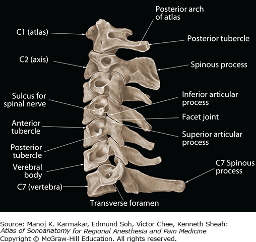
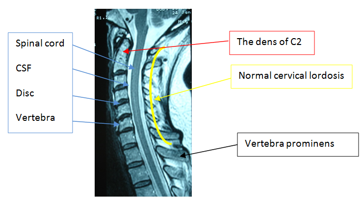
2 posterior arch of c1. The cervical spine has 7 stacked bones called vertebrae labeled c1 through c7.
 Thoracic Spine An Overview Sciencedirect Topics
Mri of the cervical spine sagittal t2 weighted image.
Cervical spine anatomy mri. Spinal anatomy encompasses the anatomy of all osseous and soft tissue structures of the spine the spinal cord and its supporting structures. Use the mouse scroll wheel to move the images up and down alternatively use the tiny arrows on both side of the image to move the imageson both side of the image to move the images. Use the mouse scroll wheel to move the images up and down alternatively use the tiny arrows on both side of the image to move the images.
4 spinous process of laxis. 1 lateral mass of c1 atlas. This mri cervical spine c spine cross sectional anatomy tool is absolutely free to use.
This module of human anatomy is dedicated to residents and students who wish to learn the basics of the anatomy of the cervical spine in mri on a 15 tesla device. 1 vertebral foramen cerebrospinal fluid. Mri is the modality of choice for the assessment of extra osseous injuries such as epidural haematomas and ligamentous disruption in patients with negative ct studies but a high index of suspicion for injury.
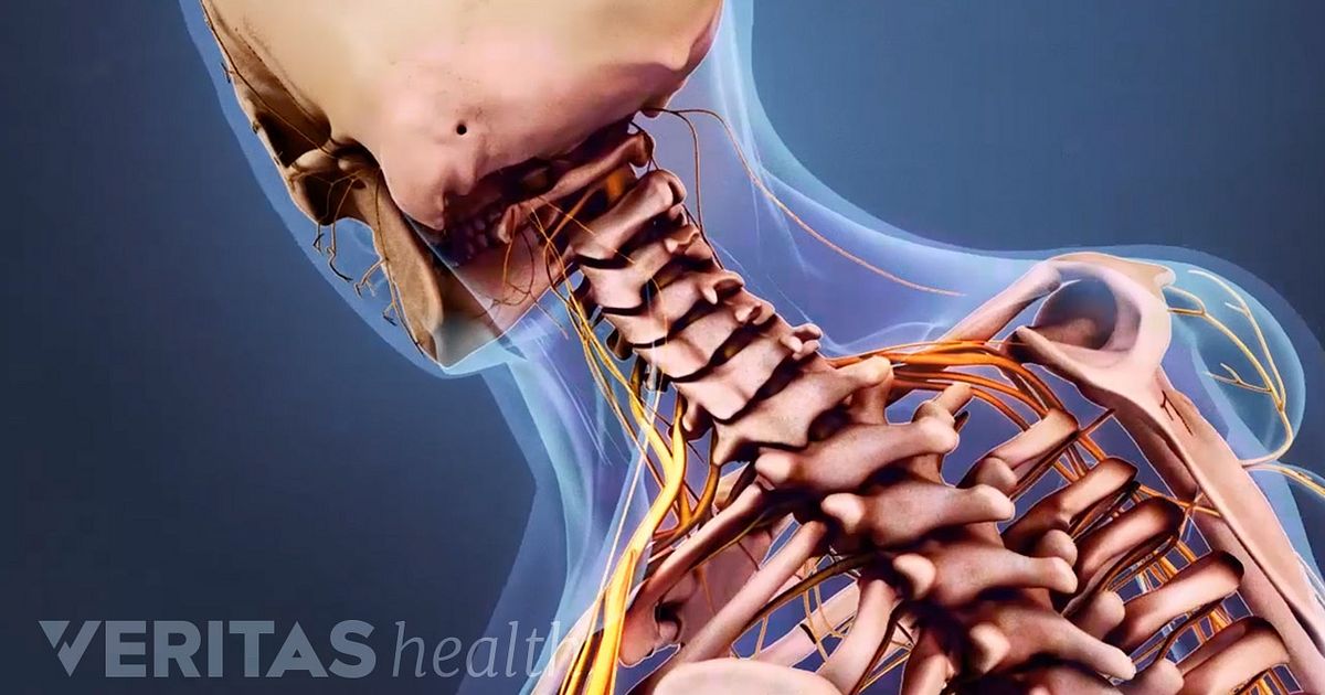
The top of the cervical spine connects to the skull and the bottom connects to the upper back at about shoulder level. Mri is a critical follow up study in patients with severe trauma to the cervical spine. Instead mris utilize strong magnetic fields that when coupled with specialized computer software generate in depth images of your body.
3 vertebral foramen with cerebrospinal fluid. As viewed from the side the cervical spine forms a lordotic curve by gently curving toward the front of the body and then back. The main difference is that mris use no radiation.
Mri of the cervical spine sagittal t2 weighted image. Mri of the cervical spine. This mri cervical spine sagittal cross sectional anatomy tool is absolutely free to use.
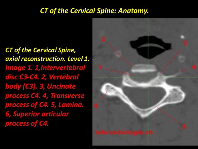
Cervical radiculopathy workup mri the american college of radiology recommends routine mri as the most appropriate imaging study in patients with chronic neck pain who have neurologic signs or symptoms but normal radiographs. Mri has become the method of choice for imaging the neck to detect significant soft tissue pathology such as disc. Except for the first and the second cervical vertebrae the vertebrae share a similar structure including a vertebral body containing trabecular bone.
This anatomy section promotes the use of the terminologia anatomica the global standard for correct gross anatomical nomenclature. The spine is composed of seven cervical twelve thoracic and five lumbar vertebrae as well as the fused sacrum and coccyx vertebral elements. 2 posterior arch of c1.
Anatomy of the cervical spine in magnetic resonance imaging mri cervical vertebrae spinal cord ligaments joints. A cervical spine mri is very different from an x ray although both are imaging techniques.

 Cervical Spine Mri
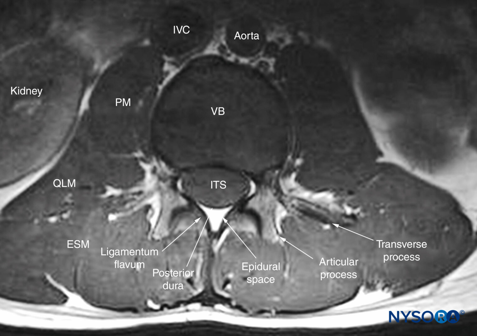
 Sonoanatomy Relevant For Ultrasound Guided Injections Of The
 Presentation1 Pptx Normal Spinal Anatomy
 Cervical Spine Imaging Normal Anatomy And Degenerative
 Radiology Basics Head Anatomy


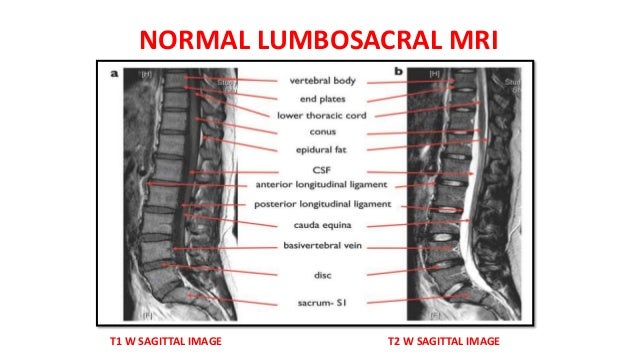
 Understanding Basic Mri Of The Spine
 Cervical Spine Anatomy Video
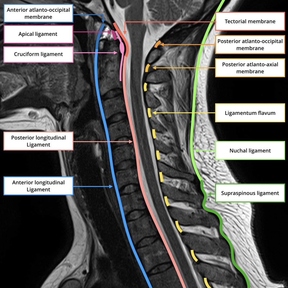 Frank Gaillard On Twitter Quick Ligaments Of The Spine
 How To Read An Mri Of The Thoracic Spine Spine Anatomy
 Nonoperative Management Of Cervical Radiculopathy American

 Materials Free Full Text Materials For The Spine
 Guidelines For The Conduct Of Clinical Trials In Spinal Cord
 Cervical Spine Anatomy Everything You Need To Know Dr Nabil Ebraheim
 Cervical Spine Mri
 Cervical Radiculopathy Spine Orthobullets
 Spinal Sonography And Applications Of Ultrasound For Central
![]() Cervical Spine Anatomy Ligaments Nerves And Injury Kenhub
Cervical Spine Anatomy Ligaments Nerves And Injury Kenhub
 Post Mva Cervical Spine Anatomy High Impact Visual
 Mri Spine Anatomy
 Spine Clinical Gallery Vantage Titan 1 5t Magnetic

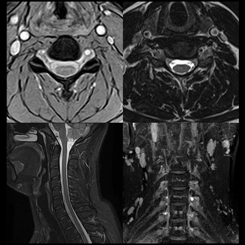
 Realtime Mri Of Cervical Spine


Posting Komentar
Posting Komentar