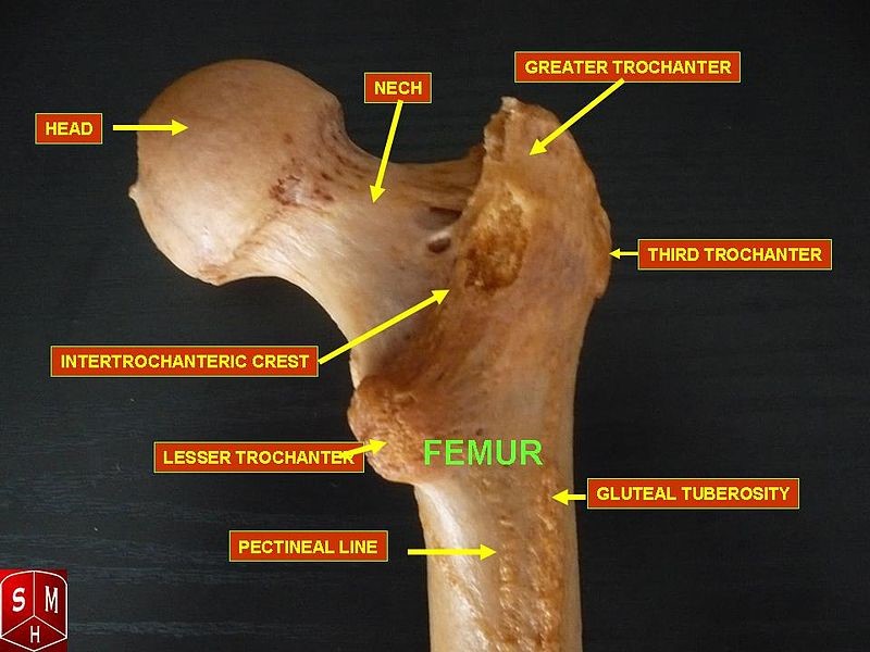
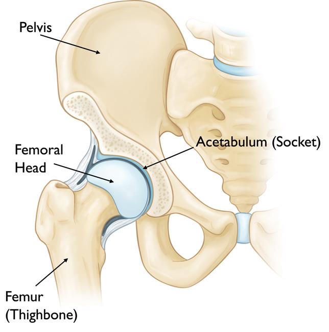
The femur is the thighbone. Organs located above the right hip.
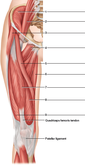
 Left Illustration And Right Photograph Of Lateral View Of
The hip joint is one of the largest joints in the body and is a major weight bearing joint.
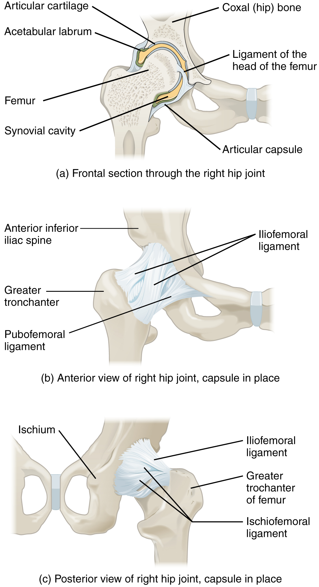
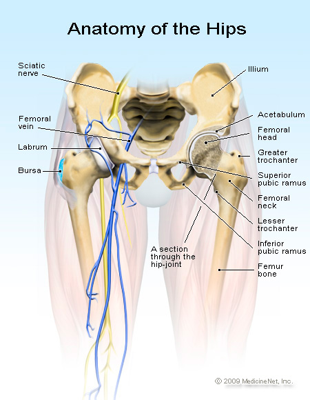
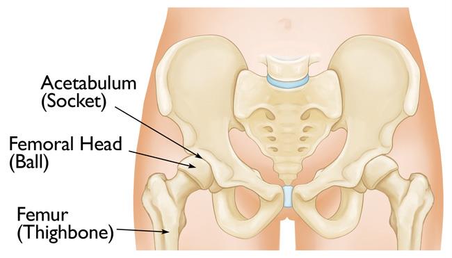
Right hip anatomy. First and foremost the hip is the joint where the acetabulum of the pelvic bone and the head of the femur meet. Adductor muscles on the inside of your thigh. It bears our bodys weight and the force of the strong muscles of the hip and leg.
Rectus femoris muscle one of the quadriceps muscles on the front of your thigh. The adult os coxae or hip bone is formed by the fusion of the ilium. Hip anatomy function and common problems.
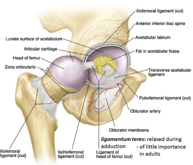
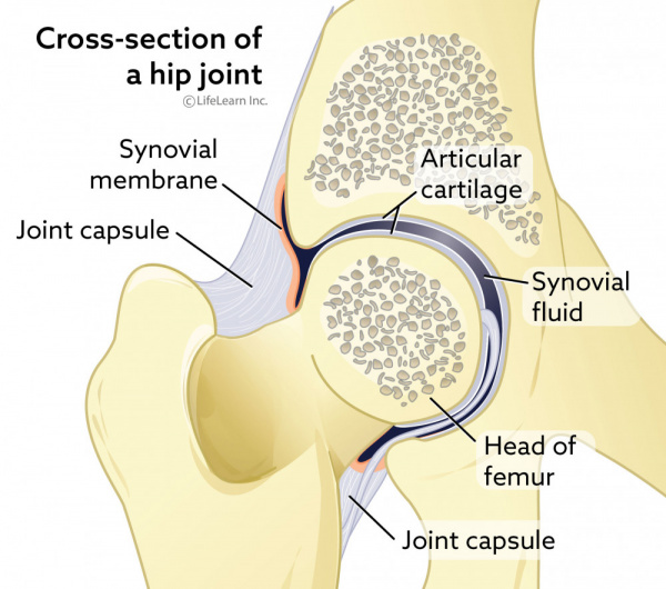
Sechrest md narrates an animated tutorial on the anatomy of the hip joint. The hip joint is reinforced by four ligaments of which three are extracapsular and one intracapsular. The hip joint is the uppermost part of the leg where the head of the thigh bone femur fits into the socket of the pelvis.
Left hip joint from within pelvis with the acetabular floor removed left. In this episode of eorthopodtv orthopaedic surgeon randale c. Right hip joint with capsule removed anterior aspect right.
Iliopsoas muscle a hip flexor muscle that attaches to the upper thigh bone. Weight bearing stresses on the hip during walking can be 5 times a persons body weight. Yet the hip joint is also one of our most flexible joints and allows a greater range of motion than all other joints in the body except for the shoulder.
A healthy hip can support your weight and allow you to move without pain. The hip joint is one of the most important joints in the human body. Hip muscles the hip joint is surrounded by several muscles including.
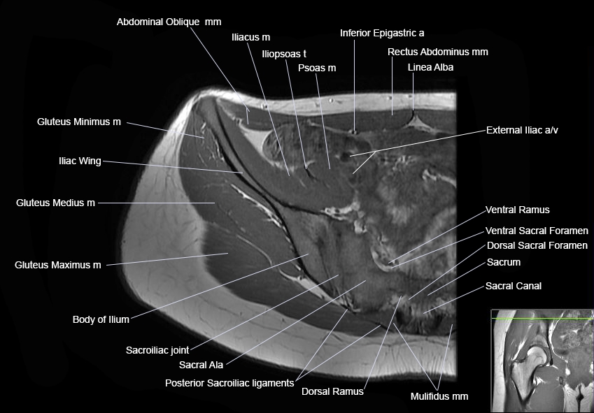
The iliopsoas muscle which extends from the lower back to upper femur. The adductor muscle on the inner thigh. Mri of the hip.
This webpage presents the anatomical structures found on hip mri. Gluteal muscles located on the back of the hip buttocks. Click on a link to get t1 axial view t1 coronal view.
The hip joint is the articulation of the pelvis with the femur which connects the axial skeleton with the lower extremity. Some of the other muscles in the hip are. We point this out because most people refer to their pelvic bones as being their hip or specifically the upper portion called the ilium.
Quadriceps a group of four muscles that comprise the. Hip pain may result from inflammation degeneration or injury to structures and tissues within the hip joint. Hip pain may be due to a variety of common causes including fractures sprains strains arthritis and bursitis.
It allows us to walk run and jump.
 Issues Around The Hip From Tendonitis To Bursitis Beacon
 Hip Anatomy Yoga Understanding The Hips For Yoga Jason
 Summit Medical Group
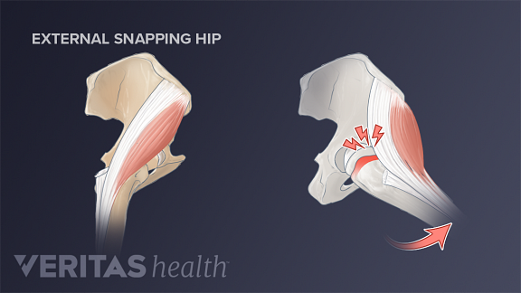 3 Types Of Snapping Hip Syndrome
 Right Hip Bone Medial Views Kidney Anatomy Human
 Figure 12 1
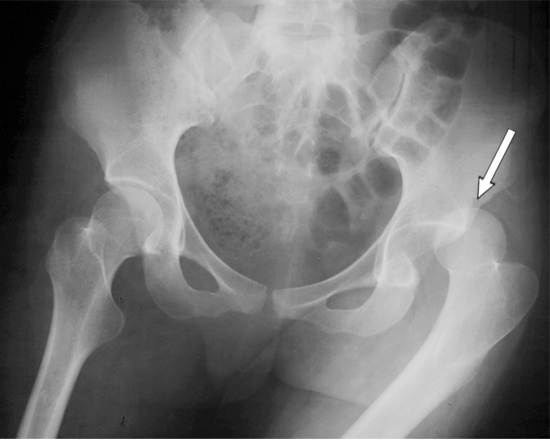 Dislocated Hip Symptoms Diagnosis And Treatments Hss
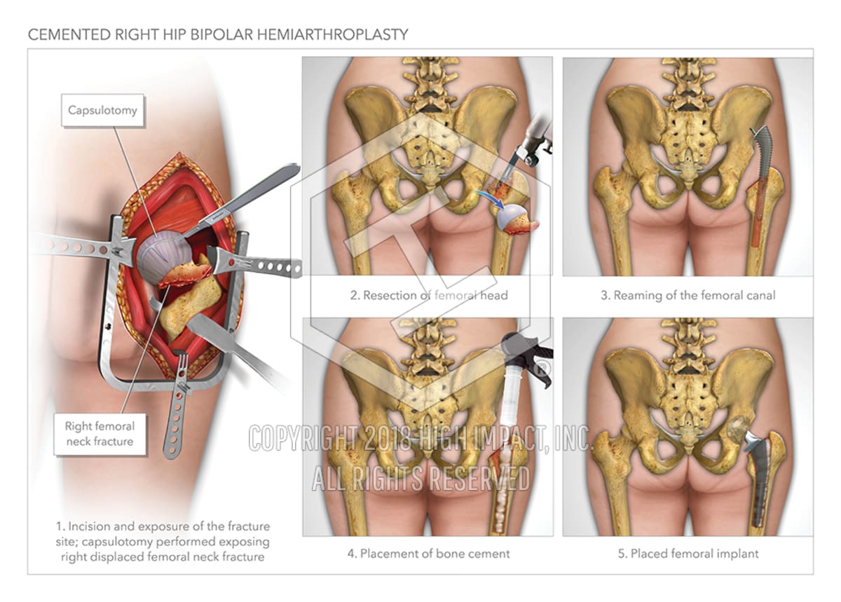 Cemented Right Hip Bipolar Hemiarthroplasty High Impact
 Hip Dislocation And Post Operative Care In Cats Vca Animal
 Joints Ligaments And Connective Tissues Advanced Anatomy
 Hip Pain The Bodyworks Clinic Marbella Spain
 Bones Of The Arm And Hand And Right Hip Bone Black And Whi
 Pelvis Hip Anatomy
 Necrosis Of The Right Hip Medical Illustration Human
 Chapter 25 Solutions Laboratory Manual For Human Anatomy
 Startradiology
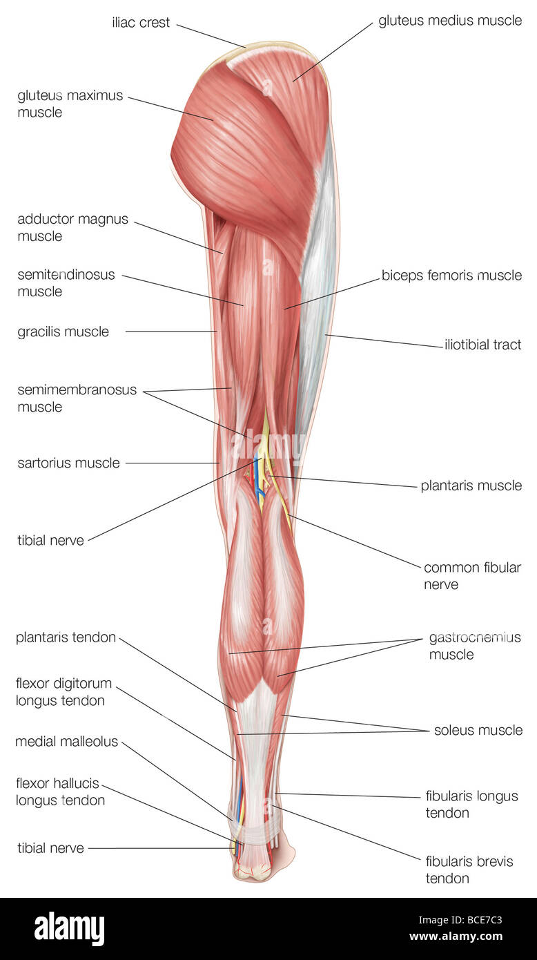 Posterior View Of The Human Right Leg Showing The Muscles

Hip Labral Tear Cleveland Clinic
 Right Hip Normal Anatomy Stock Trial Exhibits
Hip Anatomy Recon Orthobullets
 Hip Picture Image On Medicinenet Com
 Acetabular Fractures Orthoinfo Aaos
 Muscles Of The Hips And Thighs Human Anatomy And
 Pelvis Hip Anatomy
 Muscle Lab 23 Figure 23 1 Muscles Of The Anterior Right Hip
 Total Hip Replacement Orthoinfo Aaos
 The Truth About Cracking Popping Joints Yoga Journal


Posting Komentar
Posting Komentar