Weakest of the lateral ligaments. Retinacula tendons and their synovial sheaths vessels and nerves.
 Anatomy S M
Anatomy extends from the anteroinferior border of the fibula to the neck of the talus origin is 10mm proximal to tip of fibula.

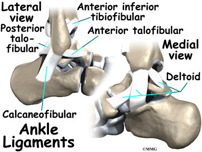
Ankle ligament anatomy. There are 3. Soft tissues of the foot and ankle ligaments. The calcaneofibular ligament cfl which connects the calcaneus or heel bone to.
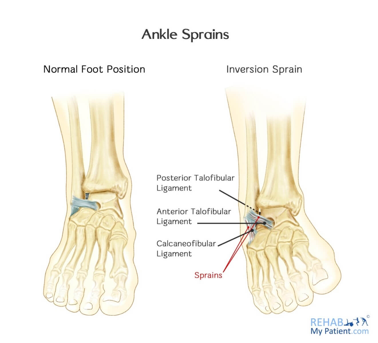
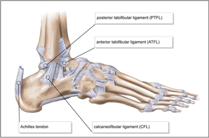
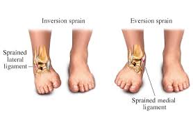
Three ligaments on the outside of the ankle that make up the lateral ligament complex as follows. Anterior talofibular ligament atfl function primary restraint to inversion in plantar flexion. It usually occurs via excessive inversion to a plantarflexed and weight bearing foot.
Ankle ligament injury is the most frequent cause of acute ankle pain. Ligaments are strong thick fibrous bands that connect bone to bone and hold them together. Fascia is a broad fibrous.
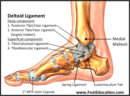
The deltoid ligament is rarely injured by itself and it is usually associated with fractures. Resists anterolateral translation of talus in the mortise. An ankle sprain refers to partial or complete tears in the ligaments of the ankle joint.
The classic ankle sprain involves the anterior talofibular ligament atfl which is also the most commonly injured ligament during inversion sprains. Ligaments are strong dense and flexible bands of fibrous connective tissue. Bone on bone abutment beyond this range protects the anterior and posterior ankle capsular ligaments from injury.
Chronic ankle pain often finds its cause in laxity of one of the ankle ligaments. There are many muscles that help to move and support the ankle and foot. Most sprained ankles occur in the lateral ligaments on the outside of the ankle.
The ligaments in the ankle help to keep the bones in proper position and stabilize the joint. Another ligament that can be injured in a severe ankle sprain is the calcaneofibular ligament. Ligaments are strong fibrous tissues that connect bones to other bones.
They are a really important part of ankle anatomy as they are the primary stabilisers of the ankle. Tendons are elastic tissues made up of collagen. It usually occurs via excessive inversion to a plantarflexed and weight bearing foot.
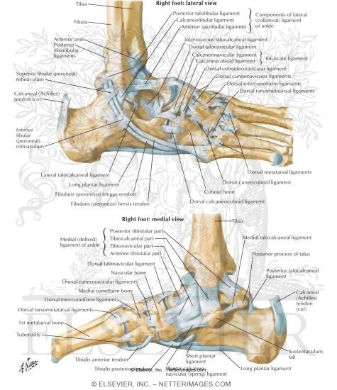
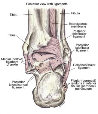
There are eleven ligaments around the ankle connecting the various different bones of the hindfoot and midfoot. The deltoid ligament is the main stabilizer of the ankle joint during the stance phase. Anatomy of the lateral ankle ligamentous complex and related structures.
The combined movement in the dorsiflexion and plantarflexion directions is greater than 100. The anterior talofibular ligament atfl which connects the front of the talus bone to the fibula or shin bone. In this pictorial essay the ligaments around the ankle are grouped depending on their anatomic orientation and each of the ankle ligaments is discussed in detail.

Arthritis Of The Foot And Ankle Orthoinfo Aaos
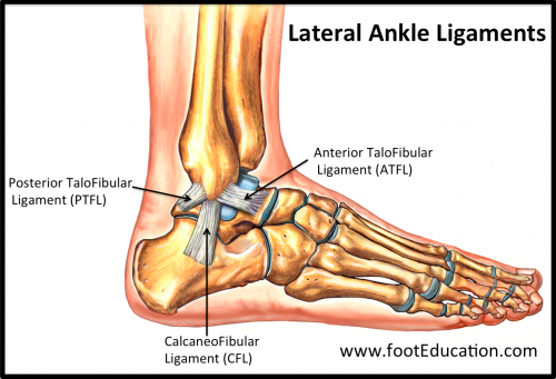 Ligaments Of The Foot And Ankle Overview Footeducation
 Anatomy Of Ankle Iron Thumb

 Ankle Anatomy Eorthopod Com
 Ligaments Of The Ankle Joint Ligaments And Tendons Of Ankle
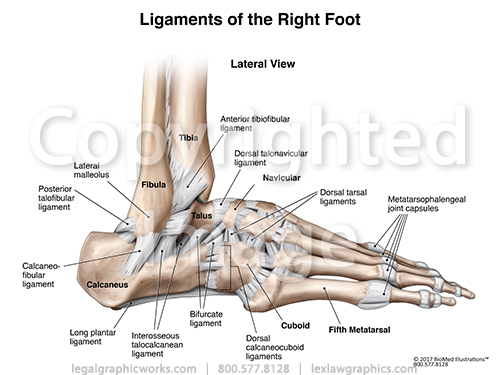 Lateral Right Ankle Ligaments
 The Foot And Ankle Practical Office Orthopedics
 Lateral Ankle Ligament Instability With Surgical
 Ankle Sprain Footcaremd
Medial Ankle Ligament Physiopedia
 Lateral Ligament Reconstruction Procedures For The Ankle
 Ankle Joint Anatomy Overview Lateral Ligament Anatomy And

 Inversion Sprain Of The Ankle Rehab My Patient
 Pin On For My Broken Overworked Body Soul
 Ankle Anatomy

 Ankle Ligaments Foot Ankle Orthobullets
 Ankle Joint Bones And Ligaments Preview Human Anatomy Kenhub
 Ligament Of Foot Ankle Anatomy Ankle Ligaments Anatomy
 Anatomy 101 Ankle Syndesmosis Distal Tibiofibular Joint


 Ligaments Of The Foot And Ankle Overview Footeducation
 Ankle Ligaments Foot Ankle Orthobullets
 Stress Tests For Ankle Ligaments Physiopedia
![]() Ankle Joint Anatomy Bones Ligaments And Movements Kenhub
Ankle Joint Anatomy Bones Ligaments And Movements Kenhub
 Ankle Anatomy
 Medial Ankle Ligament Physiopedia
 Sonography Of The Ankle The Lateral Ankle And Ankle Sprains


Posting Komentar
Posting Komentar