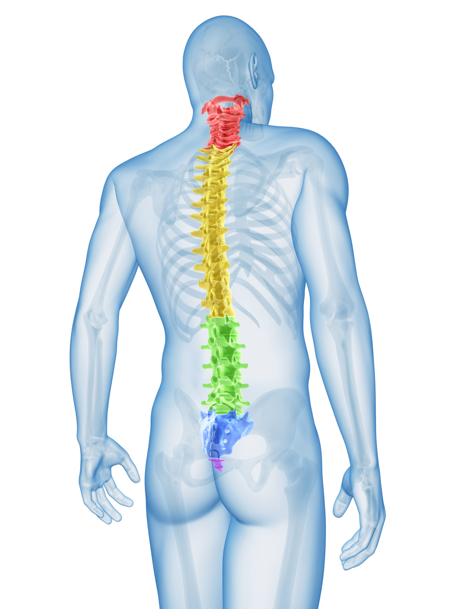
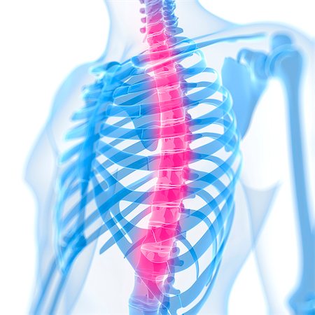
It is the only spinal region attached to the rib cage. The middle 12 vertebrae make up the thoracic spine.
 Pin On Back Torso Muscles
The upper middle back is also the one area of the body which a typical human under normal conditions might be unable to physically touch.
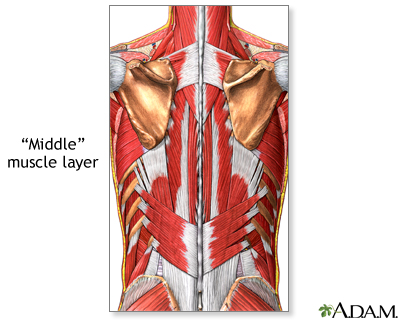
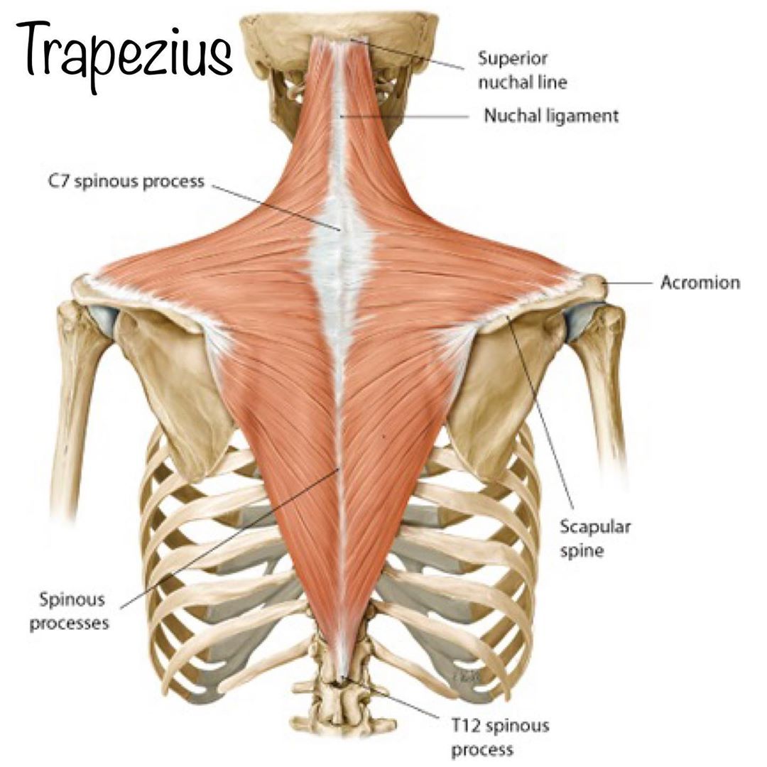
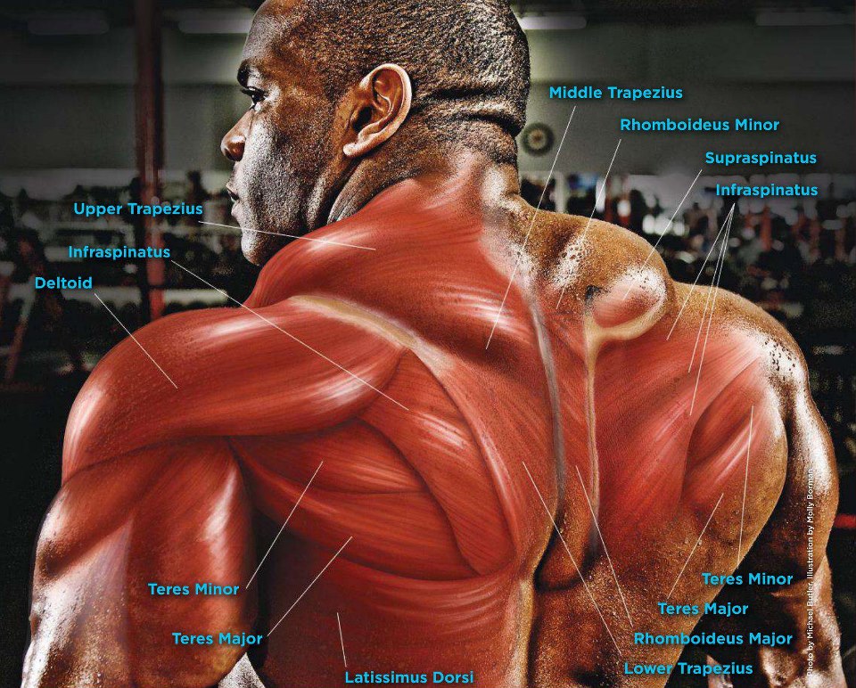
Mid back anatomy. Back muscles anatomy the surface muscles of the upper back include the trapezius muscles traps and posterior deltoids. Doctors from medicinenet say that the middle and upper part of your spine contains 12 vertebrae that are attached to your rib cage. Connecting with the cervical spine above and the lumbar spine below the thoracic spine runs from the base of the neck down to the abdomen.
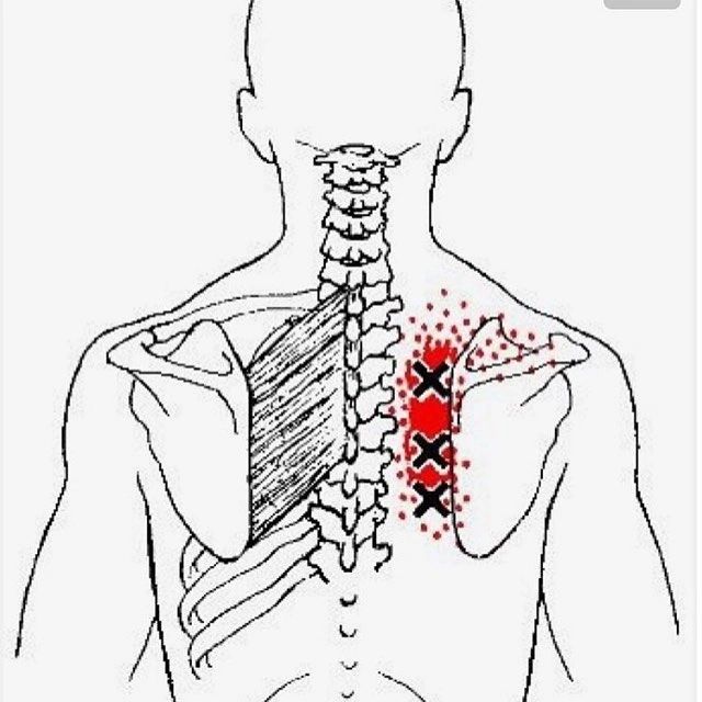
Pain originating from these muscles characteristically produces a dull generalized ache. The lats and traps. The kyphosis is shaped like a c with the opening in front.
Doctors often refer to these vertebrae as t1 to t12. Middle back anatomy the region of the middle back is called the thoracic spine. The thoracic spine is located below the cervical spine and above the lumbar spine and is comprised of 12 bony vertebrae.
The thoracic spine curves outward. An outward curve as in the thoracic spine is called kyphosis. The back anatomy includes the latissimus dorsi trapezius erector spinae rhomboid and the teres major.
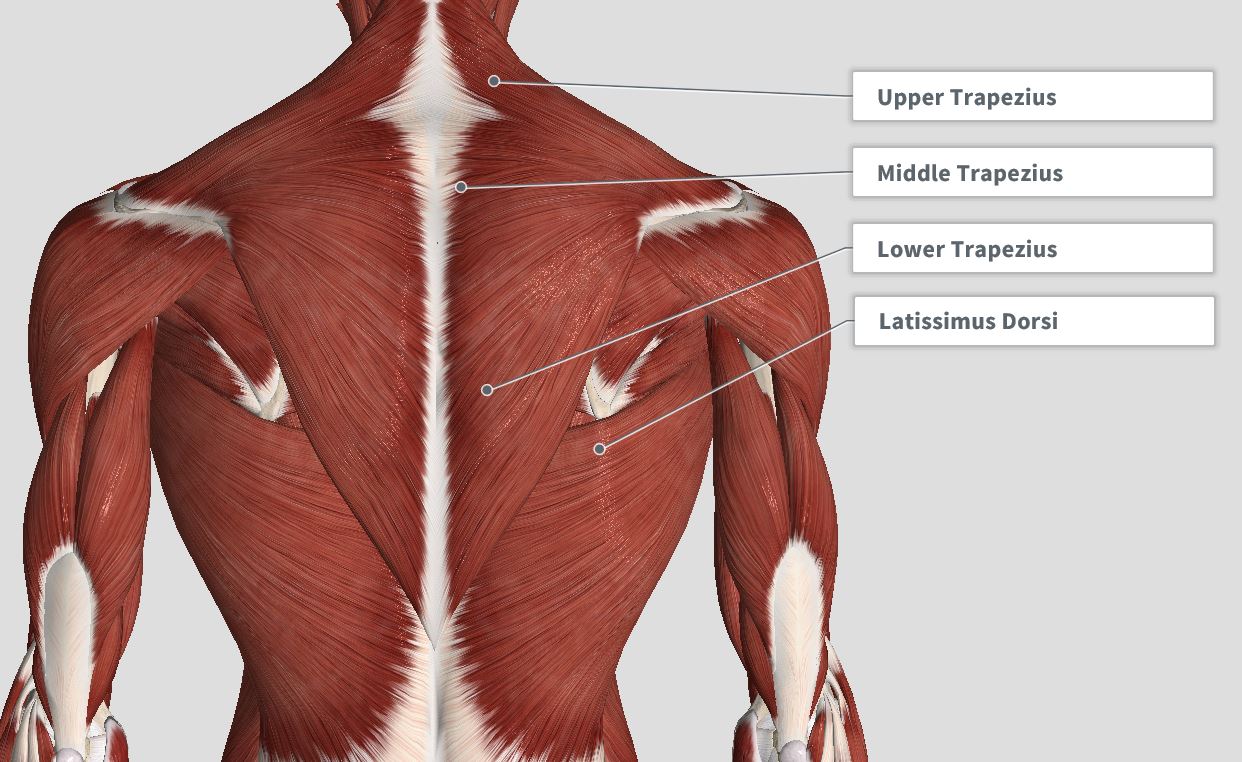
The thoracic spine is the longest region of the spine and by some measures it is also the most complex. Over exertion of the muscles from lifting and pulling and poor posture are the major contributors in mid back strains. The mid back muscles include the latissimi dorsi lats rhomboids and teres major.
Thoracic back anatomy and right side rib pain the thoracic back describes the area of your back from just below the base of your skull to about 5 inches below the lower part of your shoulder blades. These muscles give height and breadth to back development. Back muscle anatomy the back is a complex area encompassing a large number of muscles and movements.
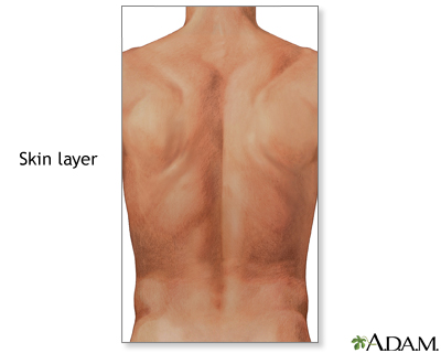
On this page youll learn about each of these muscles their locations and functional anatomy. An inward curve in the spine is called lordosis. The skin of the back is innervated by the dorsal cutaneous branches as well as the lateral abdominal cutaneous branches of intercostal nerves.
The vertebrae function to protect the spinal cord that runs through the spinal column. Well start with the two largest muscles of the back musculature. Back muscles anatomy here include the trapezius latissimus dorsi rhomboid and levator scapulae.
The low back also called the lumbar spine curves slightly inward.
 Mid Back Pain Treatment Nyc Back Pain Specialist In Manhattan
 Back Pain Spiritual Junky
 10 Of The Best Exercises For Upper Back Workouts Openfit
 Pin On Middle Back Pain Exercises
 Middle Back Pain Information Causes And Solutions For Mid

 Pin On Massage
 Mid Back Pain
 Mid Upper Back Pain Poland Chiropractic Chiropractor
 Facet Joints Ainsworth Institute
 The Elegant Outlaw Yoga

 Mid Low Back North Oakland Chiropractic Clinic
 Upper Back Pain The 14 Best Exercises And Stretches Video
 Deuk Spine Institute Laser Spine Surgery Center Blog
 Shoulder Blade Pain Causes And Treatment For Scapula Pain
 Introduction Anatomy Thoracic The Gap Physio
 My Journey Around The World Neck Middle Back Or Shoulder
 Middle Back Inflammmation Stock Illustration Illustration
 Pain Mid Section Front Back Anatomy Stock Photos Page 1
![]() Anatomy Of The Back Spine And Back Muscles Kenhub
Anatomy Of The Back Spine And Back Muscles Kenhub

 The Anatomy Of The Back Muscles Bodybuilding Wizard


Posting Komentar
Posting Komentar