They are powered by a common muscle belly shared by all the fingers which divides into 4 tendons. It is typically the longest finger.
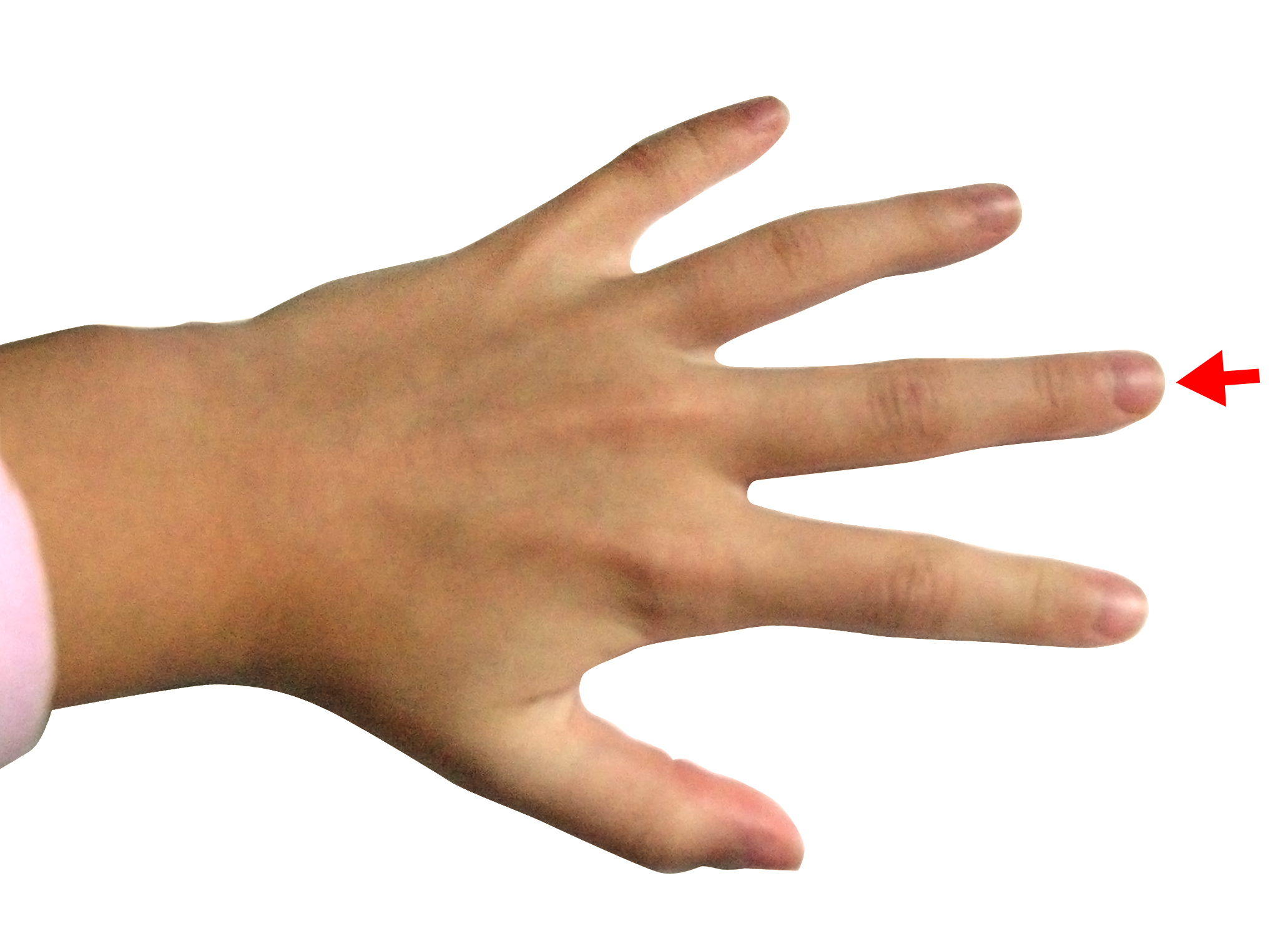
 Human Hand Show Middle Finger On White Background
The middle finger long finger or tall finger is the third digit of the human hand located between the index finger and the ring finger.

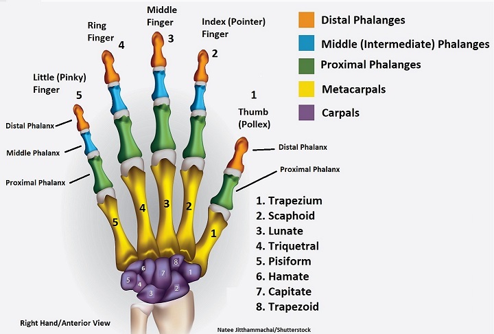
Middle finger anatomy. They travel down the forearm and within the carpal tunnel. The index finger is composed of three bones. Intermediate phalanx is the middle finger bone which is absent in the thumb.
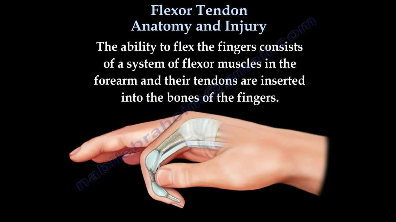
It is also called the index finger or the forefinger. Proximal phalanx is the first finger bone lying next to the palm. Ebraheims educational animated video describes the anatomy and injury of the flexor tendons of the fingers.
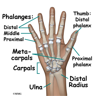
The knuckles are joints formed by the bones of the fingers and are commonly injured or dislocated with trauma to the hand. Distal phalanx is the last finger bone lying furthest away from the hand. The thumb does not have a middle phalange.
The thumb has a distal and proximal phalanx as well as an interphalangeal and mcp joint. Flexor digitorum superficialis tendons help bend the index middle ring and small fingers at the middle finger joint. They are controlled by muscles in the forearm.
This finger often possesses the largest amount of sensitivity and greatest dexterity of any of the fingers. For this reason the index finger is also known as the pointer. This finger has practical applications of both sensory touch and grasp but it is often used for expressive purposes as well.
The ability to flex the fingers consists of. Basic anatomy of the finger. This usually takes the form of non verbal hand gestures.
The first and largest knuckle is the junction between the hand and the fingers the metacarpophalangeal joint mcp. Each phalanx in a finger is named according to its location. The index middle ring and fifth digits have proximal middle and distal phalanges and three hinged joints.
In anatomy it is also called the third finger digitus medius digitus tertius or digitus iii. When fingers joints straighten they are being pulled by the extensor tendons. The pointer finger is the second digit and first finger of the human hand.
Extensor tendons connect to muscles in the middle of the forearm then extend through the wrist and hand to each finger where they form the extensor hood. This picture also contains other parts such as interossea recurrens arcus palmaris profundus dorsal carpal network palmar carpal network r. Distal interphalangeal dip proximal interphalangeal pip and metacarpophalangeal mcp.
Perforans right middle finger palmar view and so on. The distal phalanx intermediate phalanx and proximal phalanx. We are pleased to provide you with the picture named hand vascular anatomy artery diagram.
Along with the thumb and middle finger it is one of the most often used digits.
 Trigger Finger
 Hang Right Part 3 Healing Nagging Finger Injuries
Why Does Bending The Ring Finger Cause The Middle Finger And
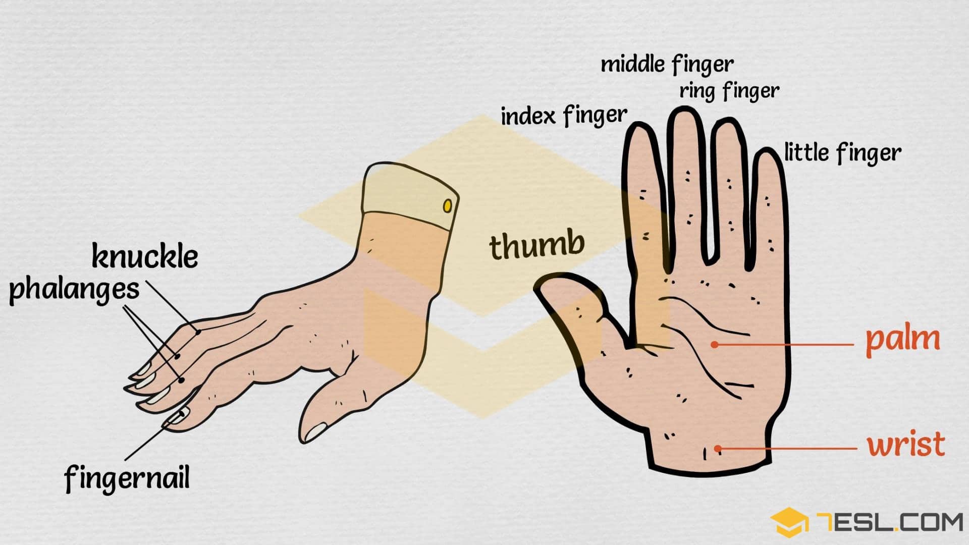 Parts Of The Hand Useful Hand Parts Names With Pictures 7
 Gifts Delight Laminated 36x24 Inches Poster Hand Middle Finger X Ray Radiation Finger Gesture Anatomy Bone Finger Thumb Index Finger Pinkie Finger
 Wrist Hand Atlas Of Anatomy
Forearm Muscles Anatomy Support Movement
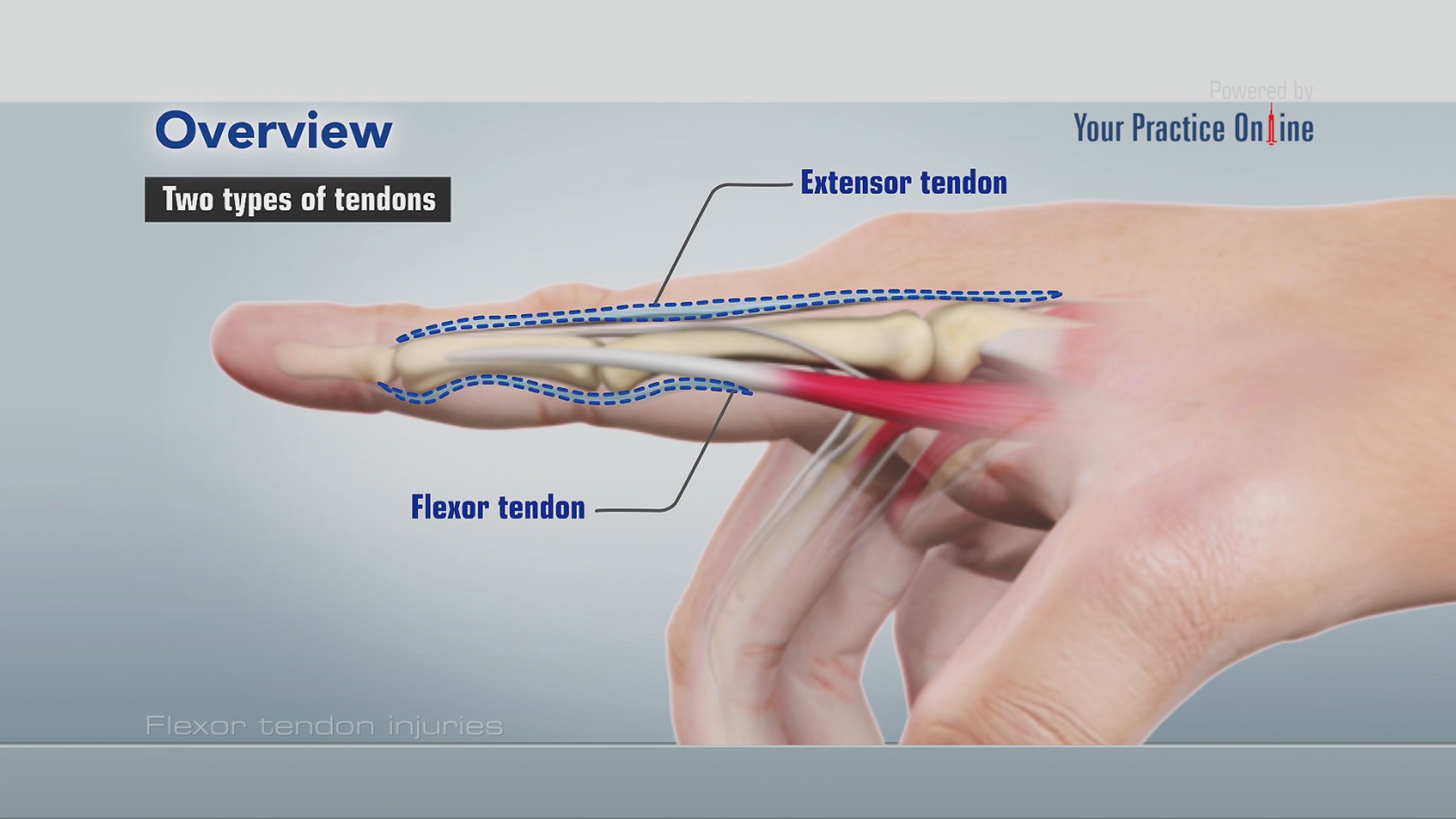 Flexor Tendon Injuries
 Ligaments Of The Fingers Hand Orthobullets
 Fingers 2 3 4 And 5 Fingers
 Rheumatoid Arthritis Disease Progression And Symptoms An
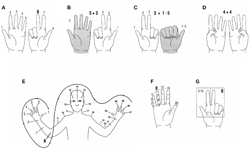 Frontiers Fingers As A Tool For Counting Naturally Fixed

 Finger Wikipedia
 Trigger Finger Stenosing Tenosynovitis Surgery Remedies
 Trigger Finger
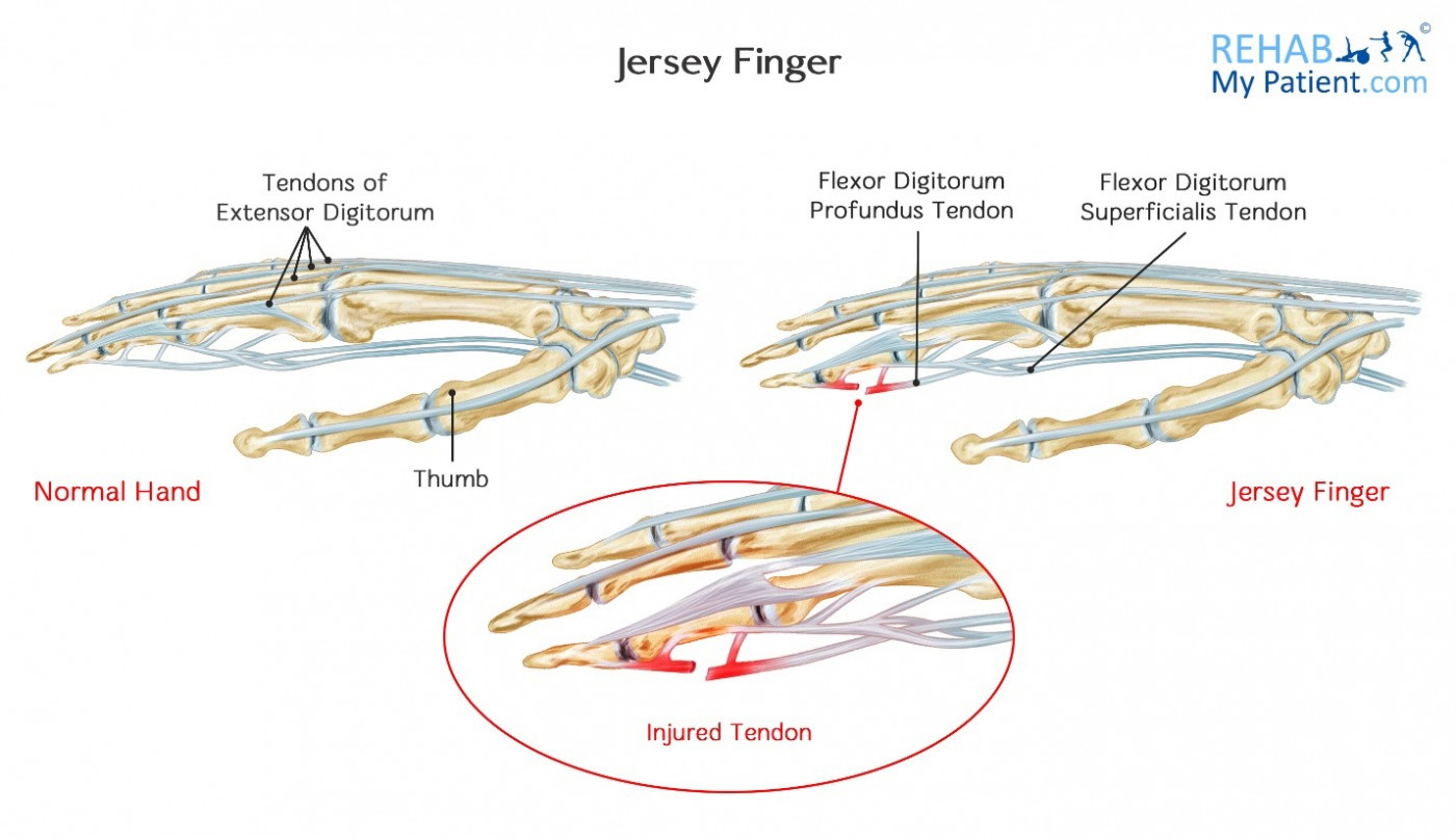 Jersey Finger Rehab My Patient
 Hand Bones And Wrist Bones Mnemonics Anatomy And Physiology
 Finger Fractures
 Hand And Finger Bones Kirkland Wa Evergreenhealth
 Odogaron S Double Hand Anatomy It Has Spurs On Its Middle
 Middle Finger Wikipedia
 Flexor Tendon Anatomy And Injury Everything You Need To Know Dr Nabil Ebraheim
 Finger Fracture An Overview Sciencedirect Topics
 Hand Anatomy Eorthopod Com


Posting Komentar
Posting Komentar