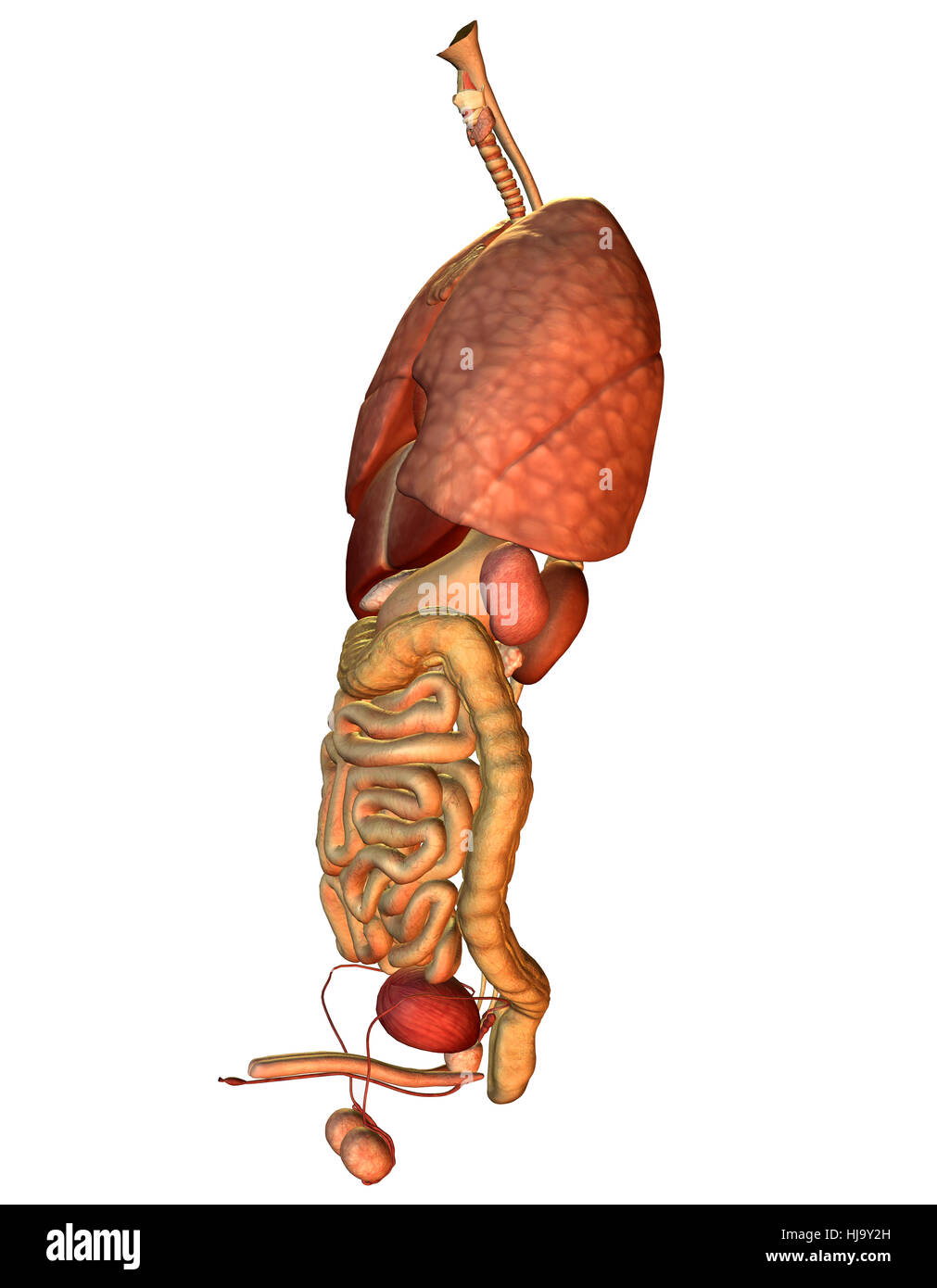
The region occupied by the abdomen is called the abdominal cavity. At the level of the pelvic bones the abdomen.
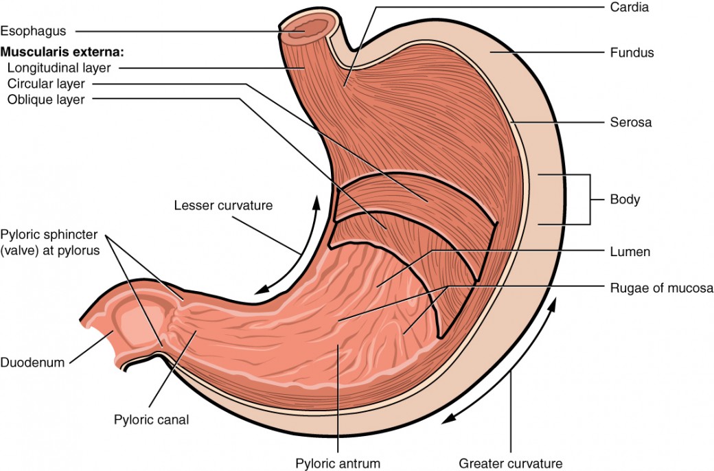
The cardia fundus body and pylorus.

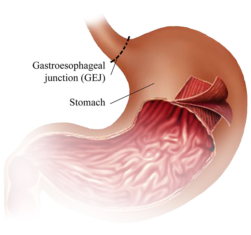
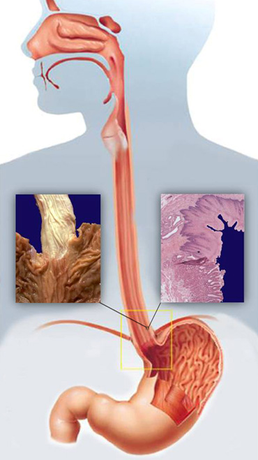
Belly anatomy. The cardia is where the contents of the esophagus empty into the stomach. As food reaches the end of the esophagus it enters the stomach through. Cardia surrounds the superior opening of the stomach at the t11 level.
Fundus the rounded often gas filled portion superior to and left of the cardia. In classical anatomy the human stomach is divided into four sections beginning at the cardia each of which has different cells and functions. Its characteristic shape is well known.
The body is the main central region of the stomach. The abdomen is the front part of the abdominal segment of the trunk. The stomach is a muscular organ located on the left side of the upper abdomen.
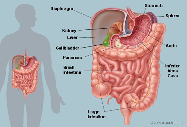
The fundus from latin meaning bottom is formed in the upper curved part. The stomach is located in the upper left area of the abdomen below the liver and next to the spleen. The majority of these organs are encased in a protective membrane termed the peritoneum.
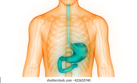
The diaphragm forms the upper surface of the abdomen. Its main function is to store and break down the foods and liquids that we consume before those. The stomach receives food from the esophagus.
The stomach has four main anatomical divisions. Anatomy of the stomach the stomach is an organ of the digestive system. The abdomen commonly called the belly is the body space between the thorax chest and pelvis.
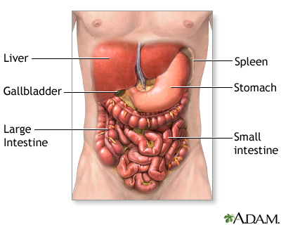
Belly definition the front or under part of a vertebrate body from the breastbone to the pelvis containing the abdominal viscera. The abdomen colloquially called the belly tummy or midriff is the part of the body between the thorax chest and pelvis in humans and in other vertebrates. Abdomen anatomy the abdomen is comprised primarily of the digestive tract and other accessory organs which assist in digestion the urinary system spleen and the abdominal muscles shown below.
It is an expanded section of the digestive tube between the esophagus and small intestine.
![]() Anatomy Of The Abdomen And Pelvis A Journey From Basis To
Anatomy Of The Abdomen And Pelvis A Journey From Basis To
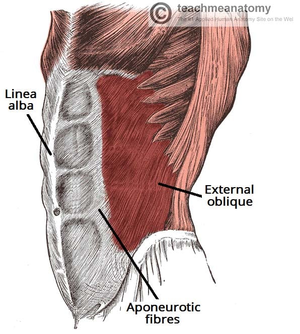
 Linea Alba And Abdominal Fascia Separation Q A With Tom

![]() Blood Vessels Of Abdomen And Pelvis Anatomy Overview Kenhub
Blood Vessels Of Abdomen And Pelvis Anatomy Overview Kenhub
 The Stomach S Not Connected To The Uterus But Some Kids And
 How Your Body Changes In Pregnancy Video
The Muscles Of The Trunk Human Anatomy And Physiology Lab

 The Abdomen Teachmeanatomy

 Stomach Anatomy Photos 52 086 Stomach Stock Image Results
 Abdominal Exploration Series Normal Anatomy Medlineplus
 Stomach Gallbladder And Pancreas Interactive Anatomy Guide
 The 9 Regions Of The Abdomen
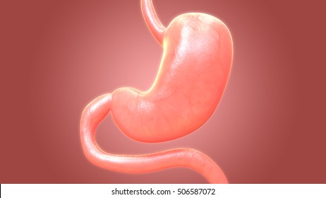
 The Stomach Anatomy And Physiology Ii
 Stomach Anatomy Photos 52 086 Stomach Stock Image Results
 Watch Your Organs Move To Make Room For Baby Every Week Of
 Anatomical Planes Of Body What Are They Types Position
 Four Abdominal Quadrants And Nine Abdominal Regions
 The Abdomen Human Anatomy Picture Function Parts
 Human Body Anatomy Pregnancy Belly Band Stock Vector
 Human Stomach Anatomy Diagram Human Anatomy Body Picture
 Abdominal Muscle Britannica
 Human Human Being Belly Tummy Biology Anatomy


 Abdominal Pain Reasons For Stomach Aches Cramps Discomfort
 Image Jpg Anatomy And Physiology Lab Unit 1 Practical 1
 Umbilical Hernia Intestine Human Anatomy Belly Vector
 Anatomy Of Human Salivary Glands With Labels White
 Abdomen Wikipedia


Posting Komentar
Posting Komentar