The most common ligament injuries are acl tears mcl tears pcl tears and knee sprains which occur when the ligaments are overstretched. The knee consists of three bones.
 Knee Physiopedia
The knee is the largest joint in the body and one of the most easily injured.

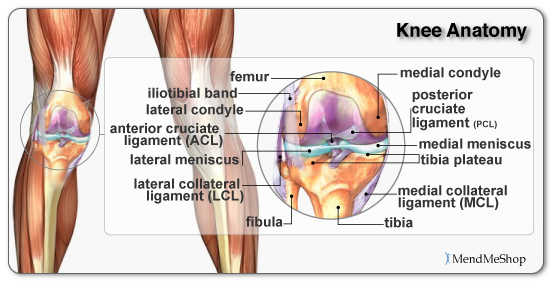
Knee injury anatomy. Participation in sports and recreational activities are risk factors for knee injury. It is made up of four main things. The largest joint in the body the knee moves like a hinge allowing you to sit squat walk or jump.
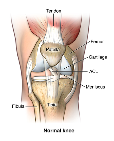
The knee is the largest and most complex joint in the body. Fast facts on knee anatomy. Damage to the acl such as a tear is a common knee injury among athletes.
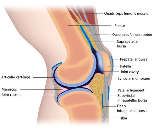
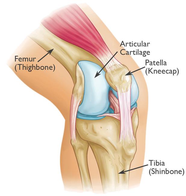
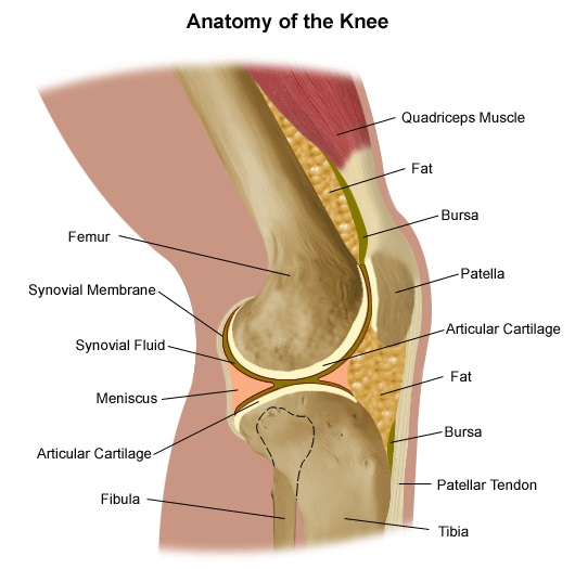
Each part of the anatomy needs to function properly for the knee to work. Knee anatomy function and common problems the knee joint is a synovial joint which connects the femur thigh bone the longest bone in the body to the tibia shin bone. Bones cartilage ligaments and tendons.
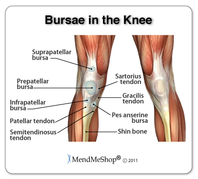
Collection of fluid in the back of the knee. Acute injury or trauma as well as chronic overuse may cause inflammation and its accompanying symptoms of pain swelling redness and warmth. The knee is a synovial joint meaning it contains a fluid filled capsule.
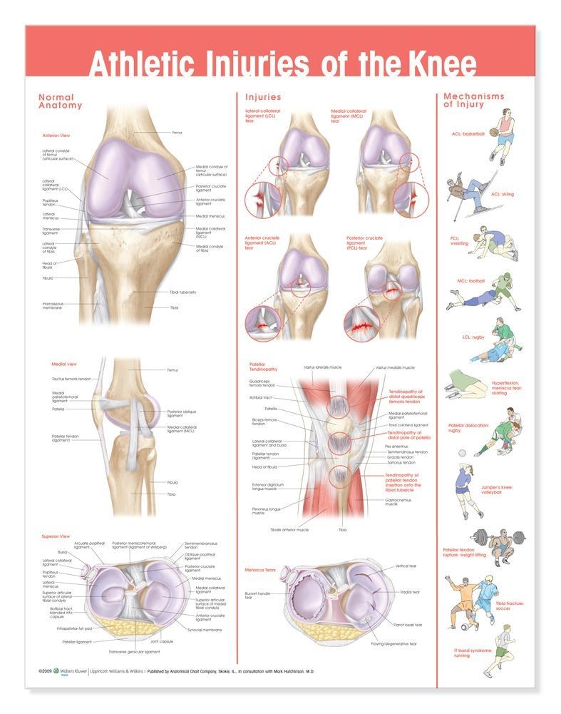
Pain swelling and warmth in any of the bursae of the knee. Knee injuries chart is an informative chart showing common injuries of the knee. The central images of normal knee anatomy are finely detailed and clearly labeled.
Three bones meet to form your knee joint. The knee is the joint where the bones of the lower and upper legs meet. Another common sporting injury is pulling or straining the hamstring tendons two groups of string like connective tissues at the back of the knee and thigh that connect some of the major muscles of the knee.
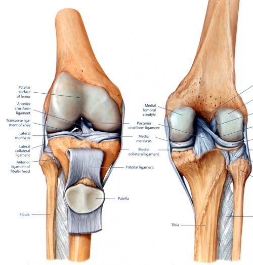
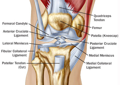
1 the tibiofemoral joint where the tibia meet the femur 2 the patellofemoral joint where the kneecap or patella meets the femur. In knee joint anatomy they are the main stabilising structures of the knee acl pcl mcl and lcl preventing excessive movements and instability. Your thighbone femur shinbone tibia and kneecap patella.
Bursitis often occurs from overuse or injury. Femur the upper leg bone or thigh bone. The knee joins together the thigh bone shin bone fibula on the outer side of the shin and kneecap.
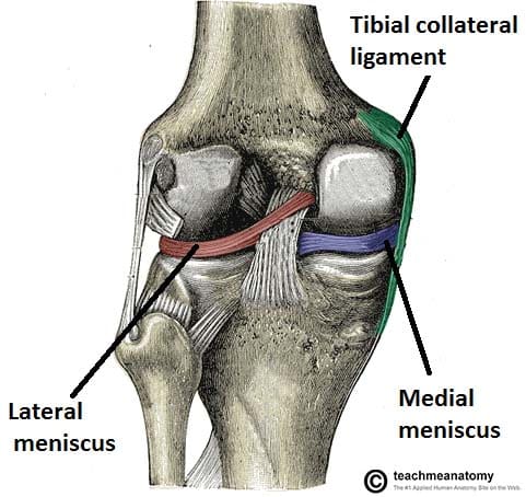
Tibia the bone at the front of the lower leg or shin bone. Anterior view of a normal knee with the patella removed oblique view of normal knee anatomy posterior view of normal knee anatomy. There are two main joints in the knee.
 Anterior Cruciate Ligament Acl Injury Orthopedics

 Acute Knee Injuries Explained Nps Medicinewise
 5 Simple Knee Injury Prevention Exercises Stack
 True Knee Patellar Dislocations Core Em
 Patellar Fractures Broken Kneecap Orthoinfo Aaos
 Guide Knee Injuries In Bjj Fighters Market
 The Knee Joint Articulations Movements Injuries
 How To Deal With Knee Injury Symptoms Lcr Health
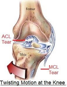 Twisted Knee Common Injuries Treatment Knee Pain Explained
 Ccl Injury And Treatment Vosm
 Injuries To Posterolateral Corner Of The Knee A
 Knee Ligament Injuries Stanford Health Care
 Pcl Injury Knee Sports Orthobullets

/188058334-crop-56aae7425f9b58b7d0091480.jpg) What Is Causing Your Knee Pain
What Is Causing Your Knee Pain
Common Knee Injuries Orthoinfo Aaos
Anatomy Of The Knee Baxter Regional Medical Center
 Lcl Injury Of The Knee Knee Sports Orthobullets
 Left Knee Anatomy Google Search Knee Injury Anatomy
 Mcl Knee Injuries Knee Sports Orthobullets
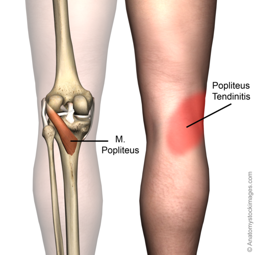 Popliteus Tendinopathy Physiopedia

 Figure Anatomy Of The Right Knee Download Scientific Diagram


Posting Komentar
Posting Komentar