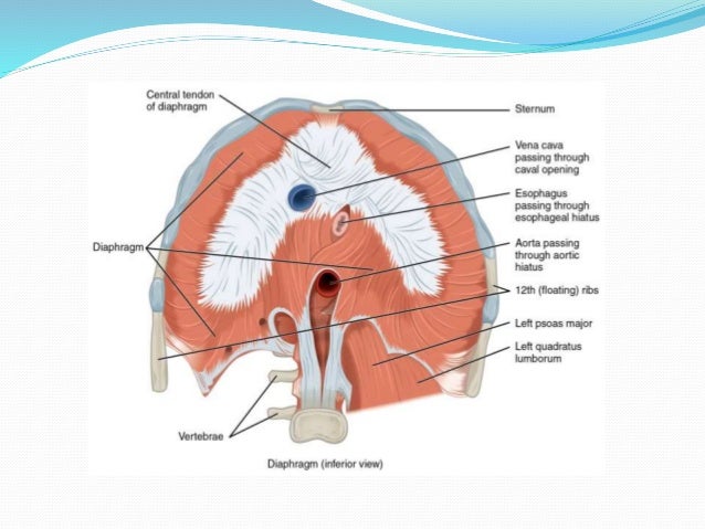
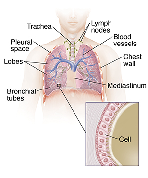
The lungs are covered by a thin tissue layer called the pleura. Thomas r gest phd more.
 Chest Muscle Injuries Strains And Tears Of The Pectoralis
Abdomen duodenum and pancreas.

Anatomy of the chest. The pectoralis minor is a thin triangular muscle that. It contains organs including the heart lungs and thymus gland as well as muscles and various other internal structures. This thoracic and pulmonary anatomy tool is especially designed for students of anatomy medical and paramedical studies.
Navid pourtaheri md phd ms. The core of the matter anatomy of the chest and abdomen. Anatomy of the chest abdomen and pelvis introduction.
Mediastinum and great vessels. Chest the epidermis is the outermost layer that provides a protective waterproof seal over the body. The model features 33 individually identified parts including a two piece larynx.
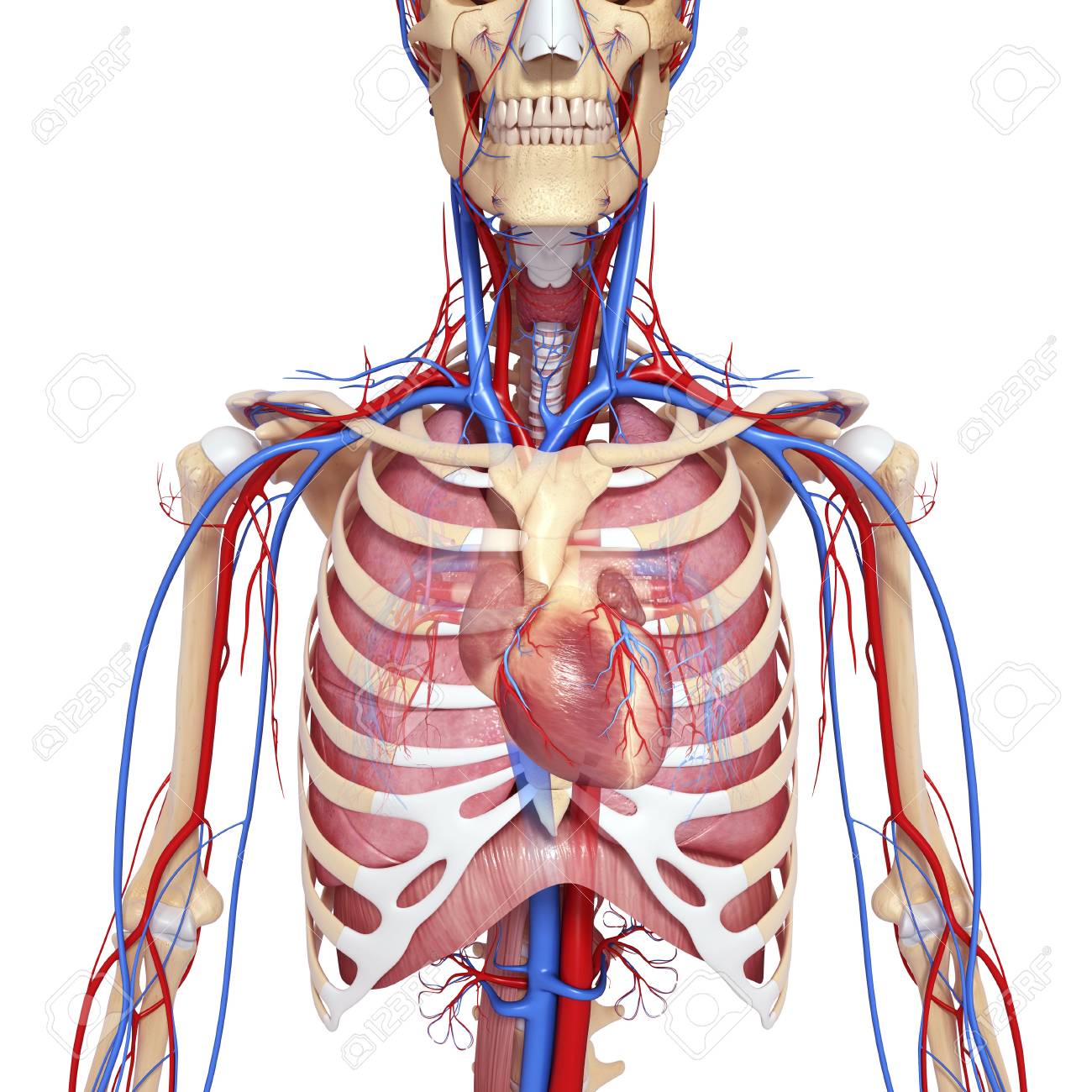
The thorax or chest is a part of the anatomy of humans and various other animals located between the neck and the abdomen. The chest is the area of origin for many of the bodys systems as it houses organs such as the heart esophagus trachea lungs and thoracic diaphragm. The circulatory system does most of its work.
All about the chest muscles function of the chest muscles. The pectoralis major is a large substantial fan shaped muscle. A great option for studying them up close in a tactile manner is the lungs with heart anatomy model.
The same kind of thin tissue lines the inside of the chest cavity also called pleura. Anatomical illustrations this e anatomy module presents an illustrated anatomy of the lungs trachea bronchi pleural cavity and pulmonary vessels. Abdomen mesenteric vessels retroperitoneum and kidneys.
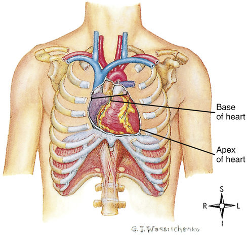
Anatomy of the heart. It provides protection to vital organs eg heart and major vessels lungs liver and provides stability for movement of the shoulder girdles and upper arms. The chest wall is comprised of skin fat muscles and the thoracic skeleton.
Sternum in the anatomy of tetrapods four limbed vertebrates elongated bone in the centre of the chest that articulates with and provides support for the clavicles collarbones of the shoulder girdle and for the ribs. The dermis is the under layer that contains sweat glands hair follicles blood vessels and more. Two of the major anatomical structures in this area are the heart and lungs.
Anatomy of the chest and the lungs. The thorax includes the thoracic cavity and the thoracic wall. Its origin in evolution is unclear.
The chest is part of a larger group of pushing muscles found in. Abdominal cavity largest hollow space of the body. A thin layer of fluid acts as a lubricant.
Anatomy of the abdomen.
 Anatomy Of The Chest Wall And The Pleura Thoracic Key
Nerves Of The Chest And Upper Back
 Male Chest Anatomy Stock Photos Male Chest Anatomy Stock
 Male Chest Anatomy Diagram Ipad Mini 2 Case
 Chest Anatomy Heart And Lungs
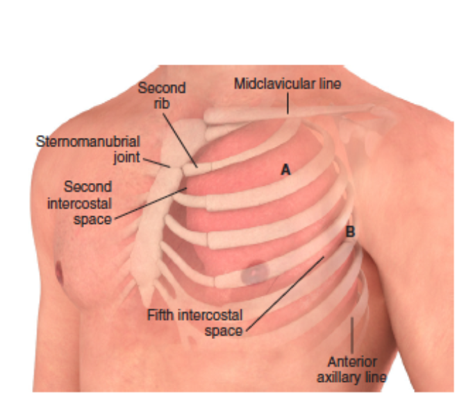
 Chest Wall Pain Costochondritis Articles Mount Nittany
 Muscles In Chest Area Human Chest Muscles Pectoral Muscles
 Human Chest Anatomy Images Stock Photos Vectors
 Normal Anatomy Of The Chest Thoracic Cavity Medical Art
 Chest Tube Insertion Series Normal Anatomy Medlineplus
 Surgical Anatomy Of The Chest Wall Springerlink
 The Muscles Of The Chest And Upper Back Anatomy Medicine Com
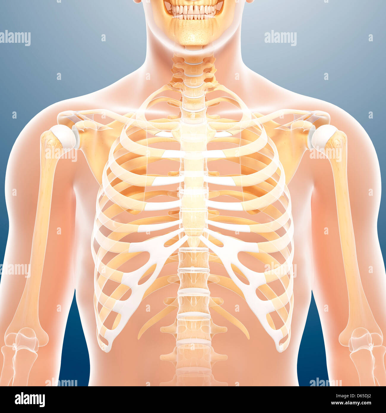
 Chest Anatomy Artwork
 Traumatic Hemothorax Core Em
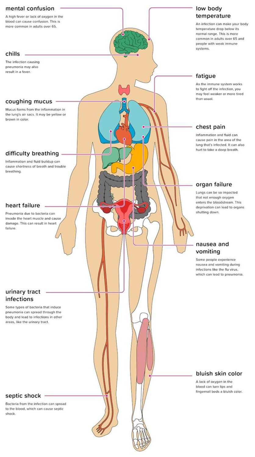 The Effects Of Pneumonia On The Body
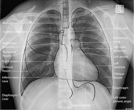 Anatomy Of A Chest X Ray Radiologypics Com
 Radiological Anatomy Of Chest Including Lungs Mediastinum
 Extrinsic Chest Muscles Functional Anatomyintegrative
 Chest Anatomy Artwork
 Thoracic Cavity Anatomy Britannica

 Chest Wall Neupsy Key
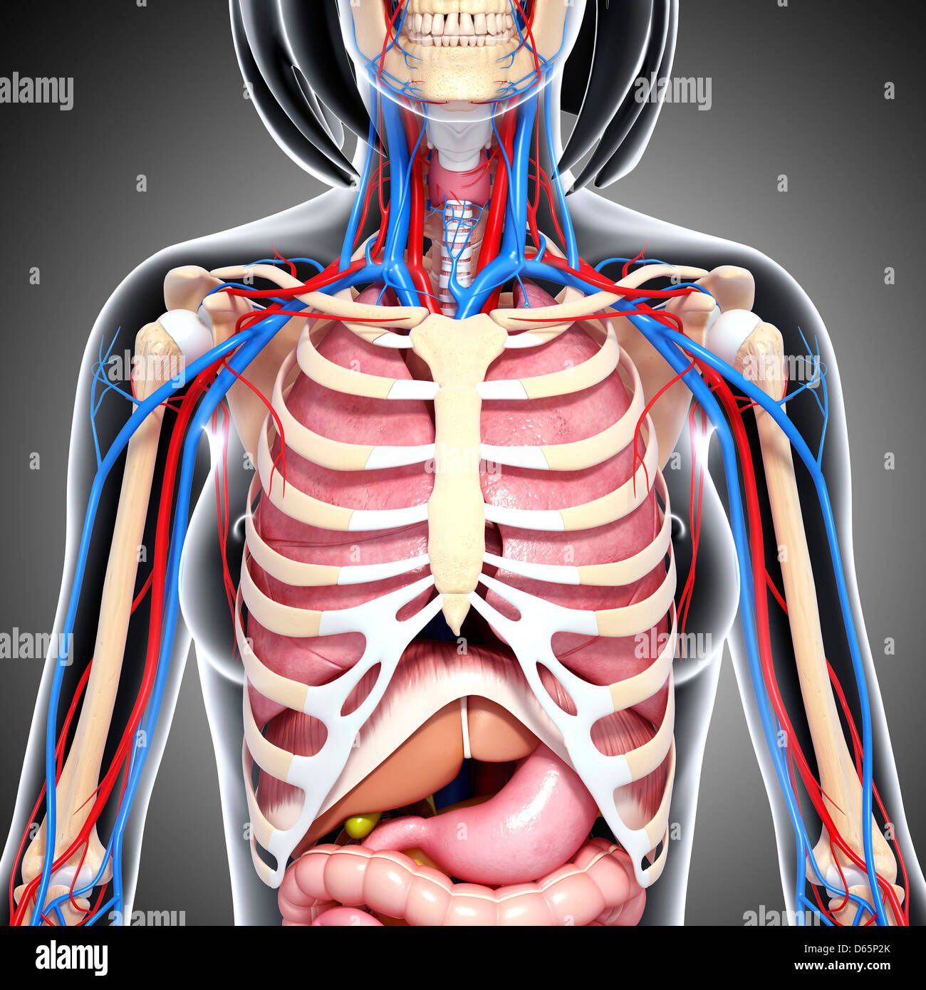 Female Chest Anatomy Stock Photos Female Chest Anatomy
 Functional Anatomy Of The Cardiovascular System Clinical Gate


Posting Komentar
Posting Komentar