Blood supply of left kidney and suprarenal gland. Information is provided about the anatomical features and landmarks for conducting a physical examination collecting biological samples making injections of therapeutic and experimental materials using imaging modalities and performing surgeries.
 Human Heart Model
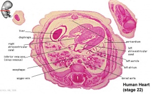
The anatomy of the postnatal heart in mouse and human the basic anatomical features of the postnatal heart in the human and mouse are very similar fig.

Mouse anatomy heart. The latter is formed from the proximal part of the left cranial caval vein lccv webb et al. Left lateral aspect of skull. Basic anatomy of the heart below is a 3d model of the heart which is fully interactive.
This is a work in progress and for demonstration purposes. It is appropriate for all strains of mouse. Blood supply of testis.
Abbreviated title page foreword introduction externals 4. Thus in both species the heart has four chambers. Mus musculus lac grey strain.
1996 1998. These pages were put together as a pilot demonstration by with the collaboration of oak ridge national laboratories duke university and gary henderson and university of california davis. Male organs deflected to left to show blood supply.
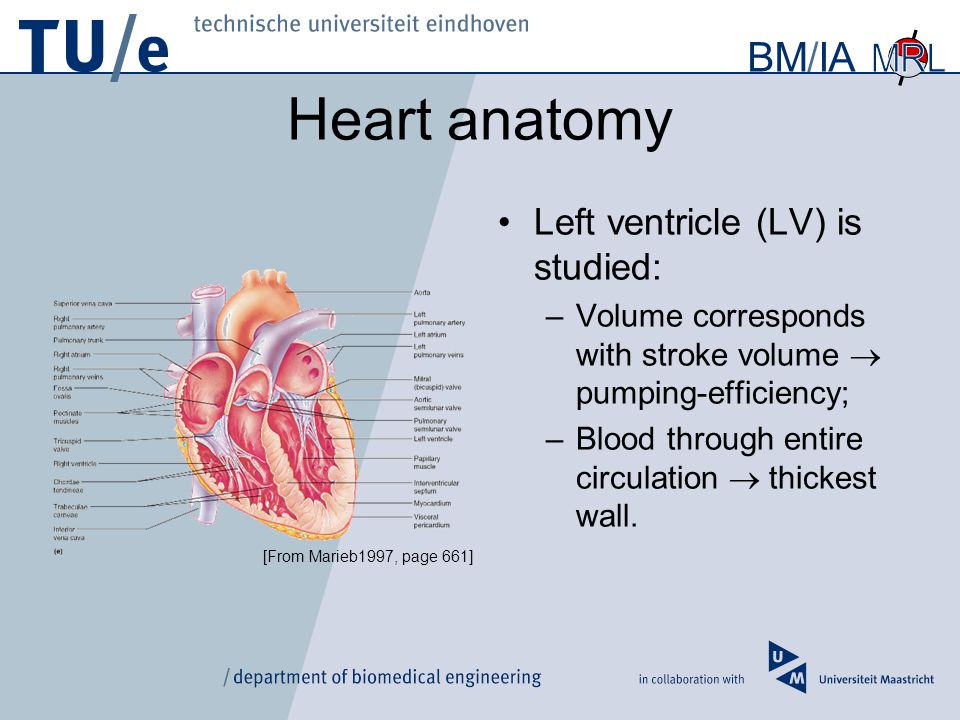
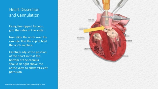
Your comments and suggestions will be helpful in. Blood samples are taken from the heart preferably the ventricle which can be accessed either via the left side of the chest through the diaphragm from the top of the sternum or by performing a thoracotomy. A color atlas and text provides detailed comparative anatomical information for those who work with mice and rats in animal research.
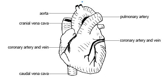
They empty to the right atrium or to the coronary sinus. Dorsal aspect of skull. Two atria separated by an interatrial septum ias and two ventricles separated by an interventricular septum ivs.
Explore the model using your mouse pad or touchscreen to understand more about the heart. Comparative anatomy of the mouse and rat. In mice the cardiac veins run on the surface of the heart within the subepicardium draining the myocardium of the left and the right ventricles as well as the left atrium.
The anatomy of the laboratory mouse margaret j. 01 1 ml of blood can be obtained depending on the size of the mouse and whether the heart is beating. Skeleton of lac grey mouse.
 Cow Heart Cell Diagram Reading Industrial Wiring Diagrams
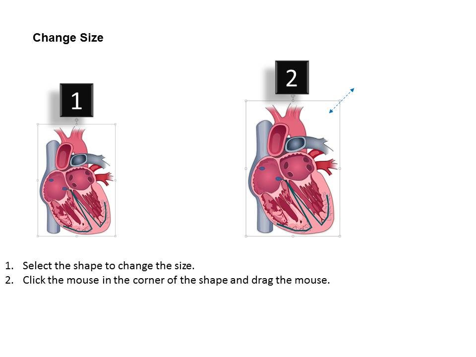
 Representation Of The Location Of The Incisions To Remove
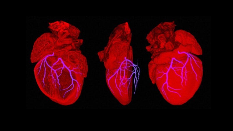
 3d Shape Variability Of The Healthy And Infarcted Mouse
![]() Anatomy And Physiology Of The Adult Mouse Heart A A
Anatomy And Physiology Of The Adult Mouse Heart A A
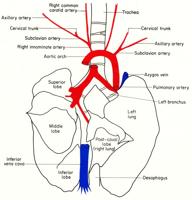
 Anatomy And Physiology Of Animals Cardiovascular System The

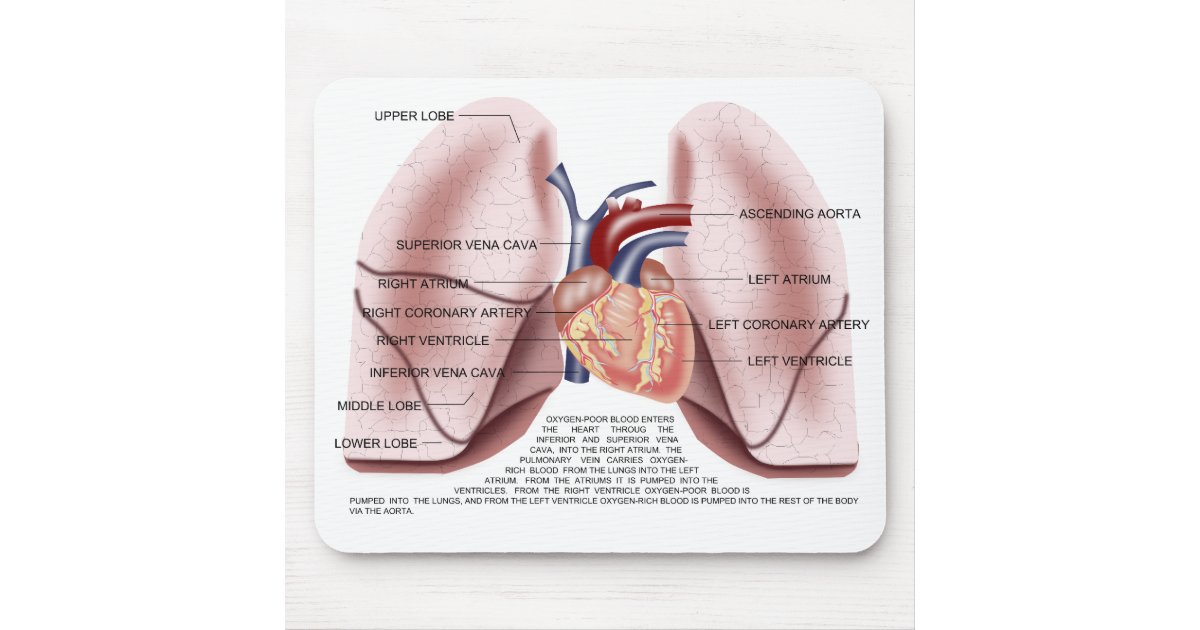 Chest Anatomy Mouse Pad Zazzle Com
 An Ode To The Right That Little Ventricle That Could The
 Techniques And Best Practices For Cardiomyocyte Isolation
 Mouse Dissection
 Cardiovascular System Heart Development Embryology
 Black Anatomical Heart Mouse Pad
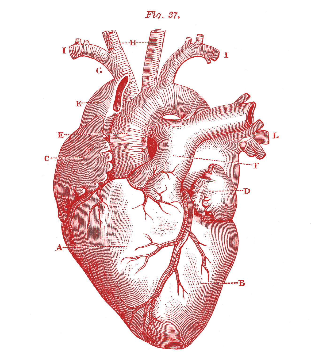
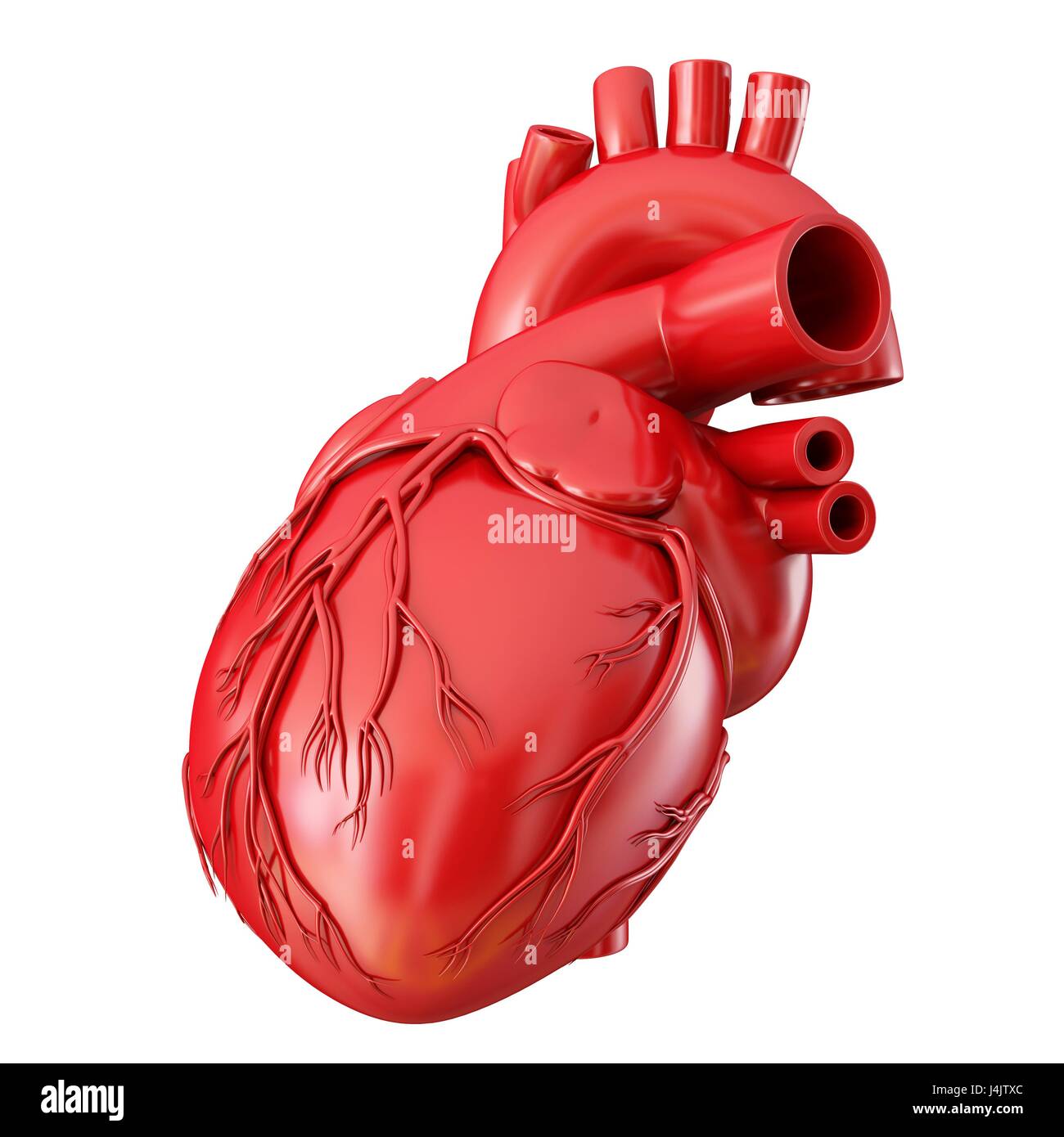 Illustration Of Human Heart Anatomy Stock Photo 140556260
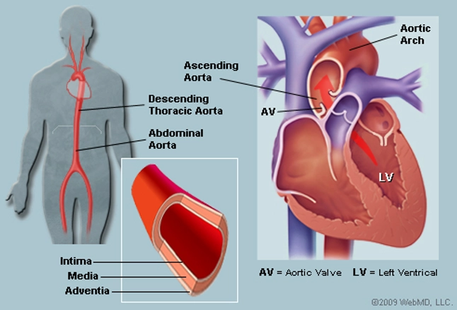 The Aorta Human Anatomy Picture Function Location And
 Anatomical Heart Mouse Pads Cafepress
 Heart Anatomy Left Atrium 3d Anatomy Tutorial
 Amazon Com Semtomn Gaming Mouse Pad Human Heart Anatomy
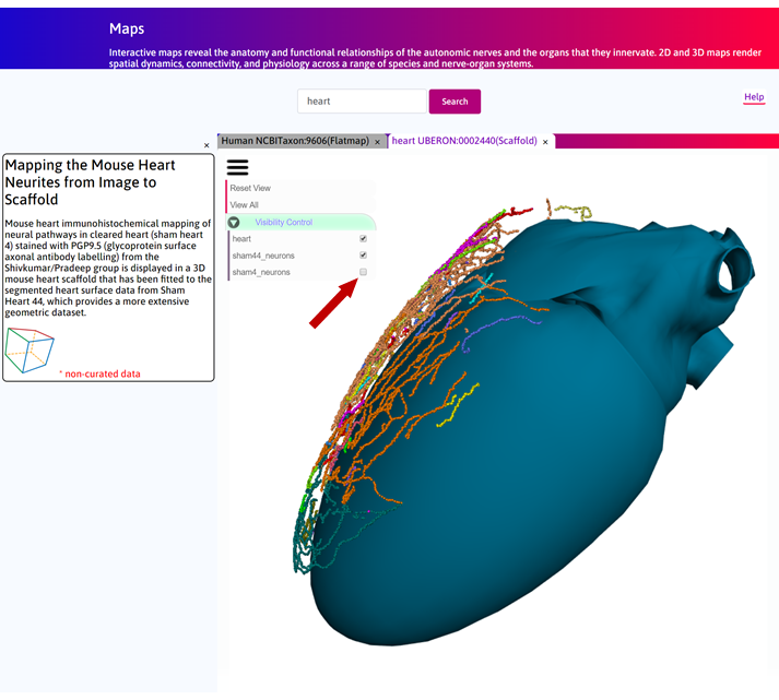 Mapping The Mouse Heart Neurites From Image To Scaffold
 Research Agenda Examine Mouse Anatomy Choose Organ
 Amazon Com Boszina Mouse Pads Chamber Human Heart Anatomy
 Human Heart Anatomy Mousepad
 Black And White Tattoos Tattoo Heart Octopus Sketch Snake
 Anatomy And Physiology Of Animals Cardiovascular System The
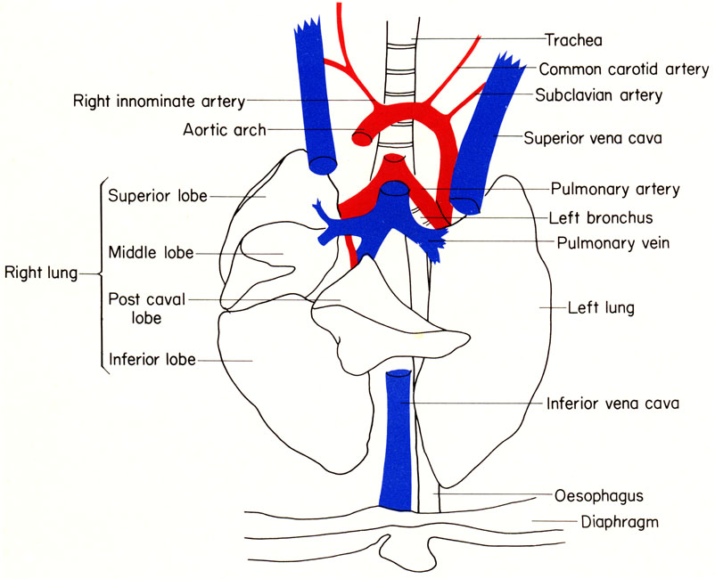
 Abdominal And Thoracic Organs In The Mouse These Organs Are
 Cardiac Structure Cardiac Magnetic Resonance Imaging A
 Anatomy Of The Laboratory Mouse In Vivo Imaging Atlas On A
 0514 Heart Anatomy Medical Images For Powerpoint
 Breakthrough Scientists Build A Beating Mouse Heart With

 Anatomy And Physiology Of Animals Cardiovascular System The
 Amazon Com Wknoon Gaming Mouse Pad Cardiovascular Human
 5 Anatomical Heart Pictures The Graphics Fairy


Posting Komentar
Posting Komentar