It sits in the centre of the sole sandwiched between the plantar aponeurosis and the tendons of flexor digitorum longus. The plantar fascia is a complex structure that extends from the medial calcaneal tubercle the heel bone to the proximal phalanges of the toes the bone at the base of the toe at the metatarsophalangeal mtp joints.
 Foot And Ankle Musculoskeletal Key
A transverse also known as horizontal plane is an x z plane parallel to the ground which in humans separates the superior from the inferior or put another way the head from the feet.

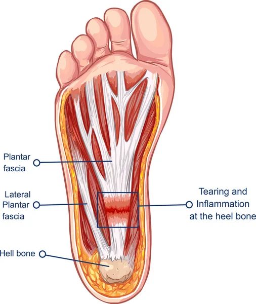
Plantar foot anatomy. Doctors once thought bony growths called heel. This image shows the anatomy of the plantar foot and is labeled with corresponding identification tags. Plantar fasciitis is inflammation of the thick band of tissue also called a fascia at the bottom of your foot that runs from your heel to your toes.
Originates from the medial tubercle of the calcaneus and the plantar aponeurosis. The plantar aponeurosis also known as the plantar fascia is a strong layer of white fibrous tissue located beneath the skin on the sole of the foot. Calcaneocuboid ligament the ligament that connects the calcaneus and the tarsal bones and helps.
Towards the front of the foot at the. Foot anatomy and biomechanics blood supply to the foot layers of the plantar foot nerves of the foot foot muscle forces deformities. Plantar calcaneonavicular ligament a ligament of the sole of the foot that connects.
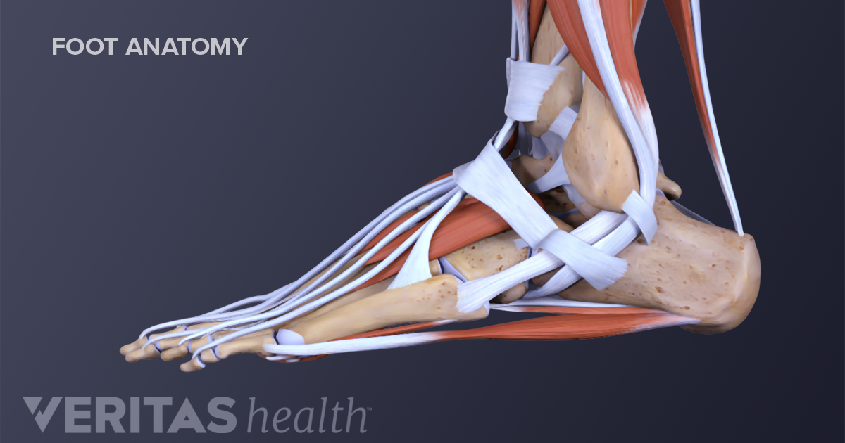
The plantar fascia or plantar aponeurosis forms part of the deep fascia of the sole of the foot and provides a strong mechanical linkage between the calcaneus and the toes. Arising predominantly from the calcaneal tuberosity the plantar fascia attaches distally through several slips. Plantar fascia the longest ligament of the foot.
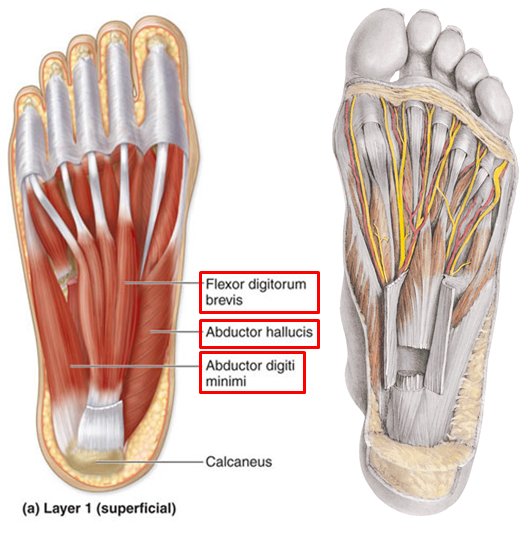
Anatomy of the plantar fascia. Muscles of the plantar foot are divided into four layers. Ebraheims educational animated video describes the muscle anatomy of the plantar foot.
It attaches to the middle phalanges of the lateral four digits. Anatomy of the plantar fascia. The main ligaments of the foot are.
As it progresses from the heel bone to the toes it breaks into five sections.

 Medivisuals Plantar Anatomy Of Left Foot Medical Illustration
 Plantar Interossei Lpn Anatomy Orthobullets
 Anatomy Of The Right Foot Plantar View Medical Illustration
Plantar Fasciitis And Bone Spurs Orthoinfo Aaos
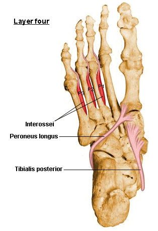 Layers Of The Plantar Foot Foot Ankle Orthobullets

Plantar Foot Anatomy Nerves Fa07 Foot Anatomy Human

 Plantar Foot Muscles Diagram Quizlet
 Muscle Anatomy Of The Plantar Foot Everything You Need To Know Dr Nabil Ebraheim
 Plantar Fasciitis Symptoms
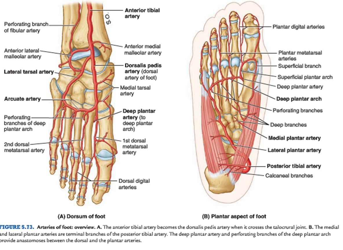
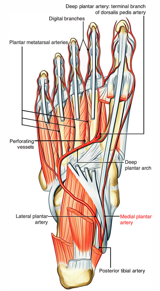 Arteries Of Foot Earth S Lab
 ᐈ The Bottom Of Your Foot Stock Pictures Royalty Free
 Plantar Surface Of The Foot Layer 1 A Layer 2 B Layer
 The Plantar Fascia Is A Thick Band Of Connective Tissue That
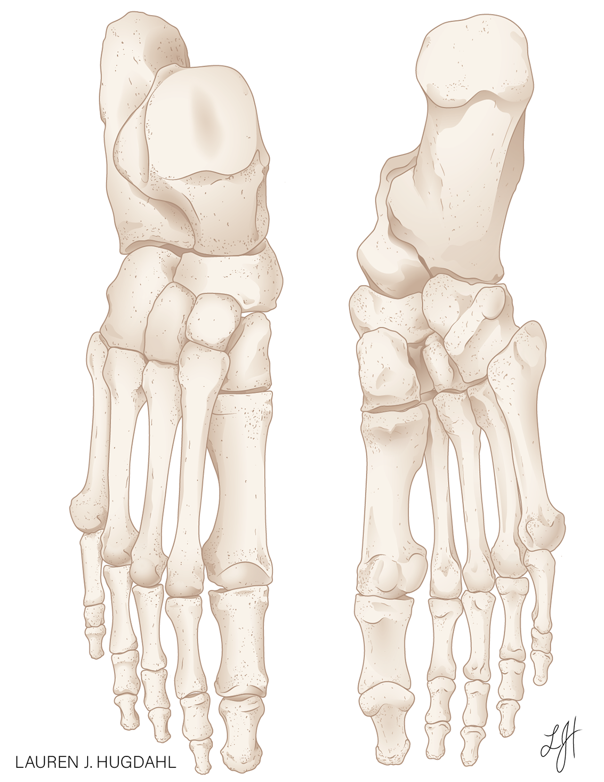 Dorsal And Plantar Skeletal Foot Anatomy On Behance
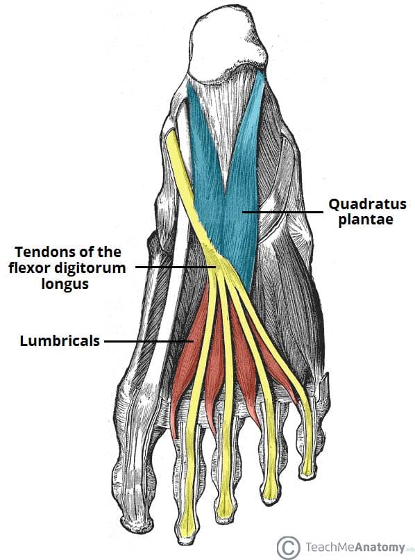 Muscles Of The Foot Dorsal Plantar Teachmeanatomy
 Plantar Fasciitis Fleet Feet Delray
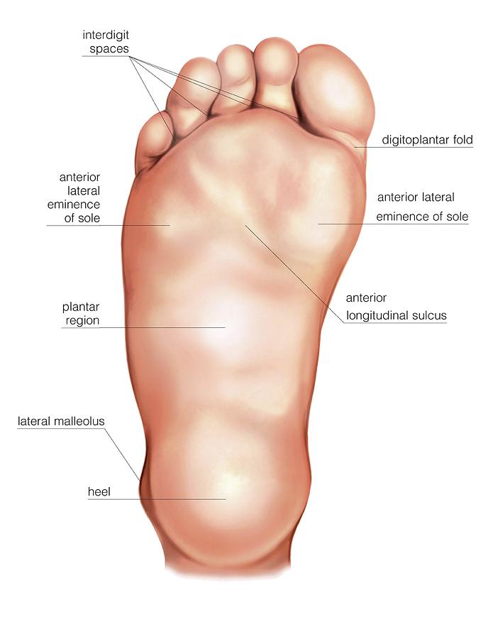 Anatomy Regions Of The Right Foot
 Orthosleeve Fs6 Plantar Fasciitis Compression Foot Sleeves Pair
 Plantar Fasciitis Overcoming Heel And Arch Pain Naturally


Posting Komentar
Posting Komentar