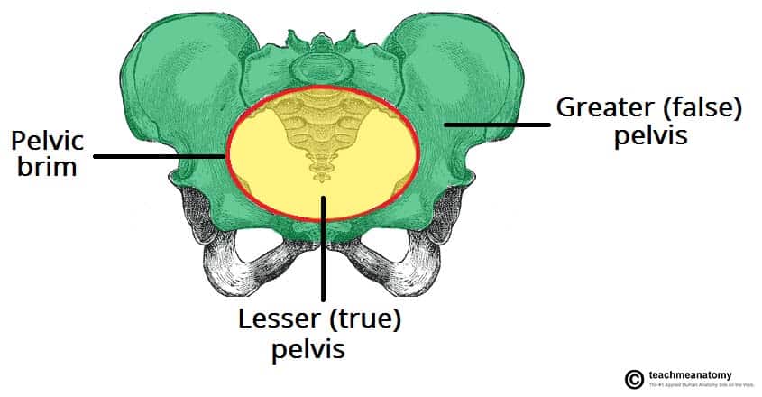
The female sacrum is wider shorter and less curved and the sacral promontory projects less into the pelvic cavity thus giving the female pelvic inlet pelvic brim a more rounded or oval shape compared to males. The interior walls are straight the subpubic arch wide the sacrum shows an average to backward inclination and the greater sciatic notch is well rounded.
 Female Pelvis Bone Anatomy Canvas Print
Filmed at the college of medicine university of sharjah uae.

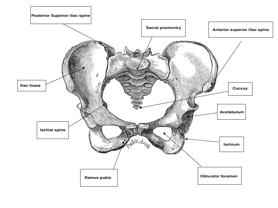
Female pelvic bone anatomy. Presented and edited by drakram jaffar phd using a plastic model. The pelvis is a bony structure that can be found in both male and female skeletons. It is therefore of great importance to determine the diameter of this canal and therefore the childbearing capacity of the mother.
Its inlet is either slightly oval with a greater transverse diameter or round. The exception to this compound structure when compared to all other bones is that it has differences that are classified by sex both for functional and general developmental reasons. They also include the vagina which is the entryway to the uterus.
Assessment of the female bony pelvis the lesser pelvis is the bony canal through which the fetus has to pass during childbirth. There are two hip bones one on the left side of the body and the other on the right. Filmed by mrnasser zahra lab technician.
Also check related. The pelvic outlet in male pelvis is narrower whereas the pelvis outlet in female pelvis is wider. The gynaecoid pelvis is the so called normal female pelvis.
A male pelvic bone is heavier taller and much thicker while a female pelvic bone is thinner and denser. The pubic arch or space under the base of the pelvis is also wider for this reason. The female pelvic organs include the egg producing ovaries and the uterine tubes that carry the eggs into the uterus for potential fertilization by male sperm.
This is so a baby can pass through the pubic outlet the circular hole in the middle of the pelvic bones during childbirth. In male pelvis the obturator foramen is round while in female pelvis the obturator foramen is oval. The female pelvic bones are typically larger and broader than a males.
Together they form the part of the pelvis called the pelvic girdle. The rest of the human skeleton differs only in size which is genetically determined and is usually slightly larger in males than in females.
 The Pelvis Human Anatomy And Physiology Lab Bsb 141
 One Half Of Female Pelvic Bone Diagram Wiring Diagram Page
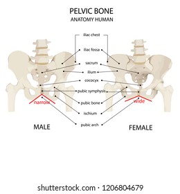
 Male Vs Female Pelvis 12 Major Differences Plus Comparison
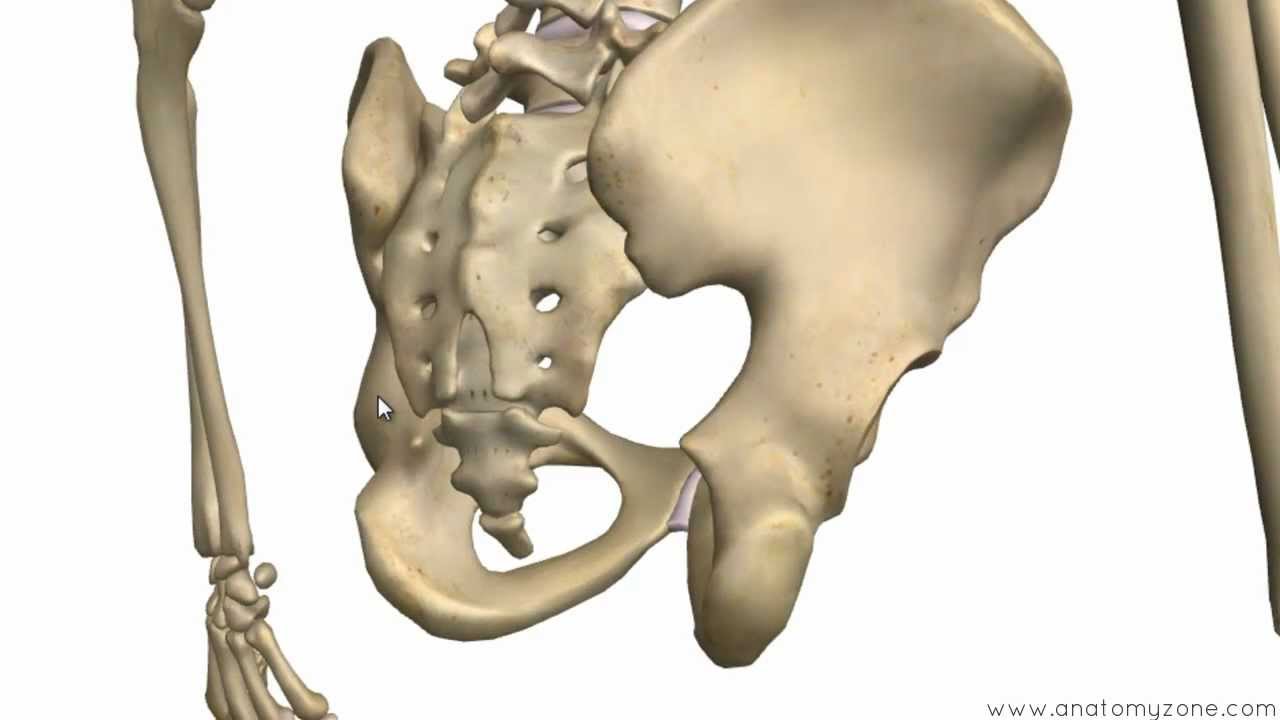 Bones Of The Pelvis Hip Bones Anatomy Tutorial
 Anatomy Gross Anatomy Physiology Cells Cytology Cell
 Female Pelvis Bone Anatomy Clipart K42330930 Fotosearch
 Pelvic Cavity Wikipedia
 The Pelvic Girdle And Pelvis Anatomy And Physiology I
A
 Pelvic Bone Images Stock Photos Vectors Shutterstock
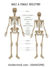
 Female And Male Pelvis Bones Anatomy Print Black And White
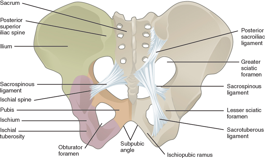 8 3 The Pelvic Girdle And Pelvis Anatomy And Physiology
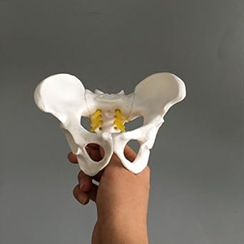 Airgoesin Female Pelvis Skeleton Bone Model Anatomical Human Medical Anatomy Small Size Teaching
 Pelvis Hip Anatomy
 Female Pelvis Bone Anatomy Canvas Print
 Male Vs Female Pelvis Google Search Anatomy Bones Women
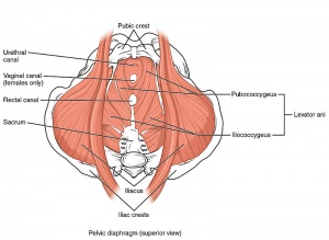 Pelvic Floor Anatomy Physiopedia
 Female Pelvic Anatomy Ppt Video Online Download
 Female Pelvis Bone Anatomy
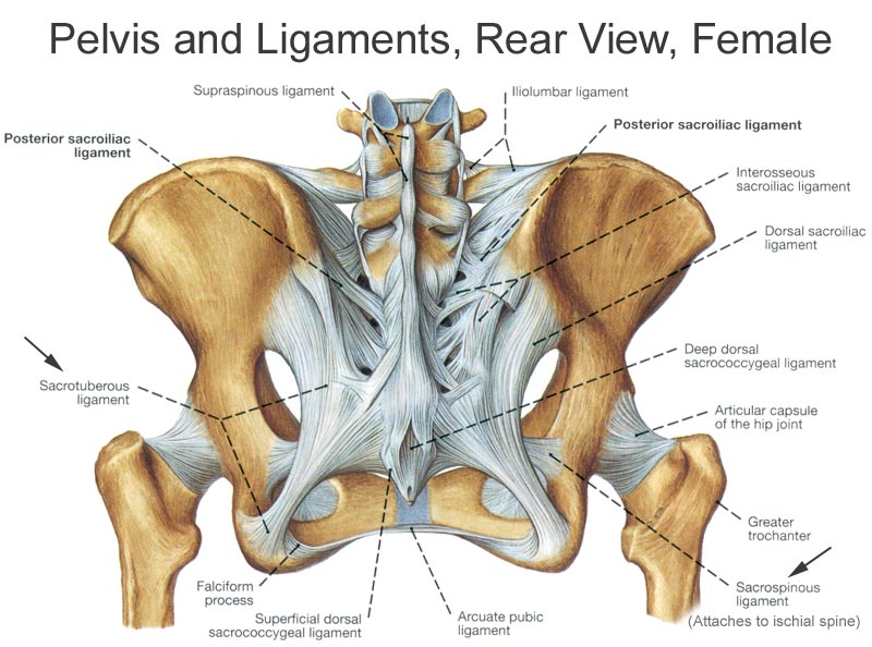 Bony Pelvis Anatomy Bone And Spine
 The Pelvic Girdle Structure Function Assessment

 Female Pelvis Bone Anatomy
 Male Pelvis Images Stock Photos Vectors Shutterstock
 The Pelvis Anatomy Images Pelvic Floor Connective Tissues


Posting Komentar
Posting Komentar