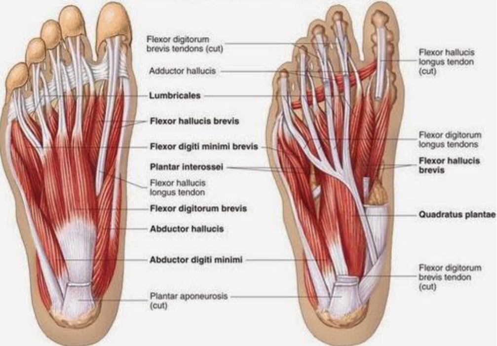
Tendons are elastic tissues made up of collagen. Soft tissues of the foot and ankle ligaments.
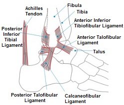
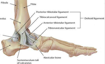 Medial Ankle Ligament Physiopedia
In this article we shall look at the anatomy of the ankle joint.

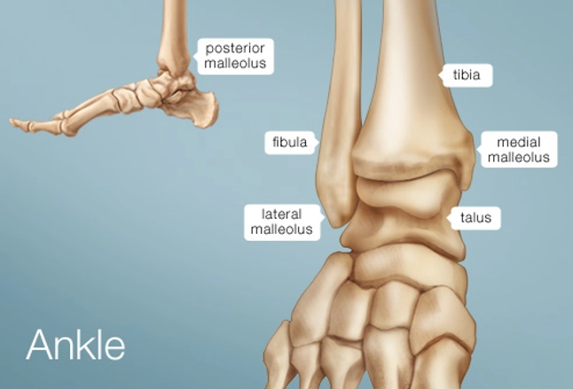
Foot anatomy ankle. The ankle is the joint that is located between the leg and the foot. Rehabilitation can prevent pain and swelling from becoming a long term problem. The talus bone supports the leg bones tibia and fibula forming the ankle.
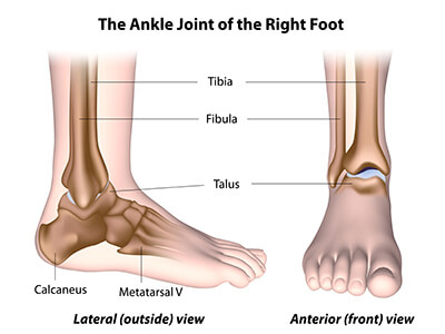
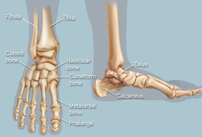
Foot and ankle anatomy. The calcaneous bone is the largest bone in your foot while the talus bone is the highest bone in your foot. Footeducation is committed to helping educate patients about foot and ankle conditions by providing high quality accurate and easy to understand information.
These all work together to bear weight allow movement and provide a stable base for us to stand and move on. The hindfoot forms the heel and ankle. Foot and ankle anatomy is quite complex.
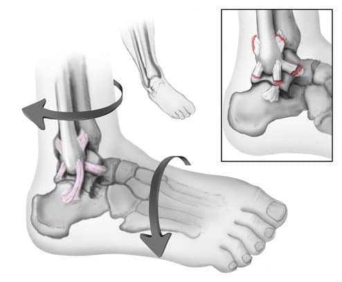
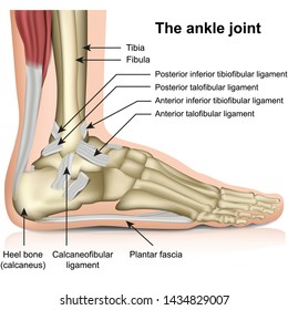
Upper ankle joint tibiotarsal talocalcaneonavicular and subtalar joints. The ankle joint also known as the talocrural joint allows dorsiflexion and plantar flexion of the foot. The hind foot consists of the talus bone or ankle bone and the calcaneous bone or heel bone.
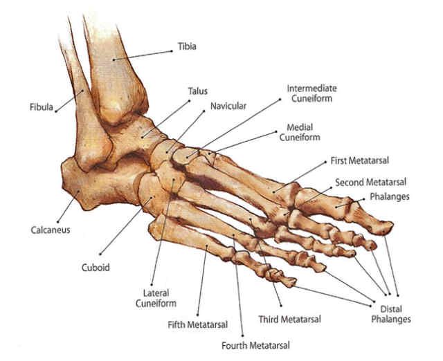
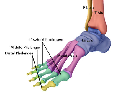
The foot can be divided into three anatomical sections called the hind foot mid foot and forefoot. The articulating surfaces ligaments movements and any clinical correlations. The calcaneus heel bone is the largest bone in the foot.
The ankle joint or talocrural joint is a synovial joint formed by the bones of the leg and the foot the tibia fibula and talus. This joint is a main contributor of stability in the lower limbs and it allows humans to perform actions such as running jumping and walking 1 2. Use our anatomy tools to learn about bones joints ligaments and muscles of the foot and ankle.
Ligaments are strong dense and flexible bands of fibrous connective tissue. It is made up of three joints. There are many muscles that help to move and support the ankle and foot.
Damage to one of the ligaments in the ankle usually from an accidental twist or turn of the foot. Fascia is a broad fibrous. The foot consists of thirty three bones twenty six joints and over a hundred muscles ligaments and tendons.
 Anatomy Of An Ankle Sprain Bouldercentre For Orthopedics
 A1f563ae839981eee950c076f74038fc Ankle Anatomy Foot Anatomy
 Ankle Images Stock Photos Vectors Shutterstock

 Foot And Ankle Anatomy Bones Muscles Ligaments Tendons
 Anatomy Of The Foot North Arkansas Podiatry
![]() Ankle And Foot Anatomy Bones Joints Muscles Kenhub
Ankle And Foot Anatomy Bones Joints Muscles Kenhub
 Ankle Foot Anatomy
 Foot Ankle Foot Anatomy Ankle Anatomy Leg Anatomy
 Ottawa Foot And Ankle Rule Anatomy Of The Right Foot And
 Ankle Human Anatomy Image Function Conditions More
 Foot Ankle Preservation Baltimore Md Towson Orthopaedics
 Foot And Ankle Anatomy Bones Muscles Ligaments Tendons
 Developing Strength Stability In The Foot Ankle And
 Layers Of The Plantar Foot Foot Ankle Orthobullets
 Foot Ankle Skeleton Elastic Mounted 3b Smart Anatomy
 Foot Bones Anatomy Conditions And More
Sprained Ankle Orthoinfo Aaos
Ankle Foot Atlas Of Anatomy
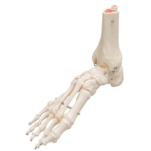
 Foot And Ankle Skeleton Model
 Understanding And Caring For Your Feet Breaking Muscle
 Anatomy Of The Ankle Maxeffortmuscle Com
 Foot Surgery Vienna Va Ankle Surgery Herndon
 Anatomy And Injuries Of The Foot And Ankle Chart 20x26 Clinicalposters
 Feet Human Anatomy Bones Tendons Ligaments And More
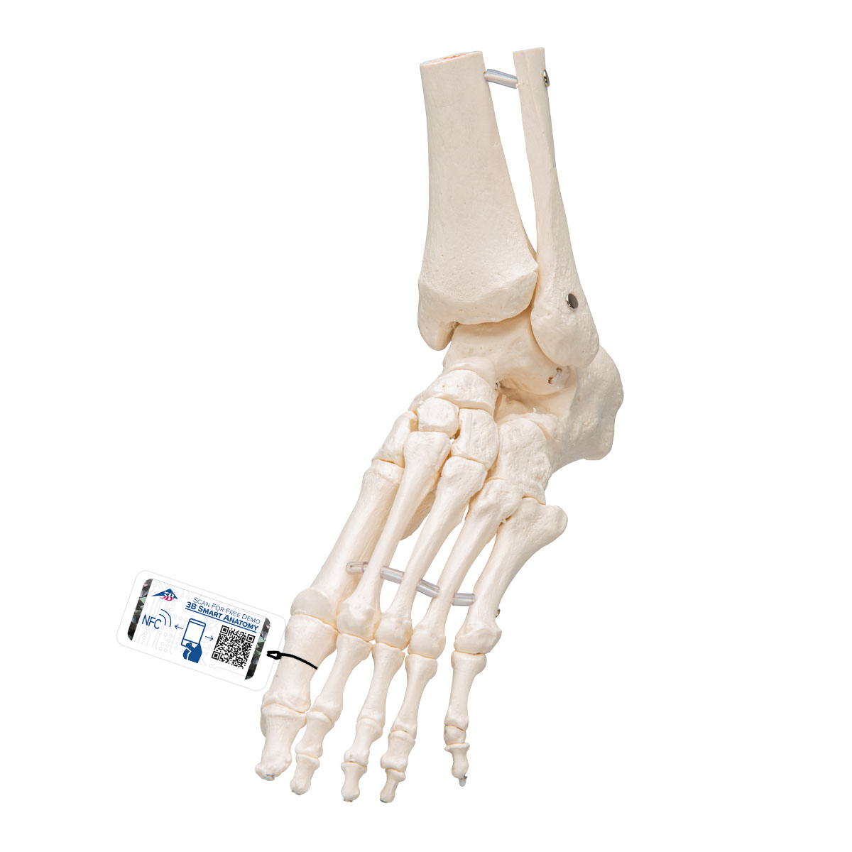
 Human Foot Ankle Skeleton Wire Mounted 3b Smart Anatomy

 Anatomy Of The Ankle Maxeffortmuscle Com
 Foot And Ankle Issues Northern Arizona Healthcare

 Get To Know The Ankle Joint Yoga Journal
 Ankle Foot Anatomy
Anatomy Ankle Joint Clinicals Medicine Tr051 Studocu
 Ankle Foot Atlas Of Anatomy
Posting Komentar
Posting Komentar