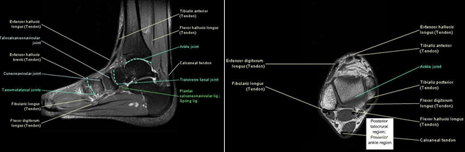
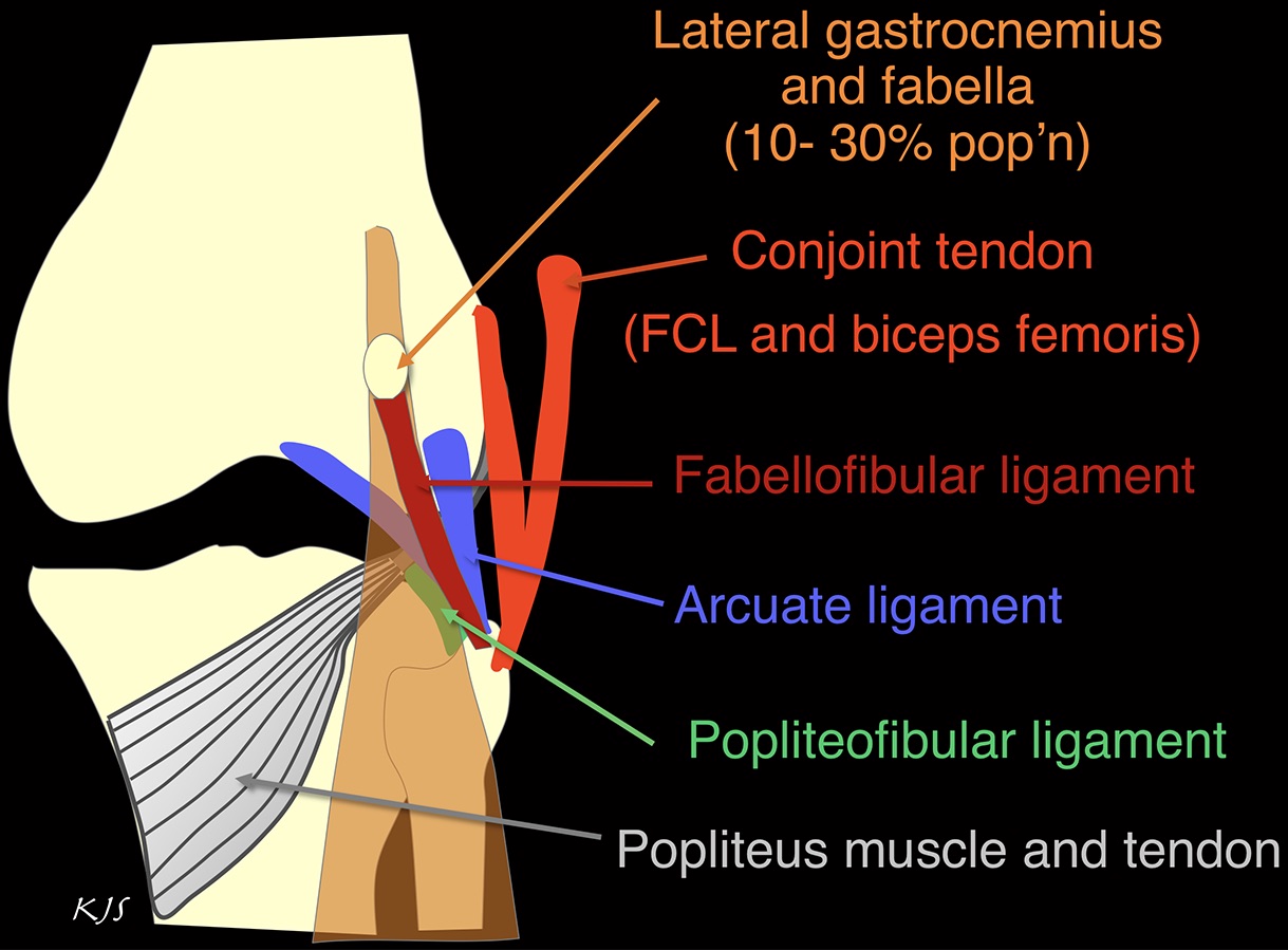
Three ligamentous groups support the ankle joint. It is also a fundamental communication tool to teach patients anatomy and pathology.
 A Maximally Damaged Ankle And Surprising Results Regenexx
This joint is a main contributor of stability in the lower limbs and it allows humans to perform actions such as running jumping and walking 1 2.

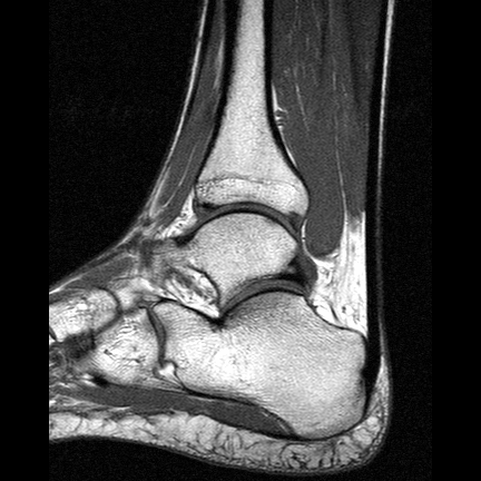
Mri ankle anatomy. This webpage presents the anatomical structures found on ankle mri. Once you have studied the bones scan the joints for effusion. Routine ankle mr imaging is performed in the axial coronal.
Racsuq advanced surgical anatomy course upper and lower limbs. Rmhalf msk ankle. Mri of the ankle.
Internal derrangements of joints. On mri the ligaments appear as thin linear low signal intensity structures connecting adjacent bones usually delineated by high signal intensity fat. Scroll through the image stack for the.
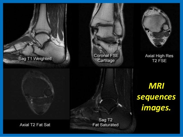
Mri of the ankle and feet. Start your exam with fatsat images of the bones to screen for edema. Click on a link to get sagittal view t1 axial view t2fatsat coronal view t2fatsat sagittal view t2fatsat.
Normal mri ligament anatomy. 663 3 normal extremity. Screen on fatsat images for bone marrow edema.
Ankle mri examination systematic approach. 37 magnetic resonance imaging mri the ankle is the joint that is located between the leg and the foot. The past 15 years have witnessed an explosion of information regarding the role.
Uq med year 1 gafradiographic anatomy lower limb. This module is a comprehensive and affordable learning tool for medical students and residents and especially for physicians anatomists rheumatologists orthopaedic surgeons and radiologists. Use the mouse to scroll or the arrows.
Magnetic resonance mr imaging has opened new horizons in the diagnosis and treatment of many musculoskeletal diseases of the ankle and foot. The ligamentous groups that support the ankle joint include the lateral complex the medial complex deltoid ligaments the ankle syndesmosis and the spring calcaneonavicular ligament complex. Knee shoulder shoulder arthrogram ankle elbow.
It demonstrates abnormalities in the bones and soft tissues before they become evident at other imaging modalities. Mr imaging of the ankle and foot introduction.
 Mri Ankle Google Search Anatomy Images Radiology Ankle
 Shoulder Mri Approach To Msk Mri Series
 Courses Mri Online

Mri Imaging Of Soft Tissue Tumours Of The Foot And Ankle

Mri Of The Achilles Tendon A Comprehensive Review Of The
 Mri Ankle Unidad Especializada En Ortopedia Y Traumatologia
 Mri Anatomy Of Ankle
 Knee Anatomy Mri Knee Coronal Anatomy Free Cross
 Mri Ankle Anatomy Ankle Anatomy Anatomy Human Anatomy

 Mri Sliders Mri Anatomic Imaging Of The Ankle 1 Mr Tip Com
 Mri Anatomy Of Ankle
 Presentation1 Pptx Ankle Joint
 Sinus Tarsi Syndrome Eurorad
 Mri Anatomy Of Ankle Radiology Case Radiopaedia Org

 The Radiology Assistant Ankle Mri Examination

Ankle Ligaments On Mri Appearance Of Normal And Injured
 Ankle Mri


Posting Komentar
Posting Komentar