Mri of the elbow. Racsuq advanced surgical anatomy course upper and lower limbs.
 Presentation1 Pptx Mri Of Elbow Joint
Elbow mri mri technique.

Mri elbow anatomy. To facilitate accurate diagnosis a concise structured approach to evaluation of the elbow by mri is presented. Normal elbow mri for reference. Gross anatomy articulations the elbow joint is made up of three articulations 23.
Detailed anatomy 1 ulna. Normal elbow mri for reference. Use the mouse to scroll or the arrows.
3 extensor carpi ulnaris muscle. The elbow is a complex synovial joint formed by the articulations of the humerus the radius and the ulna. Internal derrangements of joints.
This atlas of anatomy is suited especially for radiologists surgeons rheumatologists and physicians specialising in musculoskeletal imaging. The elbow is a complex joint and commonly injured in athletes. Stanford bone tumor bayesian network issssr msk lectures for residents ocad msk cases from around the world stanford msk mri atlas has served almost 800000 pages to users in over 100 countries.
Promoted articles advertising play add to share. About anatomy mri magnetic resonance imaging is particularly well suited for the medical evaluation of the musculoskeletal msk system including the knee shoulder ankle wrist and elbow. Copyright c 2005 2019 alex freitas md.
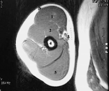
Anatomy of the elbow mr cross sectional imaging and 3d medical pictures this anatomy module deals with the radioanatomy of the elbow in mri and 3d reconstructions. Capitellum of the humerus with the ra. This mri elbow cross sectional anatomy tool is absolutely free to use.
Evaluation of the elbow by magnetic resonance imaging mri is an important adjunct to the physical examination. Injuries such as anterior cruciate ligament meniscus and rotator cuff tears are all easily diagnosed when there is a firm understanding and knowledge of human anatomy. Uq med year 1 gafradiographic anatomy upper limb.
It contains 260 mri slices 60 3d reconstruction images with 155 labelled anatomical structures. Knee shoulder shoulder arthrogram ankle elbow wrist hip contact. Use the mouse scroll wheel to move the images up and down alternatively use the tiny arrows on both side of the image to move the images.
 Mri Elbow Anatomy Dr Ahmed Eisawy
 Mri Scan Of The Elbow Showing Normal Neurovascular Anatomy
 Radiology Images
 Anatomy Of The Elbow Ct Arthrography
 Elbow Mri Mri Technique Gross Anatomy Tendinous
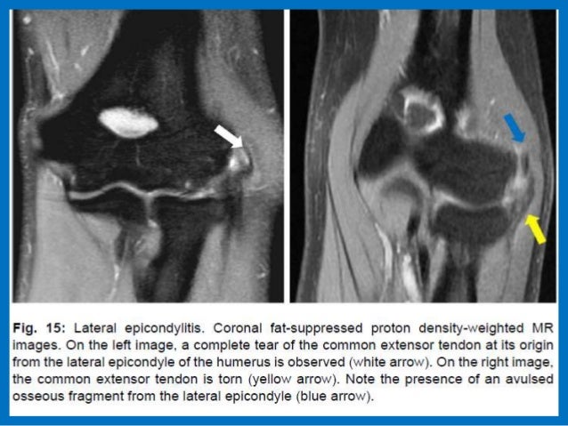
 Lateral Epicondylitis Tennis Elbow Shoulder Elbow
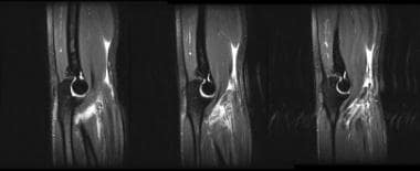
 The Radiology Assistant Elbow Mri
 Figure 11 From Magnetic Resonance Imaging Of The Elbow Part
 Mri Blog 12 09
 Knee Anatomy Mri Knee Coronal Anatomy Free Cross
 Elbow Mri

 Muscular Anatomy Of Upper Limb Mri Anatomy
 The Radiology Assistant Elbow Mri
 Normal Elbow Mri Radiology Case Radiopaedia Org
 Elbow Mri Mri Technique Gross Anatomy Tendinous
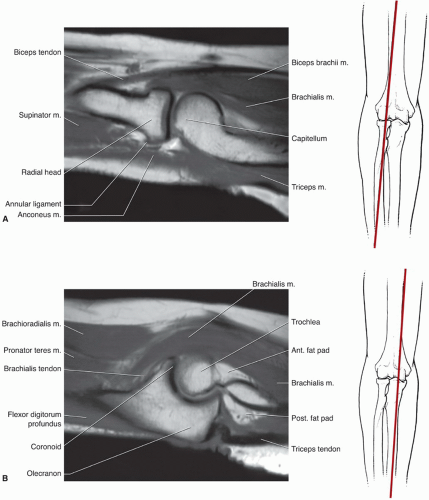
 Arun S Mri Protocols Elbow Mri Reference Lines
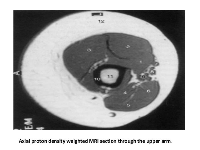
Arm Forearm And Hand Mri Of Anatomy
 Arm Forearm And Hand Mri Of Anatomy
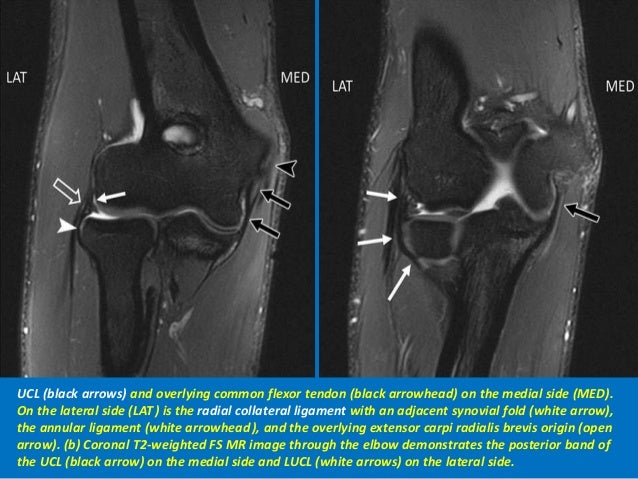
 Sonoanatomy Relevant For Ultrasound Guided Upper Extremity
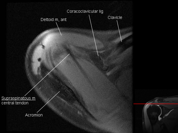 Mri Shoulder Arthrogram Anatomy
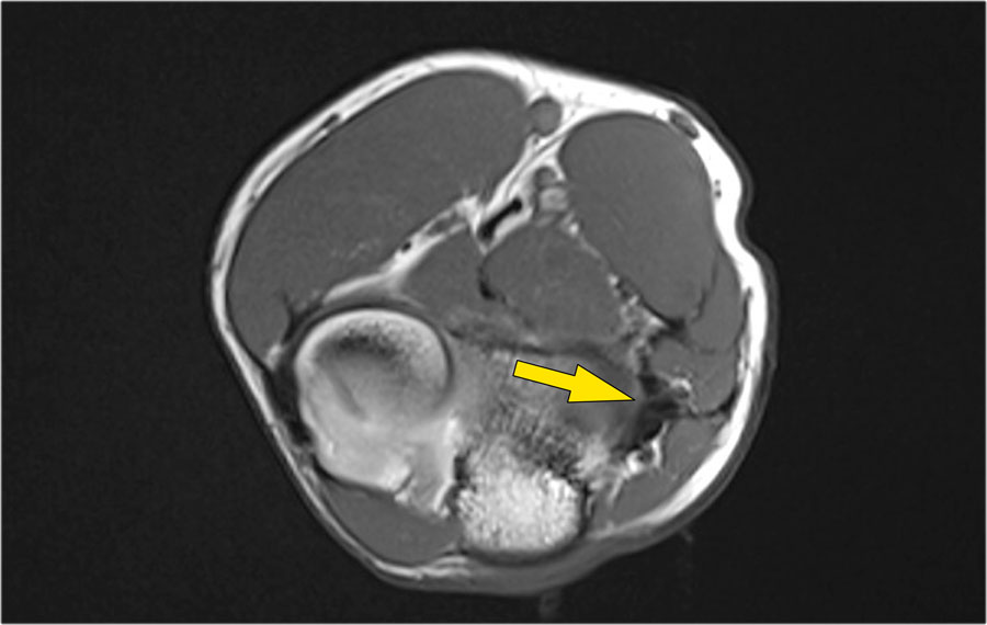
 Distal Biceps Avulsion Shoulder Elbow Orthobullets
 Elbow Mri Mri Technique Gross Anatomy Tendinous
 Presentation1 Pptx Mri Of Elbow Joint
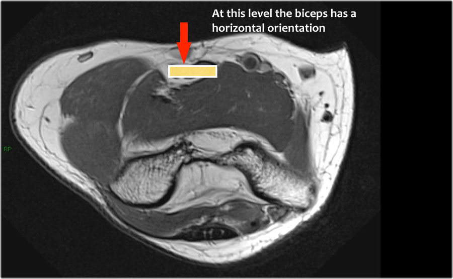
 Arm Forearm And Hand Mri Of Anatomy
 Arm Forearm And Hand Mri Of Anatomy
 Ecr 2018 C 0570 The Elbow Anatomy And Pathology Of
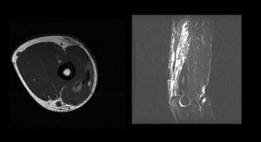
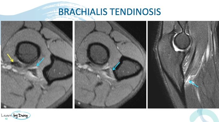 Mri Of The Brachialis Tendon Insertion Radedasia
 Elbow And Forearm Musculoskeletal Key


Posting Komentar
Posting Komentar