The anatomy of pregnancy and the fetus. The fetus is inside the membrane sac within the uterus and high within the abdomen.
 Details About Pregnant Woman Pelvis Section 4 Parts Anatomical Anatomy Model With Baby Fetus
A multiple pregnancy involves more than one offspring such as with twins.

Pregnant anatomy. As a pregnant woman or the partner of one you may be curious about the anatomy of a pregnancy the fetus and the special organs that keep it happy and healthy and connected to mom. Open the activity on the right to compare your body before pregnancy to your body at 37 weeks. The muscles of your abdomen support much of its weight.
Anatomy to better understand the changes your body goes through during the last trimester and labor it is helpful to be familiar with basic anatomy. One greys anatomy doctor is pregnant after the season 16 premiere. Examining the development of the baby in the womb and the parallel changes in the mothers body and structured to follow the process week by week the pregnant body book follows every anatomical and physiological change and tracks it in unprecedented detail.
The anatomy scan is a level 2 ultrasound which is typically performed on pregnant women between 18 and 22 weeks. Specially commissioned 3d artworks illustrations scans and photography show exactly how a baby changes and grows during pregnancy and how the female body adapts to carry it. Pregnancy can occur by sexual intercourse or assisted reproductive technology.
Preeclampsia generally develops late in pregnancy after week 20 and is characterized by a sudden onset of high blood pressure severe swelling of the hands and face and signs that some organs may not be working normally including protein in the urine. When a level 2 ultrasound is done. Have you ever wished that your belly had a little window so that you could watch how your baby grows and develops.
If youre considered high risk for preeclampsia ask your doc about low dose aspirin. Those who want to can find out the sex of the baby if desired. By the end of the 36th week of pregnancy your enlarged uterus almost fills the space within your abdomen.
Pregnancy also known as gestation is the time during which one or more offspring develops inside a woman. If you have a condition that needs to be monitored such as carrying multiples you may have more than one detailed ultrasound. Heres why you should have see the surprise coming and its full backstory.
Most anatomy scans are performed in the second trimester of pregnancy typically at 20 weeks but they can be done anytime between 18 weeks and 22 weeks. Medical anatomical pregnant human female pelvis with pregnancy 9 months baby fetus model life size with removable organs 4 parts hand painted 45 out of 5 stars 2 11255 112. Youll see how your body adjusts in amazing ways to support your growing baby.
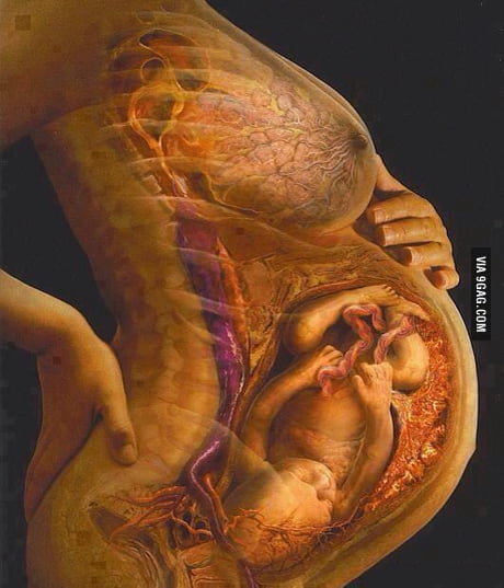 Anatomy Of Pregnant Women 9gag


Pregnant Womb Fetus Anatomy Cookie Cutter
 Anatomy Of A Pregnant Woman Vector Image 1863663
 Pregnancy Anatomy Illustration On Behance
 Pregnant Female Anatomy 3d Model 91535340 Pond5
 Pregnant Anatomy With Fetus Stock Photo Picture And Low

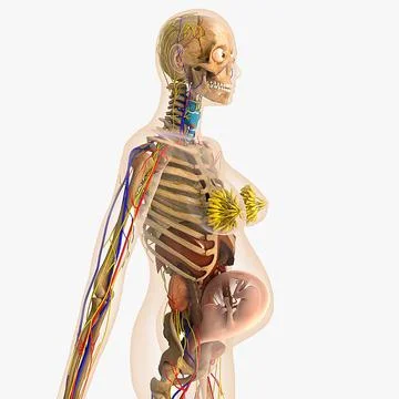
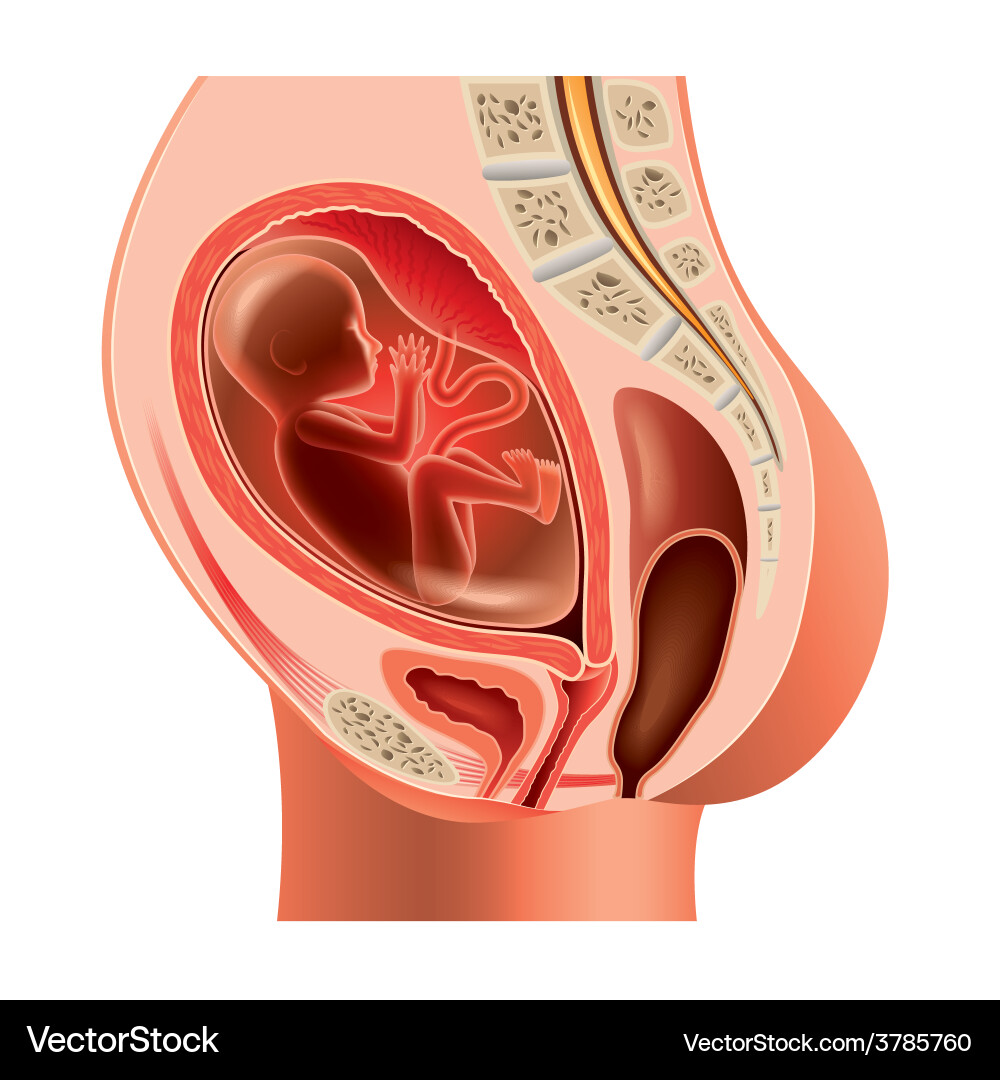
 37 Weeks Pregnant Anatomy Sectional View
 Stock Illustration
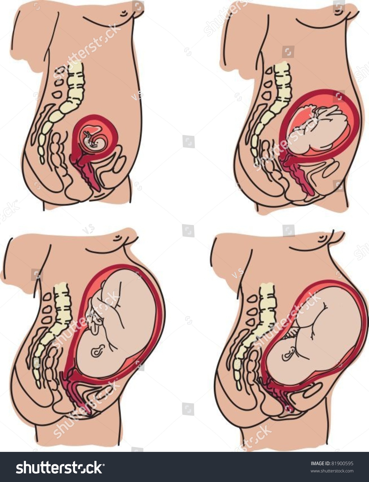 Pregnant Anatomy Fetus Stock Vector Royalty Free 81900595
 Grey S Anatomy Fans Slam Hypocrite Owen For Yelling At
 Normal Pregnant Female Anatomy Canvas Print
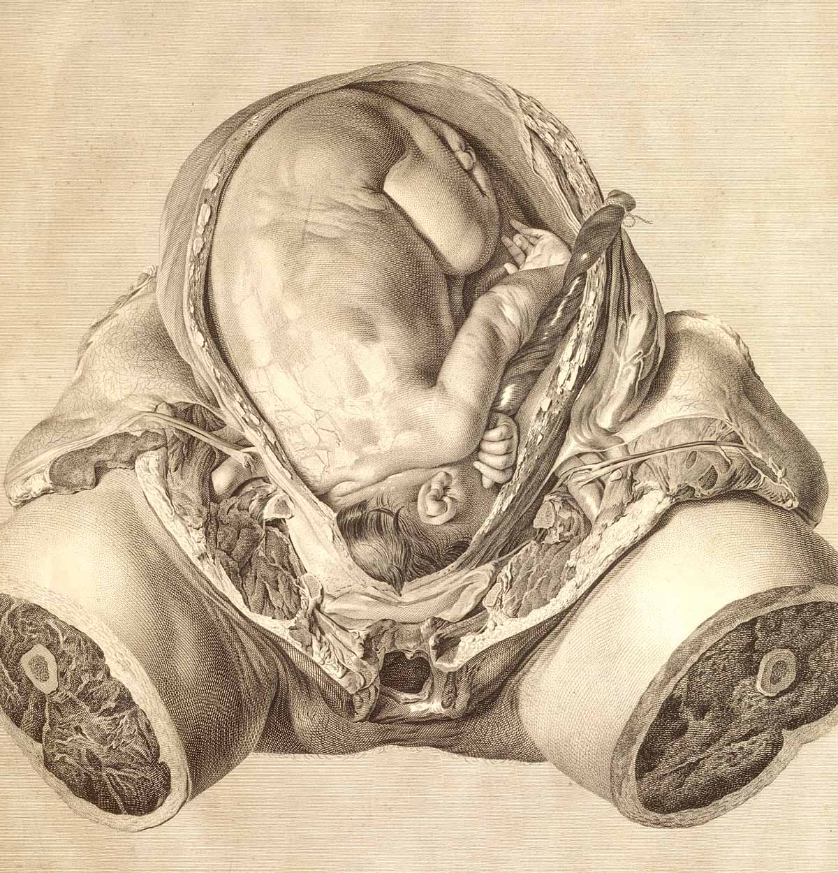 Dream Anatomy Gallery William Hunter And Jan Van Riemsdyk
 Pregnancy Anatomy
 Pregnancy And Birth Laminated Anatomy Chart
 Anatomy Of A Pregnant Person Birth Doula In Fredericksburg
 The Anatomy Of The Pregnant Woman Illustration From Fasciculus Medicinae By Johannes De Ketham
 Pregnant Woman Anatomy And Fetus Isolaed On White
 Amazon Com Ahawoso Outdoor Garden Flag 12x18 Inches
 Pregnancy Anatomy Nexj Health
 Pregnant Anatomy Belly Images Stock Photos Vectors
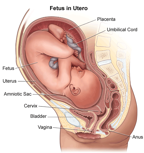 Anatomy Fetus In Utero
 Pregnant Anatomy Fetus Isolated

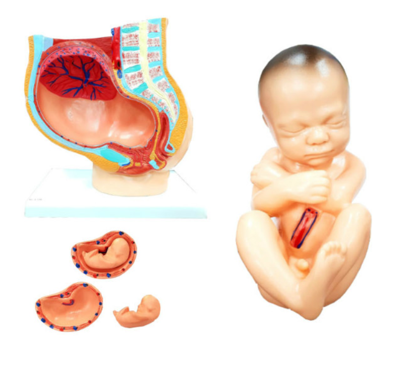 Pregnant Woman Pelvis Section 4 Parts Anatomical Anatomy


Posting Komentar
Posting Komentar