If you have a condition that needs to be monitored such as carrying multiples you may have more than one detailed ultrasound. Ultrasound means outside of the range of human hearing.
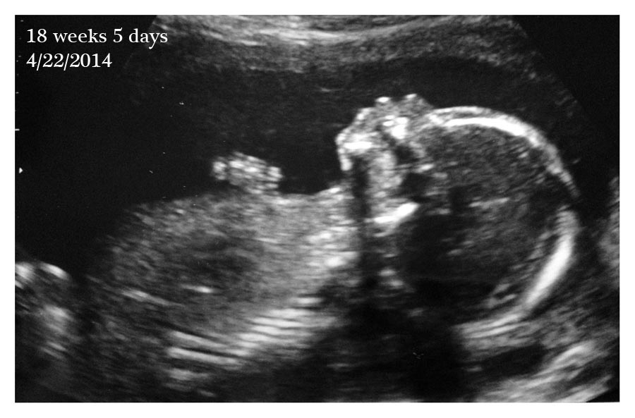
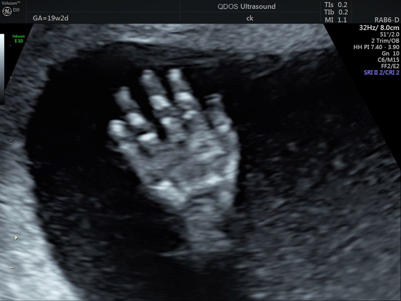
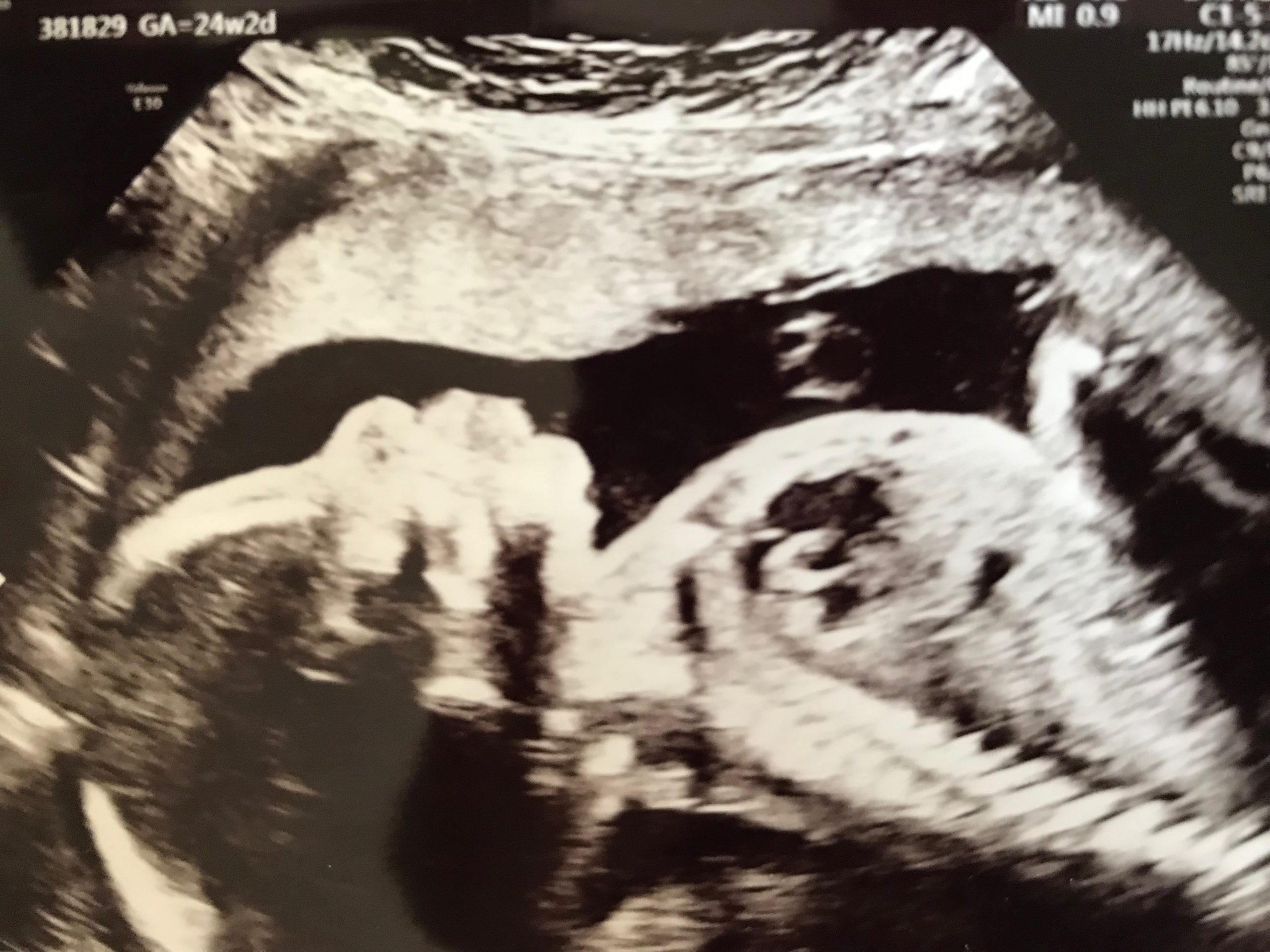
Most anatomy scans are performed in the second trimester of pregnancy typically at 20 weeks but they can be done anytime between 18 weeks and 22 weeks.
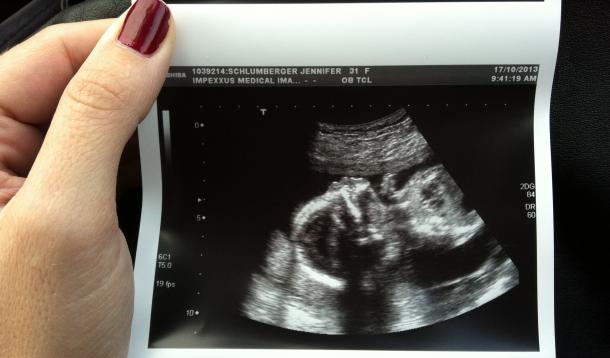
What is anatomy ultrasound. It became hugely important for me to have three sets of names going into the ultrasound so that we were. The anatomy scan is a level 2 ultrasound which is typically performed on pregnant women between 18 and 22 weeks. Like all routine medical procedures in pregnancy you can choose whether you want to have this ultrasound or not.
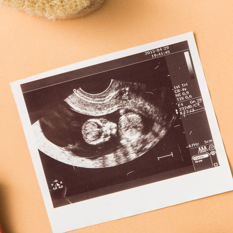
What is an anatomy ultrasound. Find out what youll see when you have yours. Description this is a detailed scan of your babybabies anatomy.
The 20 week ultrasound or anatomy scan is an eagerly anticipated ultrasound for parents. At this scan the ultrasound tech sonographer examines the baby from head to toe. The 20 week ultrasound also known as the anatomy scan is when a sonographer uses an ultrasound machine to.
Some wavelengths are intended to break up tissue but the wavelengths that we use for diagnostics imaging are not at all in the ranges that could cause significant tissue damage. When a level 2 ultrasound is done. The scan is performed transabdominally.
Check for physical abnormalities in baby check mamas uterus fluid levels and placenta. The anatomy or morphology ultrasound is a routine ultrasound that occurs between 18 and 22 weeks of pregnancy. In women at high risk for preterm delivery multiple pregnancies previous preterm birth abnormalities of the uterus or previous cervical surgery we may also carry out a transvaginal scan to measure the length of the cervix.
There are different wavelengths of ultrasounds. When the pregnancy hits the 20th week of gestation an anatomy ultrasound is often ordered. As the anatomy scan approached i began to build the what if scenarios up in my head.
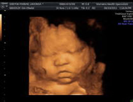
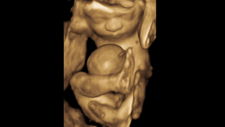
Those who want to can find out the sex of the baby if desired. This sonogram is used to determine fetal anomalies the babys size and weight and also to measure growth to ensure that the fetus is developing properly.
 High Resolution Fetal Ultrasound Children S Hospital Of
 A Gallery Of High Resolution Ultrasound Color Doppler 3d
 The Anatomy Ultrasound Everything You Should Know
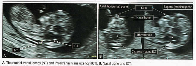
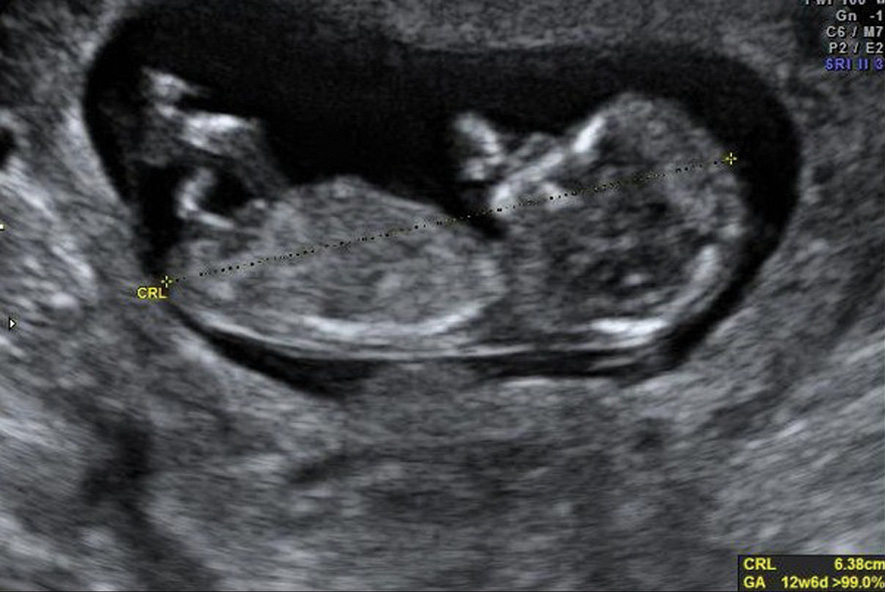 Ultrasound Assessment Of Normal Fetal Anatomy Chapter 5
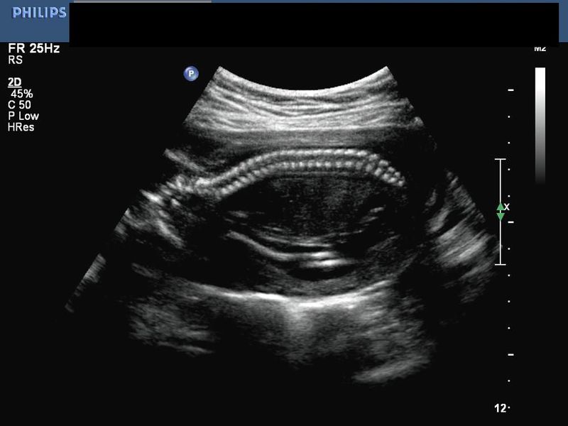
 Labelled Fetal Heart Ultrasound
 What To Ask At Your 20 Week Ultrasound Heart Savvy Momma
 Normal Obstetrical Ultrasound Obgyn Net
 Ultrasound Of Liver Segments Anatomy
 Normal Fetal Anatomy Ultrasound Services In Ernakulam
 The Anatomy Scan Ultrasound Is An Amazing Experience You Ll
 Abdominal Aortic Aneurysm Ultrasound Radiology Key
 It S A Boy Full Anatomy Ultrasound Of My Unborn Son


 Did You Find Out The Gender Of Your Baby Yummymummyclub Ca
 Pdf Optimizing Fetal Heart Screening At The Anatomy
 18 Week 3 Day Anatomy Ultrasound Chihuahuaesque Flickr
 19 Week Scan Perth Mid Trimester Ultrasound Pregnancy
 Violet S Story 20 Week Ultrasound Anatomy Scan Still

 Anatomy Scan And 4d Ultrasound In Japan Tiny Tot In Tokyo

Pregnancy Ultrasound Womens Health Specialists
 Halfway There Hobson Homestead
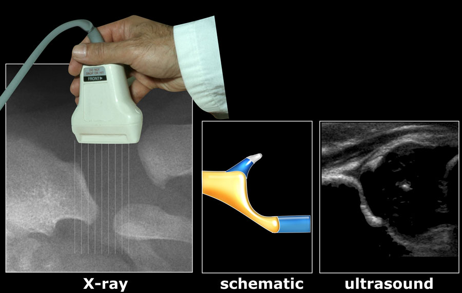 The Radiology Assistant Developmental Dysplasia Of The Hip
 20 Week Anatomy Ultrasound Youtube
 Aorta Images Emergency Ultrasonography
 Ultrasound Zygotta

 18 20 Week Anatomy Ultrasound Pictures Babycenter


Posting Komentar
Posting Komentar