The skin has two layers called the epidermis and the dermis. A person who is in shock may have pale skin and goose bumps and someone with a fever may feel warm to the touch.

A mole can be either subdermal under the skin or a pigmented growth on the skin formed mostly of a type of cell known as a melanocyte.

Mole anatomy. Anatomy of the skin. The skin also helps regulate body temperature gathers sensory information from the environment stores water fat and vitamin d and plays a role in the immune system protecting us from disease. It shields the body against heat light injury and infection.
A circumscribed stable malformation of the skin or sometimes the oral mucosa which is not due to external causes. Clinical anatomy for dummies. Warts may be treated at home with chemicals duct tape or freezing or removed by a physician.
Moles anatomy and physiology tuesday march 6 2012. When there is an irregular accumulation of melanocytes in the skin freckles appear. Beneath the two layers is a layer of subcutaneous fat which also protects your body and helps you adjust to outside temperatures.
A virus infects the skin and causes the skin to grow excessively creating a wart. You will get credit for the thoughtfulness of your response state an opinion and defend while noting something specific from one of the. Skin has two main layers.
The mole runs are in reality worm traps the mole sensing when a worm falls into the tunnel and quickly running along to kill and eat it. The high concentration of the bodys pigmenting agent melanin is responsible for their dark color. Because their saliva contains a toxin that can paralyze earthworms moles are able to store their still living prey for later consumption.
Formulate an opinion in response to the readings and post it. The dermis is the middle layer of the three layers of skin. The value of dissection read at least one opinion from each viewpoint from the links below.
Its located between the epidermis and the subcutaneous tissue. The excess or deficiency of tissue may involve epidermal connective tissue adnexal nervous or vascular elements. Moles are larger masses of melanocytes and although most are benign they should be monitored for changes that might indicate the presence of cancer.
It contains connective tissue blood capillaries oil and sweat glands nerve endings and hair follicles. The skin can be a good indicator of health. This tough layer of cells is the outermost layer of skin.
Skin has two main layers.
 Facts About Moles Live Science

 The Nose Takes A Starring Role Scientific American
 How Moles Destroy Your Lawn The Forelimb Kinematics Of
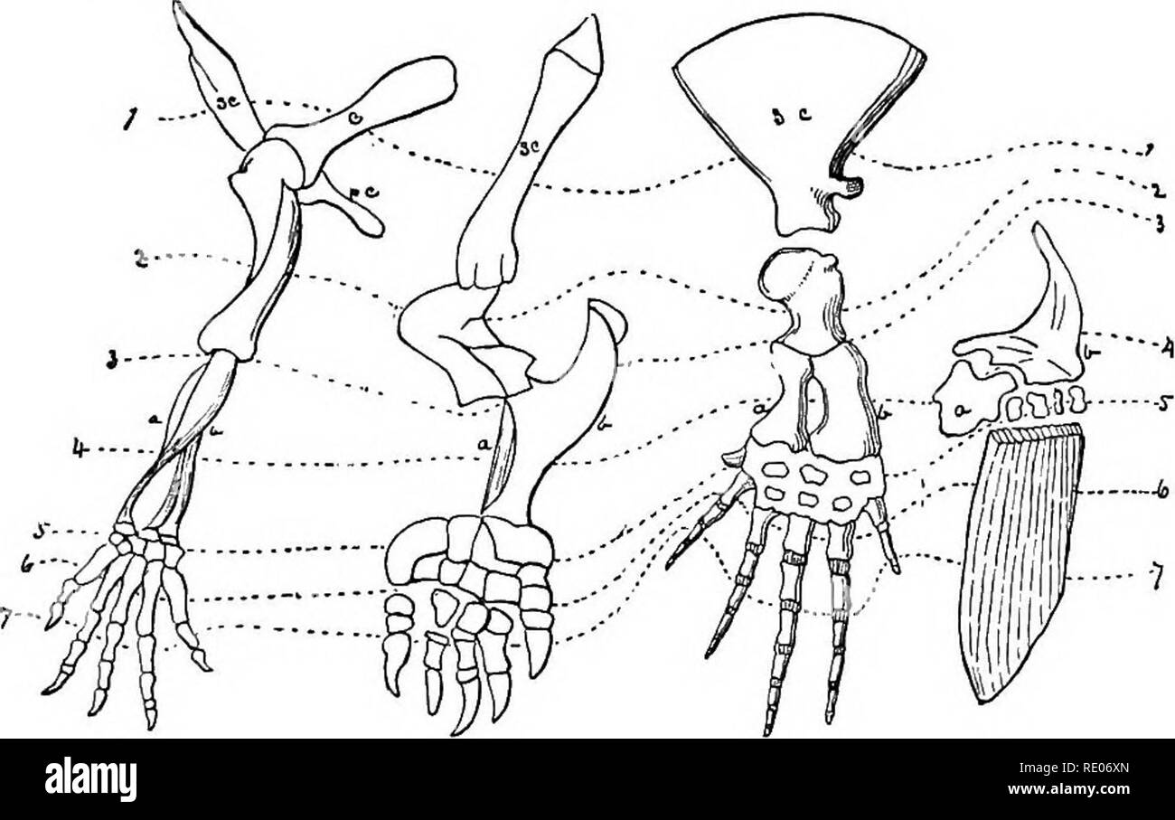
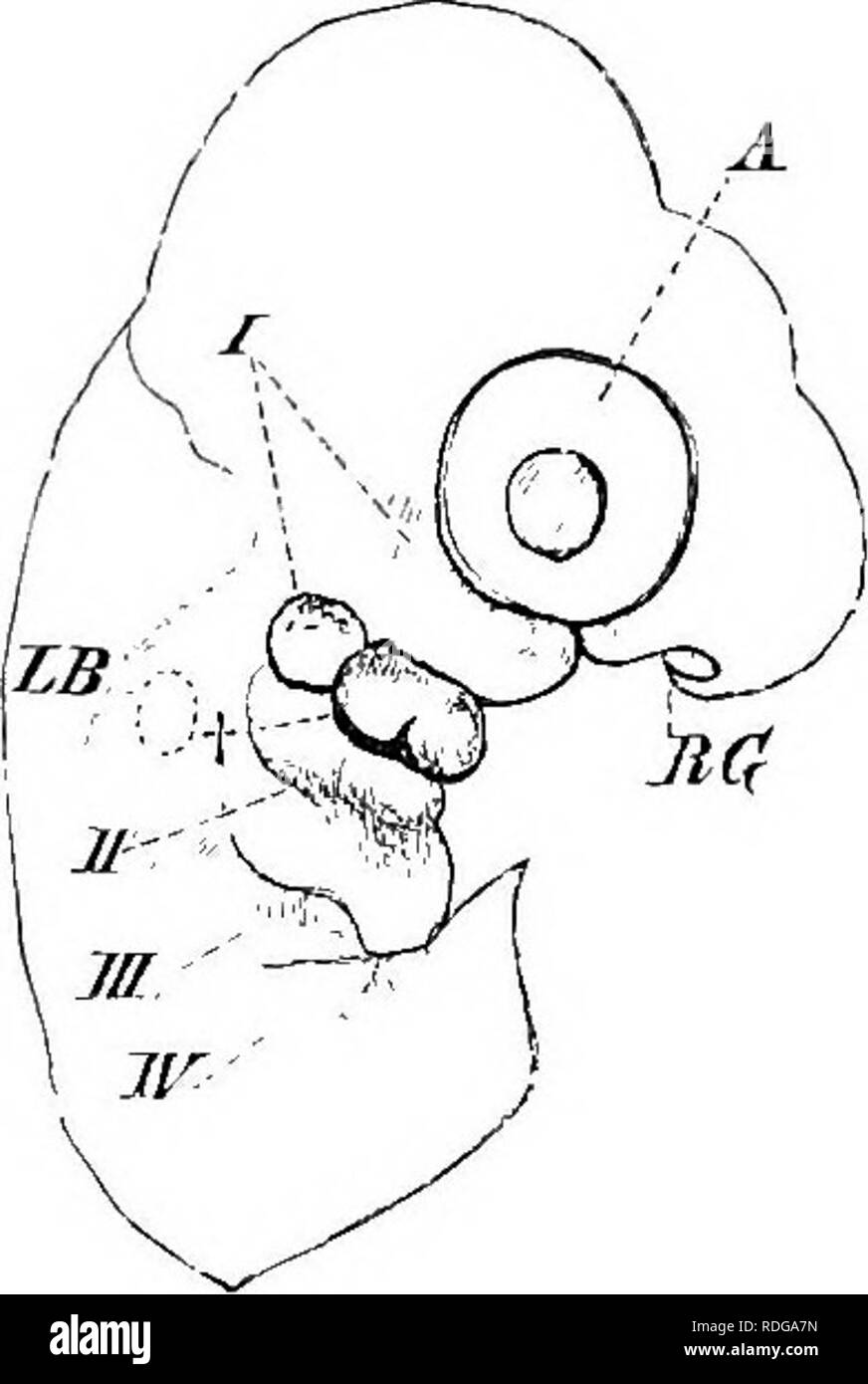
 Outlines Of The Comparative Physiology And Morphology Of
 Skin Mole Anatomy Stock Photos Page 1 Masterfile
 Anatomy Of The Reproductive Organs In A Pregnant
 Inside The Bizarre Life Of The Star Nosed Mole World S
 Anatomy Of Mole External Genitalia Setting The Record
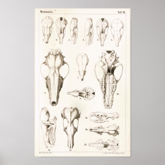
 Mole And Shrew Skulls Veterinary Anatomy Print
Star Nosed Mole Form And Function
 Differences Between Moles Voles Shrews Ehrlich Pest Control
 Scientists Shed Light On Eyesight Of Moles News The
 Skin Cancer Anatomy Headandneckcancerguide Org
 Elements Of The Comparative Anatomy Of Vertebrates Anatomy
 Anatomy Of Mole External Genitalia Setting The Record
 Extreme Anatomy Macro Close Up Of Single Brown Mole On Surface
 Amazon Com Mole W Muscle Anatomy 1767 Scarce Original
 Animal Woodland Mole Anatomy Printable Animals
 The Unusual Anatomy Of The Naked Mole Rat H Glaber A

 Comparison Of The Facial Anatomy And Trigeminal Sensory
 5 Tips For Checking Your Moles For Skin Cancer
Moles Laguna Hills Saddleback Valley Orange County Ca
 Hermit Crab Anatomy Squats Mole Anatomy
Mole Printout Enchantedlearning Com


Posting Komentar
Posting Komentar