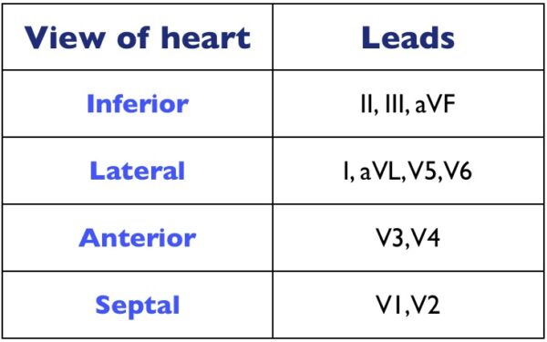
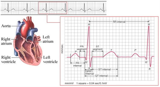
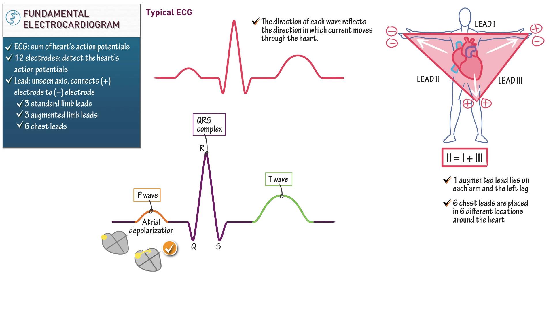
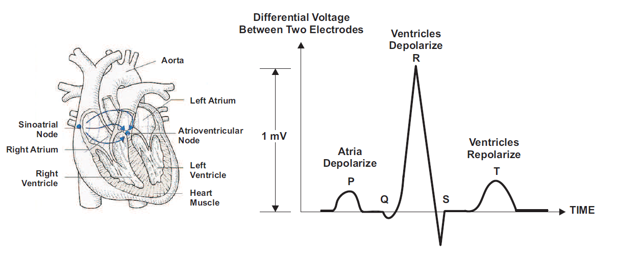
Lead refers to an imaginary line between two ecg electrodes. What is happening when the ventricles are filling.
 The Basics Of Ecg Interpretation Part 1 Anatomy And
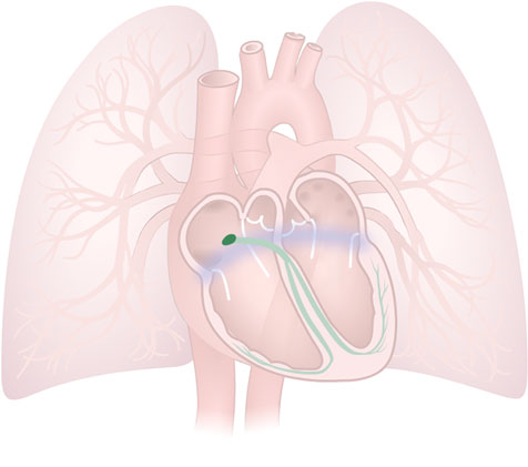
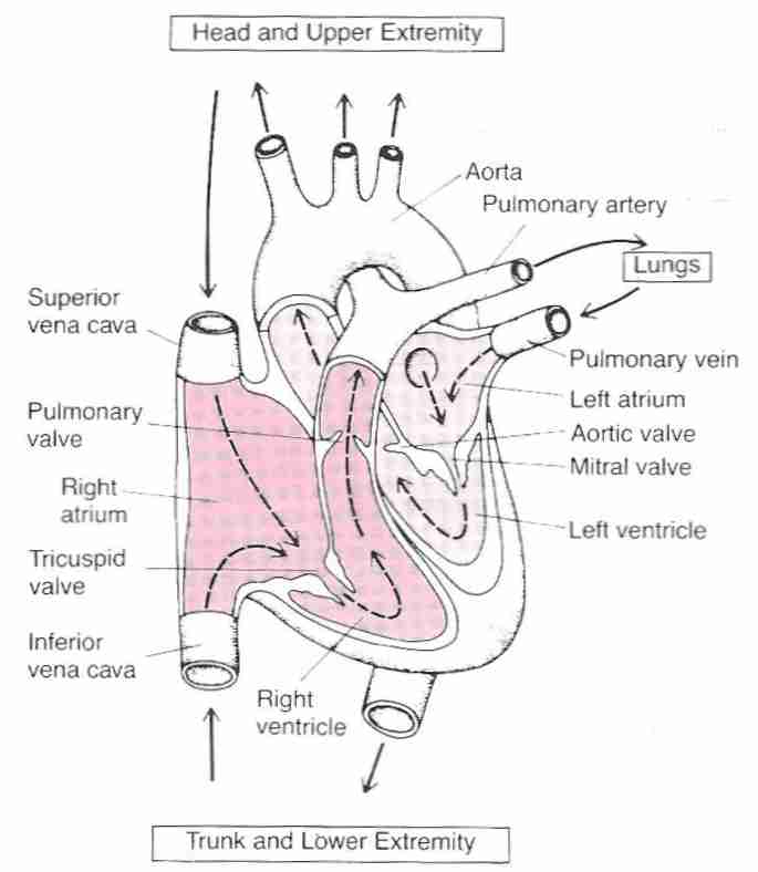
Focus topic heart anatomy and physiology the human heart is a hollow cone shaped muscular organ roughly the size of its owners fist that weighs approximately 9 to 12 oz 250 350 g size of the human heart.

Ecg anatomy. Basic cardiac anatomy anatomy of the heart introduction to electrocardiology and ecg interpretation cardiac electrophysiology. Ecg exigency and cardiovascular curveball ecg clinical cases. A quivering ventricular muscle that is unable to pump blood.
Ecg a to z by diagnosis ecg interpretation in clinical context. A double membrane that covers the outside of the heart. The 12 lead ecg.
Abnormal conduction may be apparent. A smooth layer of cells that lines the inside of the heart b. Ecg library basics waves intervals segments and clinical interpretation.
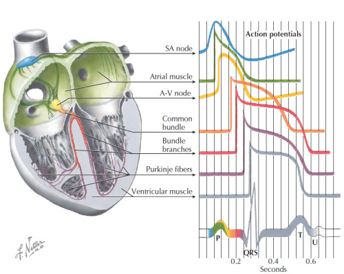
Anatomy of the heart ecg. What is an ecg. This electrical activity controls the heart beat.
Action potentials and electrical vectors. Ecg reference sites and books the best of the rest. The ecg measures the electrical activity of the heart.
Most often the ecg assessment includes the following. Thick muscular wall that separates the heart into right and l. There are 12.
These electrodes allow leads to be calculated. 100 ecg quiz self assessment tool for examination practice. Heart muscle that is irritated conducts electricity differently than heart muscle that is normal.
The letters ecg stand for electro cardiogram. Determination of the rate assessment of the rhythm evaluation of the electrical conduction patterns. An ecg is recorded by placing electrodes on the surface of the skin.
Middle layer of the heart that is made of muscle tissue. How it all works electrodes. The electrodes are wires that you attach to the patient to record the ecg.
The electrocardiogram or ecg is a simple diagnostic test which records the electrical activity of the heart over a set time period via this exam tips post helps us to understand the anatomy and physiology behind the ecg and how to interpret it.
 048 How To Read An Electrocardiogram Ecg Ekg
 Understanding An Ecg Geeky Medics
 Mi Localization Litfl Medical Blog Ecg Anatomy Basics
 Anatomy Physiology Online Cardiac Conduction System And Its Relationship With Ecg
 All About Ecg

 Heart Anatomy And Ecg Background

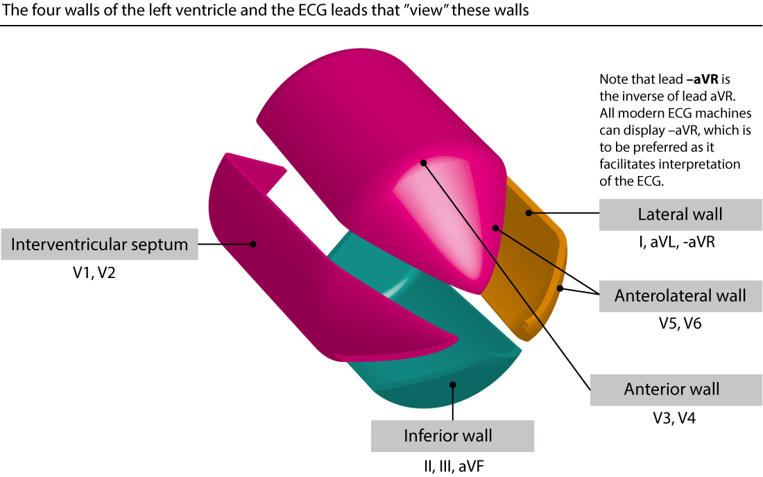
 Ecg Copy Ecg Anatomy Of The Heart The Walls Of The Heart
 Partonomy Of Anatomy For The Ecg The Lines Represent Part
 Wolff Parkinson White Syndrome Part 1 Ecg Medical Training
 Ecg Research
Schaum S Outline Of Ecg Interpretation
 Ecg Interpretation Nursing Cardiac Anatomy Physiology
 Introduction To Pediatric Neonatal Ecg Interpretation
 Ecg Localization Of Myocardial Infarction Ischemia And
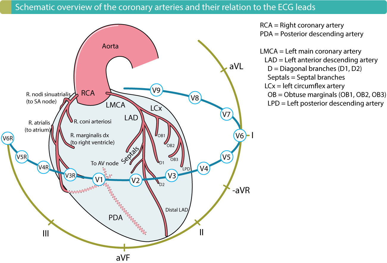
 Coronary Anatomy And Corresponding Ecg Leads Chalk Talk
 Cv Physiology Electrocardiogram Ekg Ecg
 Heart Anatomy Ecg Kaap Ppt Video Online Download
 19 2 Cardiac Muscle And Electrical Activity Anatomy And
 Electrocardiogram Ecg Ekg Definition Readings Procedure
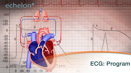 Online Courses Echelon
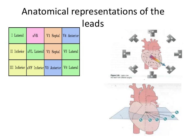
 Ekg Leads And Where They Represent You Can Understand A Lot
 Introduction To Ecg
 Electrocardiograph Ecg Ekg Interpretaion
 Smart Sensing With Ultra Low Power Mcus Part 4 Holter
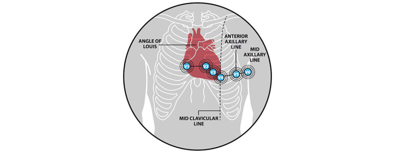
 The Ecg Leads Electrodes Limb Leads Chest Precordial
 Cv Physiology Electrocardiogram Ekg Ecg
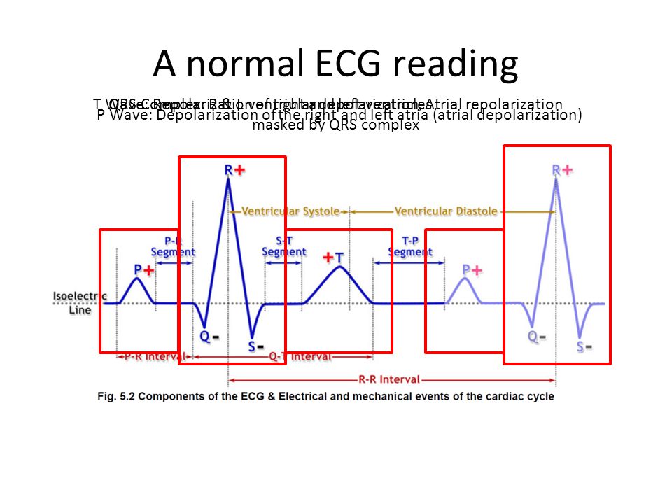
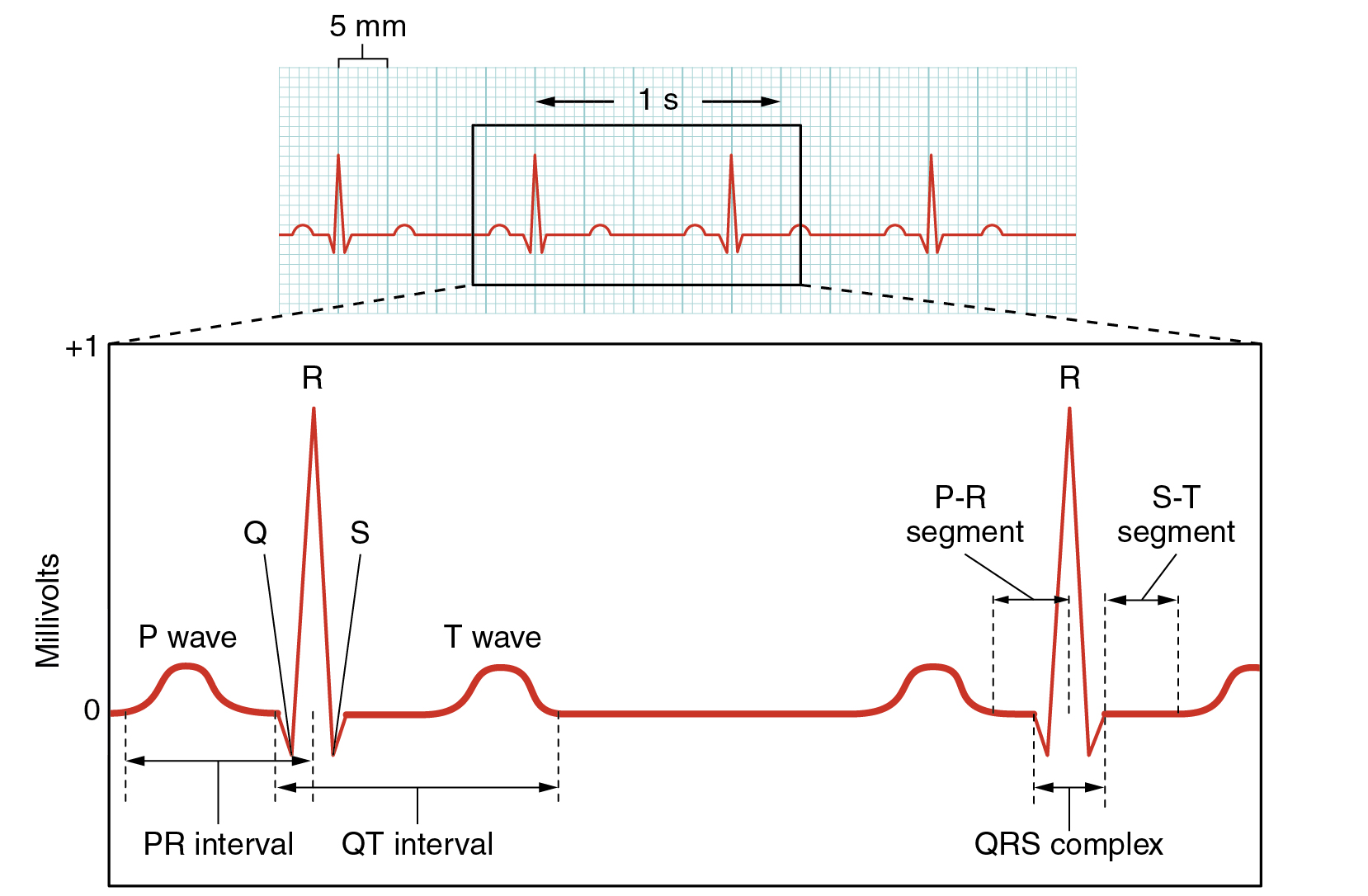
 Electrocardiogram Ekg Components And Intervals Healthlink Bc
 Electrocardiography Wikipedia


Posting Komentar
Posting Komentar