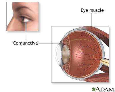
Glasses contact lenses or surgery can correct the blurry vision it causes. The choroid continues at the front of the eyeball to form the ciliary body.
 Superior Rectus Muscle An Overview Sciencedirect Topics
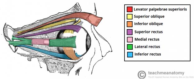
There are six eye muscles that control eye movement.

Eye anatomy muscles. Eye muscle anatomy there are six extraocular muscles that move the globe eyeball. Nerve signals that contain visual information are transmitted through the optic nerve to the brain. The two oblique muscles of the eye are responsible for the rotation of the eye and assist the rectus muscles in their movements.
Its antagonist is the lateral rectus muscle that abducts the eye allowing it to look laterally or away from the bodys midline. Muscles of eye movement. The extraocular muscles are the six muscles that control movement of the eye and one muscle that controls eyelid elevation levator palpebrae.
The cilliary muscles are located inside the ciliary body. The eyelids are soft tissue structures that cover and protect. These muscles are named the superior rectus inferior rectus lateral rectus medial rectus superior oblique and inferior oblique.
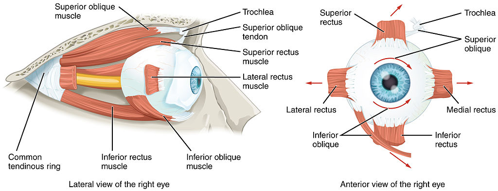
Eye anatomy bones of the orbit. One muscle moves the eye to the right and one muscle moves the eye to the left. The actions of the six muscles responsible for eye movement depend on the position of the eye at the time of muscle contraction.
A problem with the curve of your cornea. There are four recti muscles. Extraocular muscles help move the eye in different directions.
The lacrimal gland is a part of the lacrimal apparatus. If you have it your eye cant focus light onto the retina the way it should. Six extraocular muscles.
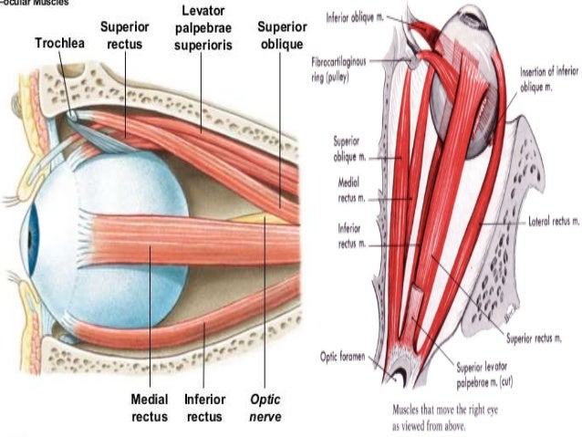
The bony orbit is made out of seven bones which include the maxilla. The four recti muscles and the two oblique muscles. The superior oblique muscle rotates the eye medially and abducts it when the eye if facing forward while the inferior oblique rotates the eye laterally and adducts it.
They can be divided into two groups. These muscles characteristically originate from the common tendinous ring. The other four muscles move the eye up down and at an angle.
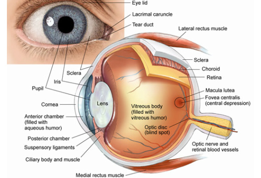
See diagram anatomy of the eye above. The eye is surrounded by the orbital bones and is cushioned by pads of fat within the orbital socket. Superior rectus inferior rectus medial rectus and lateral rectus.
These are the muscles that continuously change the shape of the lens for near and distant vision. There are six muscles involved in the control of the eyeball itself. Swelling and discoloration bruise around your eye caused by an injury to the face.
Anatomy of the eye.
 The Eye Teachmeanatomy
 Orbits And Eyes Anatomical Illustrations
 Eye And Extraocular Muscles Illustrations Radiology Case

 Extra Ocular Muscles Anatomy
 Eye Anatomy And Function
 Extrinsic Eye Muscles Diagram Quizlet
 Eye Anatomy Blood Supply Orbit Extraocular Muscles
 Learn How Your Eye Works And Why Eye Muscles Is The Missing
 Eye Anatomy And Structure Muscles Nerves And Blood Vessels
 Anatomy Of The Eye American Association For Pediatric
 Human Eye Ball Anatomy Physiology Diagram
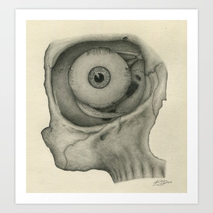 Anatomy Muscles Of The Eye Art Print By Hoffmananatomy
 Eye Opener Anatomy Muscles Of The Eye
 Eye Muscle Repair Series Normal Anatomy Medlineplus
 Anatomy Of The Human Eye A Superior View Of The Human Eye
 Rectus Muscle Anatomy Britannica


 Anatomy Of The Eye American Association For Pediatric
 Extra Ocular Muscles Recti Acland S Video Atlas Of Human
 Extraocular Muscles Pic An0002
 Human Eye Anatomy Diagram Stock Vector Illustration Of
What Are The 6 Extrinsic Muscles Of The Eye And Their
 Lateral Rectus Muscle An Overview Sciencedirect Topics


Posting Komentar
Posting Komentar